Archives of Hepatitis Research
Prevalence of hepatitis B virus infection among persons with hepatitis D virus and diabetes mellitus in Pakistan, 2019-2021
Ahmad Raza1, Muhammad Waqar Mazhar2*, Saira Saif2, Shanzab Noor3, Mudasara Sikandar2,Iram Shahzadi4, Wajeeha Iram2, Hira Tahir4 and Fatima Mazhar5
2Department of Bioinformatics and Biotechnology, Government College University, 38000Faisalabad, Pakistan
3Department of Physiology, Government College University, 38000Faisalabad, Pakistan
4University of Health Sciences, Lahore, Pakistan
5Department of Microbiology, Muhammad Nawaz Sharif University of Agriculture, Multan, Pakistan
Cite this as
Raza A, Mazhar MW, Saif S, Sikandar M, Mehmood J, et al. (2022) Prevalence of hepatitis B virus infection among persons with hepatitis D virus and diabetes mellitus in Pakistan, 2019-2021. Arch Hepat Res 8(1): 001-004. DOI: 10.17352/ahr.000031Copyright License
© 2022 Raza A, et al. This is an open-access article distributed under the terms of the Creative Commons Attribution License, which permits unrestricted use, distribution, and reproduction in any medium, provided the original author and source are credited.Introduction: The HBV virus has its enveloped protein that surrounds nucleic acid for its protection. Hepatitis B DNA virus belongs to the Hepadnoviridae family and is similar to retroviruses. Globally, HBV infected people approximately 2 billion in the world, about 350 million people were chronic carriers. One million deaths are caused by the HBV virus every year. Each year, 400,000 new cases were reported in Latin America. Among all over the World, Pakistan was considered as one of the largest chronic viral hepatitis infection countries.
Methodology: 975 samples were recoded from different districts of south Punjab. The sera of the patient’s sample were examined to analyze the LFTs, HBS serology, HBS ELIZA, anti-HBeAg, AFP, anti- HDV, HBC IgM, and HBV DNA EAL TIME PCR. An anticoagulant sample was used to analyze their prothrombin time, HB level, WBC count, PLT counts, and HBA1C. Data were analyzed by using Microsoft Excel 2019 and SPSS version 14.0.
Results: 975 samples were collected from the Multan district of Punjab. Out of which 638(65.43%) patients were detected positive for HBsAg serology, 312(32.84%) were not detected. The HBA1C test values are higher in HBV patients and its normal value is below 6.4%. In 1st group 258 patients out of 638 are HBV PCR DETECTED, bilirubin 2.1+-5.7, ALP 425.1±575.1, AST 119.6±102.6, ALT 62.0±41.6, and albumin 3.5±0.9. in the 2nd group, 149 patients out of 638 are HBV PCR detected, bilirubin 1.4±1.9, ALP 488.1±339.2, AST 143.0±117.4, ALT 78.1±53.4, and albumin 4.4±0.6.
Conclusion: The prevalence of HBV in diabetic patients is higher as compared to control diabetic patients. the patients with high serum AFP levels tend to be HBs-Ag-positive, and those who have low AFP levels are associated with cryptogenic cirrhosis. The positive HBc IgM is higher in anti-HDV-negative cases as compared to HDV-positive cases. The value of HBV DNA was higher in anti-HDV-negative cases. The HBsAg correlates with HBV DNA level and it’s a level higher in the IT phase and lowers in the IC phase.
Introduction
The inflammation of the liver is caused by the hepatitis B virus [1]. Hepatitis B is classified into two main categories acute and chronic hepatitis. The infection of acute hepatitis B was a short term that lasts for the first 6 months after the infection of the virus and symptoms occurred within 3 months. Chronic hepatitis B takes years to show symptoms and refers to lifelong infection [2,3]. Hepatitis B virus caused acute as well as chronic hepatitis in the world. The HBV virus has its enveloped protein that surrounds nucleic acid for its protection. Hepatitis B DNA virus belongs to the Hepadnoviridae family and is similar to retroviruses [4].
Globally, HBV infected people approximately two billion in the world, about 350 million people were chronic carriers. One million deaths are caused by the HBV virus every year. Each year, 400,000 new cases were reported in Latin America, and 10 to 70% of them evolve to hepatocellular carcinoma [5]. The environment was divided into high, intermediate, and low endemicity areas on the basis of HBV infection epidemiology. The infections rate higher than 8% are high, 2% to 8% are intermediate and lower than 2% are lower endemicity areas. Among all over the World, Pakistan was considered as one of the largest chronic viral hepatitis infection countries [6].
The natural course of HBV is divided into three phases: immune tolerant, inflammatory, and HBeAg seroconversion phases. The infected patients re-enter into the hepatitis reactivation, which leads towards cirrhosis and human hepatocellular carcinoma [3,7].
A subviral agent known as Hepatitis D Virus (HDV) requires a preexisting infection with HBV. The HDV has circular single-stranded RNA consisting of 1672 to 1697 nucleotides with extensive intramolecular complementarity [8]. Globally 18 million people are affected by HDV and its mostly found in western Asia, Europe, and Italy [9]. In recent studies, Turkey, India, Australia, China, and Japan had more prevalence rates in past years [10]. The HDV infection with the HBV was associated with a higher rate of fulminant hepatitis as compared to HBV infection. The individuals with chronic HBV infection and HDV superinfection caused severe progressive chronic liver disease [6]. Diabetes Mellitus causes hyperglycemia in patients and insulin secretions to become low. Diabetes Mellitus caused complications such as cardiovascular disease, neurology, retinopathy, and renal failure. The number of patients with Diabetes Mellitus is markedly increasing every year and the prevalence of HBV virus was higher in the diabetic patient as compared to control patients [11].
During embryonic life, within the first few weeks, the Alpha Feto-Protein becomes low and reaches lower than 1mg/ml. Serum AFP is elevated in some disease states, AFP screening has been suggested when the tumor becomes redactable. In non-malignant forms of liver AFP is elevated in acute chronic hepatitis, and cirrhosis [12]. The absolute level of AFP is examined to distinguish between benign and malignant causes of elevation [13].
The main reason for HBV virus transmission is through blood, blood fluids, unsterilized needles, used syringes, birth, and sexual contact [3,14]. HBV showed symptoms such as fever, fatigue, upset stomach, lack of appetite, change in urine color, pale eyes and skin, and grey color stool [3,15]. Liver damage is diagnosed by blood tests, liver biopsy, or liver ultrasound. There is no specific treatment to cure acute hepatitis. But, can be cured by using some fluids, healthy and proper nutrition, and doing rest. Hospitalization is required in some cases. Interferon, liver transplants, and antiviral medications are used for the treatment of the HBV virus. These treatments prevent the effects of temperature or can slow down [16].
Methodology
975 samples were collected from the district of south Punjab from October 2020 to February 2021. Patient information was recorded including name, age, and sex. Collected 5ml venous blood from the patient by using disposable syringes, transferred into vials containing coagulant and anticoagulant, centrifuged and plasma was separated from red blood cells for further analysis.
The serum was used for serology test, LFTs by semi-auto chemistry analyzer. Added 800ul R1 solution in the test tube. Added 200ul R2 solution in R1solution test tube and kept at R/T for 5 minutes. After this added 100ul plasma in the test tube and run on Mindray 2600DR.
Serum HBsAg levels were quantified by using HBsAg assay, based on chemiluminescent microparticle immunoassay according to manufacturer protocol. The detection ranges from 0.05-250IU/ml, and the higher value of HBsAg requires dilution.
HBA1C tests were performed on Indiko (Fully Automated) Clinical Chemistry Analyzer by Thermo Scientific; US-FDA Approved. were detected by radioimmunoassay. Anti HDV, HBV ELIZA, anti-HBe, and AFP level tests were formed on enzyme-linked immunosorbent assay. After HBV chromatography, we performed an HBV enzyme-linked immunosorbent assay and then move towards HBV REAL-TIME PCR QUANTITATIVE. The plasma of sample was analyzed for HBV virus by Real-time PCR using an HBV Quantification kit. The addition of probes in the master mixture emit light and quantified the viral load. The detection limit ranged from 30 to 1.7×10⁸IU/mL for HBV PCR. The viral load of samples with HBV DNA <30IU/ml was “undetected”.
Data were analyzed by using Microsoft Excel 2019 and SPSS version 14.0.
Results
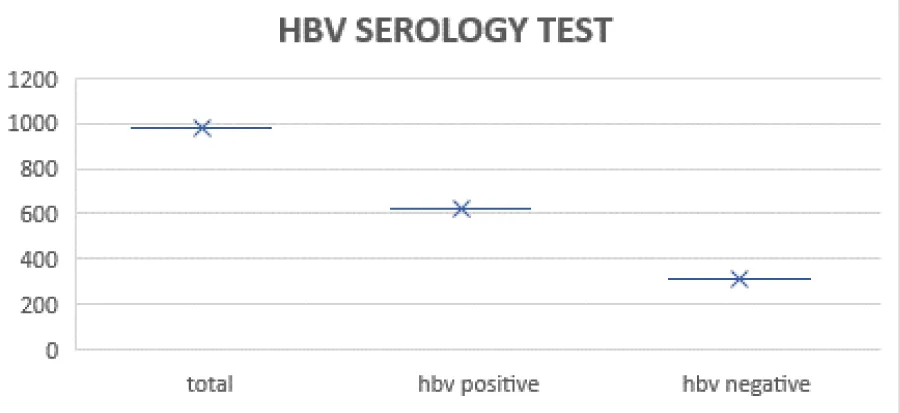
975 samples were collected from the Multan district of Punjab. Out of which 638(65.43%) patients were detected positive for HBsAg serology, 312 (32.84%) were not detected in Figure 1.
The lower limit of detection is 30IU/ml, with 100% probability. The Applied Biosystems (Step One™ and Step OnePlus™ Real-Time PCR Systems) assay detect quantitates and genotypes. The sensitivity of HBV DNA REAL-TIME PCR is 30IU/ml. Out of 975 samples, 449(46.05) were male and 526(53.94%) were female. Out of 638 samples 407(63.79%) viral load greater than 30IU/ml, the result was detected for those patients who have higher viral load than their sensitivity. positive and negative viral load mentioned in Figure 2.
The further analysis with 975 patients (Table 1). In 1st group 258 patients out of 638 are HBV PCR DETECTED, bilirubin 2.1±5.7, ALP 425.1±575.1, AST 119.6±102.6, ALT 62.0±41.6, and albumin 3.5±0.9. in 2nd group 149 patients out of 638 are HBV PCR DETECTED, bilirubin 1.4±1.9, ALP 488.1±339.2, AST 143.0±117.4, ALT 78.1±53.4 and albumin 4.4±0.6. the results show that the patients with high serum AFP levels tend to be HBs-Ag-positive, the other hand those who have low AFP levels are associated with cryptogenic cirrhosis.
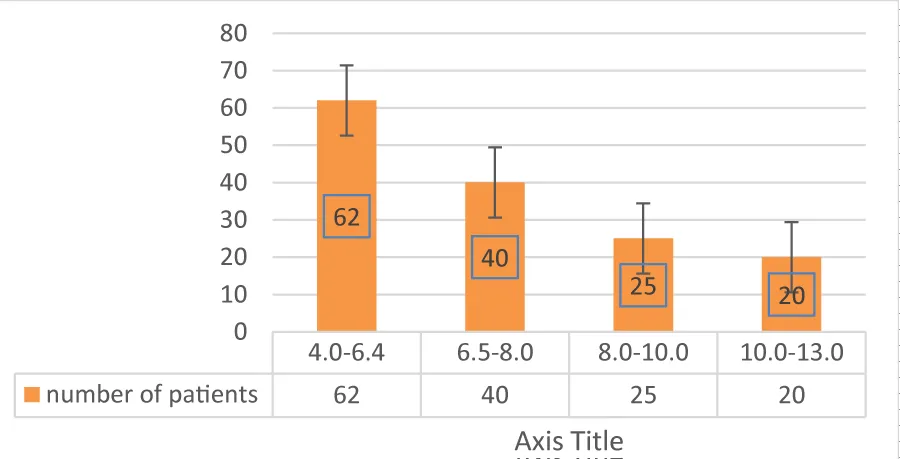
The graph shows that the HBA1C test values are higher in HBV patients and its normal value is below 6.4%. The prevalence of HBV in diabetic patients is higher as compared to control diabetic patients in Figure 3.
253 patients (90.35%) were negative for HBeAg and (73.9%) HBV DNA PCR positive cases. In anti HDV negative cases, the HBc IgM was higher as compared to HDV positive (13.68%) versus (1.78%). In anti HDV cases the value of HBV DNA was higher see in Table 2.
HBsAg and HBV DNA level in phases of chronic hepatitis B
the HBsAg and HBV DNA levels varied significantly between the patients in different phases of CHB. In different phases of CHB the HBsAg and HBV DNA level significantly different. In the immune tolerance group, the highest and lowest mean values are observed. The highest mean value was (HBV DNA: 7.41±01.01 and HBsAg: 4.07±0.83) and in the IC group the lowest was observed was (HBV DNA: 1.88±0.78 log and HBsAg: 1.98±1.42). the calculations observed in log IU/ ml units. The HBV and HBsAg shows similar variation in every phase. Higher to lowest in these order, IT group>EPH >ENH >IC group. The lowest mean value observed in IC group Figure 4.
Discussion
There are certain limitations that were determined by the present meta- analysis despite prominent efforts to test the correlation between the Diabetes Mellitus prevalence and HBV infection. Firstly, a serum marker was used to diagnose the HBV infection. These markers include the HBsAg, anti-HBc, HBV DNA or all these marker combinations in serum that make it impossible to distinguish between ongoing and past infection [17]. Moreover, hepatitis severity rate was not well known; only a few studies had indicated aminotransferase level. Secondly, meta-study involved three different studies: Case-control, cross-sectional and cohort studies. All these various types of study analysis have been partially responsible for heterogeneity [18].
Furthermore, there is no evidence provided by the two cohort studies for the association between HBV infection and Diabetes Mellitus prevalence. Thirdly, population selection for control was divided into two major types. In some studies, hospitalized patients that were not affected with HBV infection were taken as control while in other studies the healthy population was taken as control [19].
Therefore, it is concluded by the present studies that HBV infected patients have higher rate of emerging Diabetes Mellitus prevalence as compared to uninfected patients’ due heterogeneity across studies and meta-analysis studies limitations. In our study we found that the HBV prevalence in diabetic patients higher than nondiabetic.
We examined the correlation of Serum AFP level and patient’s age. Higher AFP level observed in young patients, secreting AFP in some cases. Frequently, the serum AFP levels has been higher in younger age as compared to older ones. The inverse correlation measured between AFP and patients and confirmed by universe analysis [20]. The previous study tells that younger patient linked with chronic HBV infection and generally present high HCC at more advanced stage [21]. So, it is concluded that not only age of the patients but there is also involvement of the age- related conditions, tumor infection and chronic HBV infection that are involved in an increased AFP level in HCC patients. HBV infection and inflammation not indicate by HBV DNA level.
In this study, the HBsAg levels are different during different stages of chronic hepatitis B, in the Immune Tolerant phase the level of HBsAg is higher and the lower in Internal Control phase. The HBsAg level correlates with HBV DNA levels especially in immune tolerant, HBeAg-negative hepatitis and Internal control phase.
- Mann J, Roberts M (2011) Modelling the epidemiology of hepatitis B in New Zealand. J Theor Biol 269: 266-272. Link: https://bit.ly/3uGGjmS
- McMahon BJ (2009) The influence of hepatitis B virus genotype and subgenotype on the natural history of chronic hepatitis B. Hepatol Int 3: 334-342. Link: https://bit.ly/3svQgkn
- Mazhar MW, Mahmood J, Ahmad Raza SS, Shahzadi MB, Maqsood A, et al. (2021) Molecular Analysis of HCV Real Time PCR in Multan Pakistan. J Virol Antivir Res 10. Link: https://bit.ly/3Lot6F1
- Seitz S, Habjanič J, Schütz AK, Bartenschlager R (2020) The hepatitis B virus envelope proteins: Molecular gymnastics throughout the viral life cycle. Annu Rev Virol 7: 263-288. Link: https://bit.ly/3szO5wl
- Liu JP, Lin H, Gluud C (2018) Comparison of medicinal herbs for chronic hepatitis B virus infection. Cochrane Database Syst Rev 2018: CD003182. Link: https://bit.ly/3JjdwsM
- Schweitzer A, Horn J, Mikolajczyk R, Krause G, Ott JJ (2015) Estimations of worldwide prevalence of chronic hepatitis B virus infection: a systematic review of data published between 1965 and 2013. Lancet 386: 1546-1555. Link: https://bit.ly/3rCGa1W
- Wong CH, Chan SK, Chan HL, Tsui SK, Feitelson M, et al. (2006) The molecular diagnosis of hepatitis B virus-associated hepatocellular carcinoma. Crit Rev Clin Lab Sci 43: 69-101. Link: https://bit.ly/33bzeiV
- Taylor JM (2012) Virology of hepatitis D virus. Semin Liver Dis 32: 195-200. Link: https://bit.ly/3LnHFJ0
- Abbas Z, Jafri W, Raza S (2010) Hepatitis D: scenario in the Asia-Pacific region. World J Gastroenterol 16: 554-562. Link: https://bit.ly/3Jl2rrc
- Poustchi H, Sepanlou S, Esmaili S, Mehrabi N, Ansarymoghadam A (2010) Hepatocellular carcinoma in the world and the middle East. Middle East J Dig Dis 2: 31. Link: https://bit.ly/3gItvUF
- Wong MC, Huang JLW, George J, Huang J, Leung C, et al. (2019) The changing epidemiology of liver diseases in the Asia–Pacific region. Nat Rev Gastroenterol Hepatol 16: 57-73. Link: https://bit.ly/363OQGh
- Sargiacomo C (2018) Role of NTCP in HBV infection of mesenchymal stem cells derived hepatocytes and relevance of its ontogenesis for liver diseases. 2018, UCL-Université Catholique de Louvain. Link: https://bit.ly/368kylT
- Colli, A, Nadarevic T, Miletic D¸ Giljaca V, Fraquelli M, et al. (2019) Abdominal ultrasound and alpha‐fetoprotein for the diagnosis of hepatocellular carcinoma. Cochrane Database Syst Rev 4: CD013346. Link: https://bit.ly/3uFA5Uu
- Hebo HJ, Gemeda DH, Abdusemed KA (2019) Hepatitis B and C viral infection: prevalence, knowledge, attitude, practice, and occupational exposure among healthcare workers of Jimma University Medical Center, southwest Ethiopia. Scientific World Journal 2019: 9482607. Link: https://bit.ly/3GJsK8u
- Pallavi K, Sravani D, Durga S (2017) Hepatitis review on current and future scenario. J In Silico In Vitro Pharmacol 3: 1-5. Link: https://bit.ly/3rEPh1X
- Campos-Valdez M, Monroy-Ramírez HC, Armendáriz-Borunda J, Sánchez-Orozco LV (2021) Molecular Mechanisms during Hepatitis B Infection and the Effects of the Virus Variability. Viruses 13: 1167. Link: https://bit.ly/3GEWZxA
- Kao JH (2008) Diagnosis of hepatitis B virus infection through serological and virological markers. Expert Rev Gastroenterol Hepatol 2: 553-562. Link: https://bit.ly/3gCevaZ
- Cai C, Zeng J, Wu H, Shi R, Wei M, et al. (2015) Association between hepatitis B virus infection and diabetes mellitus: A meta-analysis. Exp Ther Med 10: 693-698. Link: https://bit.ly/3HWxitT
- Dalia S, Chavez J, Castillo JJ, Sokol L (2013) Hepatitis B infection increases the risk of non-Hodgkin lymphoma: a meta-analysis of observational studies. Leuk Res 37: 1107-1115. Link: https://bit.ly/35X9AiN
- Song T, Wang L, Xin R, Zhang L, Tian Y (2020) Evaluation of serum AFP and DCP levels in the diagnosis of early-stage HBV-related HCC under different backgrounds. J Int Med Res 48: 0300060520969087. Link: https://bit.ly/34Mvepi
- Merican I, Guan R, Amarapuka D, Alexander MJ, Chutaputti A, et al. (2000) Chronic hepatitis B virus infection in Asian countries. J Gastroenterol Hepatol 15: 1356-1361. Link: https://bit.ly/3oDqXMh
Article Alerts
Subscribe to our articles alerts and stay tuned.
 This work is licensed under a Creative Commons Attribution 4.0 International License.


 Save to Mendeley
