Archives of Pulmonology and Respiratory Care
Capsular Polysaccharides, Type VI Secretion System, and Vascular Homeostasis in Klebsiella pneumoniae Pathogenesis
1Head of Marketing and Sales, Riggs Pharmaceuticals, Karachi; Department of Pharmacy, University of Karachi, Pakistan
2Assistant Professor, Department of Pathology, Dow University of Health Sciences, Karachi, Pakistan
3Professor of Pharmaceutical Chemistry, Faculty of Pharmacy, SBB Dewan University, Karachi, Pakistan
4GD Pharmaceutical Inc.; OPJS University, Rajasthan, India
5Associate Professor, Department of Pathology, Dow University of Health Sciences, Karachi, Pakistan
Author and article information
Cite this as
Haider R, et al. Capsular Polysaccharides, Type VI Secretion System, and Vascular Homeostasis in Klebsiella pneumoniae Pathogenesis. Arch Pulmonol Respir Care. 2026; 12(1): 7-22. Available from: 10.17352/aprc.000095
Copyright License
© 2026 Haider R, et al. This is an open-access article distributed under the terms of the Creative Commons Attribution License, which permits unrestricted use, distribution, and reproduction in any medium, provided the original author and source are credited.Klebsiella pneumoniae is an important opportunistic pathogen responsible for severe hospital- and community-acquired infections, with multidrug-resistant and hypervirulent strains posing an increasing therapeutic challenge. Capsular polysaccharides (CPS) and the Type VI secretion system (T6SS) are well-established virulence determinants involved in immune evasion, bacterial competition, colonization, and dissemination. Increasing experimental evidence also points to effects on vascular biology. In particular, CPS has been linked to impaired endothelial nitric oxide synthase (eNOS) signaling, while selected T6SS effectors can promote mitochondrial oxidative stress and endothelial dysfunction. These mechanisms may contribute to impaired vasodilation, vascular permeability, inflammation, thrombosis, and tissue hypoperfusion during severe infection. This narrative review examines how CPS and T6SS interact with host pathways that regulate endothelial function, including nitric oxide signaling, reactive oxygen species, inflammatory pathways, and endothelial barrier integrity. It also considers the contributions of other virulence determinants, including lipopolysaccharide, siderophores, fimbriae, and biofilm formation. Importantly, the available evidence is weighted according to its source: much of the mechanistic evidence comes from cell and animal models, whereas direct clinical validation of CPS/T6SS-mediated vascular signaling remains limited. Therapeutic strategies are therefore discussed according to their current developmental stage, distinguishing established antimicrobial treatment from experimental pathogen-directed and host-directed approaches. Anti-capsular therapies, T6SS inhibition, bacteriophage-based approaches, immunomodulation, endothelial protection, and targeted drug delivery may complement antimicrobial therapy in the future, but most remain preclinical. A clearer understanding of the temporal relationship between bacterial virulence, endothelial injury, inflammation, coagulation, and organ dysfunction will be essential for translating these findings into clinically useful interventions.
Klebsiella pneumoniae is an encapsulated Gram-negative bacterium that can cause pneumonia, bloodstream infection, urinary tract infection, liver abscess, neonatal sepsis, and other invasive diseases. Its clinical importance has increased with the spread of carbapenem-resistant and hypervirulent lineages. The 2024 World Health Organization bacterial priority pathogen list places carbapenem-resistant K. pneumoniae among the critical-priority resistant pathogens, underscoring the need for new approaches to prevention and treatment.
The pathogenicity of K. pneumoniae is not determined by a single factor. The organism uses a network of surface structures, nutrient-acquisition systems, secretion systems, and immune-evasion mechanisms. The capsular polysaccharide layer is particularly important because it limits complement deposition and phagocytic clearance and can promote persistence in host tissues. Recent work continues to demonstrate that capsule structure and abundance influence bacterial fitness and virulence, although these effects vary among strains.
The T6SS is a contact-dependent secretion apparatus with established roles in bacterial competition and host interaction. In K. pneumoniae, recent studies have strengthened the evidence that T6SS activity contributes to colonization and ecological fitness. However, the extent to which individual T6SS effectors directly drive vascular injury in humans remains uncertain. This distinction is important because many mechanistic observations have been obtained in cell culture or animal models.
The vascular endothelium is a plausible target during severe K. pneumoniae infection because it regulates vascular tone, permeability, coagulation, leukocyte trafficking, and tissue perfusion. Loss of endothelial homeostasis can contribute to leakage, microvascular thrombosis, impaired perfusion, and organ dysfunction. Nitric oxide produced by eNOS is a central component of this system, and reduced NO bioavailability is a recognized feature of endothelial dysfunction.
Experimental studies suggest that K. pneumoniae virulence factors can interfere with endothelial signaling. CPS has been associated with reduced activation of eNOS phosphorylation through phosphatase-dependent mechanisms, whereas T6SS-associated mechanisms have been linked to mitochondrial reactive oxygen species, protein kinase signaling, and inhibitory regulation of eNOS. These pathways should not, however, be interpreted as evidence of a single clinically established CPS–T6SS pathway. The two systems have distinct molecular functions, and direct evidence for coordinated regulation of their expression or a unified vascular mechanism remains limited.
Vascular injury is also shaped by lipopolysaccharide, siderophore-mediated iron acquisition, fimbriae, biofilm formation, inflammatory signaling, oxidative stress, and coagulation. These factors may act sequentially or in parallel rather than as isolated pathways. A useful framework is therefore to consider infection as a temporal cascade: colonization and immune evasion are followed by bacterial expansion and dissemination; inflammatory and endothelial activation then increase oxidative stress and barrier permeability; subsequent coagulation abnormalities and microvascular dysfunction may contribute to tissue hypoxia and organ injury.
Against this background, the present narrative review synthesizes evidence on CPS, T6SS, and vascular homeostasis in K. pneumoniae infection. Particular attention is given to the strength of evidence, the distinction between preclinical and clinical observations, interactions with other virulence factors, and the translational status of emerging therapies.
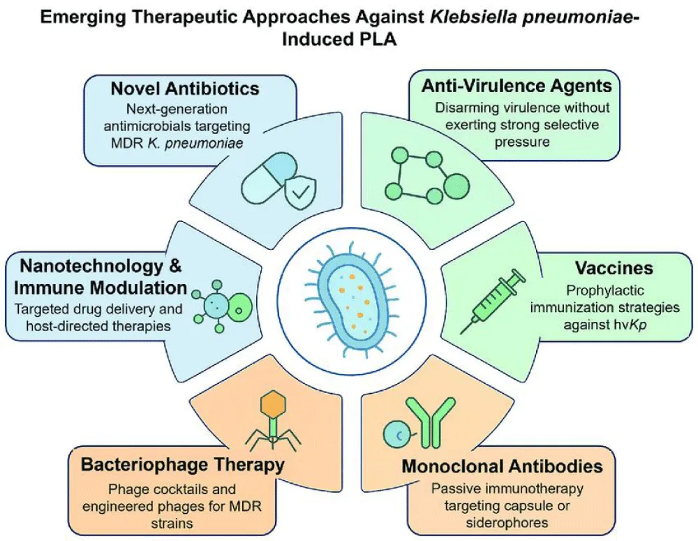
Growing recognition of the role of host-directed therapy has shifted attention from bacterial eradication alone toward preservation of endothelial function and restoration of vascular homeostasis. Strategies targeting capsular polysaccharides, inhibition of T6SS activity, modulation of oxidative stress, preservation of eNOS signaling, immunotherapy, monoclonal antibodies, anti-virulence compounds, bacteriophage therapy, and nanotechnology-based drug delivery are being explored as innovative approaches to combat multidrug-resistant K. pneumoniae. These interventions aim to reduce bacterial pathogenicity while minimizing the selective pressure that drives antimicrobial resistance.
This narrative review summarizes current knowledge regarding the molecular biology of capsular polysaccharides and the Type VI secretion system, their coordinated roles in disrupting vascular homeostasis, and the signaling pathways linking bacterial virulence to endothelial dysfunction. In addition, we discuss emerging therapeutic strategies targeting both bacterial virulence determinants and host vascular responses, highlighting future directions for translational research aimed at improving outcomes in severe K. pneumoniae infections.
Literature retrieval and evidence appraisal
This review used a structured narrative approach to identify literature on K. pneumoniae capsular polysaccharides, T6SS, endothelial dysfunction, vascular biology, antimicrobial resistance, and emerging therapies. Searches used combinations of the terms “Klebsiella pneumoniae,” “capsular polysaccharide,” “CPS,” “Type VI secretion system,” “T6SS,” “endothelial dysfunction,” “vascular,” “eNOS,” “nitric oxide,” “sepsis,” “biofilm,” “siderophore,” and “therapy.” PubMed/MEDLINE and other major biomedical databases were considered, with particular emphasis on peer-reviewed literature published from 2020 through 2026 and targeted searches for studies published during 2024–2026. Reference lists of relevant reviews were also examined to identify important earlier mechanistic studies.
Studies were screened for relevance to the central themes of the review. Experimental studies using bacterial mutants, purified virulence factors, cultured endothelial cells, organ-on-chip systems, Ex vivo vessels, or animal models were included when they provided mechanistic information. Human observational studies, clinical cohorts, guidelines, and translational reports were prioritized when discussing clinical relevance or treatment. Evidence was interpreted according to study type rather than treating preclinical observations as clinically established findings. Because this article is a narrative review rather than a systematic review, no pooled effect estimates or formal meta-analysis were performed. The principal limitation of this approach is the possibility of selection bias despite the use of predefined topic domains and targeted database searches.
Where evidence was indirect, exploratory, or limited to experimental models, the text uses qualified language such as “suggests,” “has been associated with,” or “may contribute.” Established clinical treatments are discussed separately from experimental anti-virulence and host-directed strategies. This distinction is particularly important for proposed links between CPS, T6SS, eNOS signaling, and vascular injury, for which human clinical validation remains limited.
Biology and Clinical Significance of Klebsiella pneumoniae
Klebsiella pneumoniae is a non-motile, Gram-negative, facultatively anaerobic bacillus belonging to the family Enterobacteriaceae. It is widely distributed in nature and commonly colonizes the human gastrointestinal tract and nasopharynx without causing disease in healthy individuals. However, disruption of host defenses, prolonged hospitalization, invasive medical procedures, immunosuppression, or broad-spectrum antibiotic exposure can transform this commensal organism into an aggressive opportunistic pathogen responsible for severe healthcare-associated and community-acquired infections. K. pneumoniae has become one of the most frequently isolated pathogens from bloodstream infections, ventilator-associated pneumonia, urinary tract infections, liver abscesses, surgical-site infections, and neonatal sepsis, contributing substantially to global morbidity and mortality. Recent epidemiological surveillance has demonstrated an alarming increase in multidrug-resistant (MDR), extensively drug-resistant (XDR), carbapenem-resistant (CRKP), and hypervirulent (hvK. pneumoniae) strains, thereby limiting therapeutic options and increasing healthcare costs. These developments have positioned K. pneumoniae among the most important bacterial threats identified by international public health organizations.
The remarkable adaptability of K. pneumoniae is largely attributable to its extensive repertoire of virulence factors that collectively promote colonization, immune evasion, nutrient acquisition, persistence, and dissemination within the host. These include capsular polysaccharides, lipopolysaccharide (LPS), fimbrial adhesins, siderophore-mediated iron acquisition systems, outer membrane proteins, biofilm-forming capacity, and specialized secretion systems such as the Type VI secretion system (T6SS). Rather than functioning independently, these virulence determinants interact synergistically to establish infection, evade host immune responses, and facilitate survival under diverse environmental conditions. Among these factors, the capsule remains the most important determinant of resistance to complement-mediated killing and phagocytosis, whereas the T6SS enables direct delivery of effector proteins into competing bacteria and host cells, thereby enhancing bacterial fitness and pathogenicity.
An increasing body of evidence suggests that K. pneumoniae infection extends beyond localized tissue injury and profoundly influences systemic vascular physiology. The vascular endothelium serves as a dynamic interface between circulating blood and surrounding tissues, regulating vascular tone, endothelial permeability, coagulation, inflammatory cell recruitment, and microvascular perfusion. During infection, bacterial virulence factors activate inflammatory signaling pathways, oxidative stress, mitochondrial dysfunction, and endothelial injury that collectively impair vascular homeostasis. Endothelial dysfunction is characterized by diminished nitric oxide (NO) bioavailability, increased leukocyte adhesion, platelet activation, vascular leakage, and microvascular thrombosis, all of which contribute to tissue hypoxia, organ dysfunction, and sepsis. Recent mechanistic studies have demonstrated that K. pneumoniae directly suppresses endothelial nitric oxide synthase (eNOS) activity through coordinated actions of capsular polysaccharides and T6SS-derived effectors, providing a molecular explanation for impaired vasodilation observed during severe infection.
The emergence of antimicrobial resistance has intensified interest in therapeutic strategies that target bacterial virulence rather than bacterial viability. Anti-virulence approaches directed against capsular polysaccharides, T6SS components, quorum sensing, biofilm formation, and host–pathogen signaling pathways may reduce bacterial pathogenicity while minimizing the selective pressure responsible for the evolution of antibiotic resistance. Understanding the biological characteristics of K. pneumoniae and the mechanisms by which its virulence factors disrupt vascular homeostasis therefore provides an important foundation for the development of innovative host-directed and pathogen-directed therapies.
Capsular polysaccharides: Structure, biosynthesis, and role in virulence
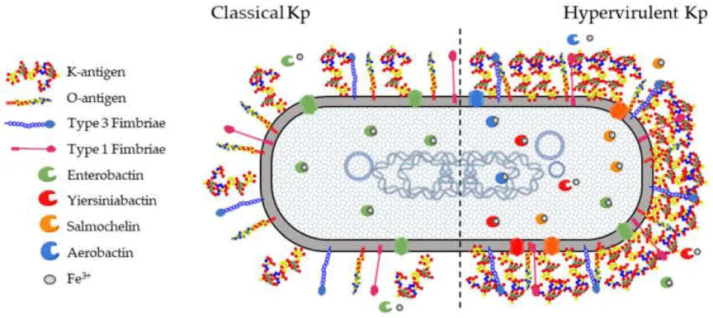
Capsular polysaccharides (CPS) constitute the outermost structural layer of Klebsiella pneumoniae and represent its most important virulence determinant. The capsule forms a hydrated, mucoid matrix that surrounds the bacterial cell, shielding it from host immune defenses while facilitating survival in diverse environmental niches. Extensive genetic diversity within the capsular polysaccharide synthesis (cps) locus has resulted in more than 180 capsule (K) types, with hypervirulent strains frequently expressing highly protective capsule phenotypes. Variations in capsule composition, chain length, and surface architecture substantially influence bacterial pathogenicity, tissue tropism, immune recognition, and clinical outcomes. Recent genomic analyses have demonstrated that horizontal gene transfer and recombination within the cps locus drive continuous capsule evolution, contributing to the emergence of multidrug-resistant and hypervirulent clones.
The biosynthesis of capsular polysaccharides is mediated by a conserved Wzy-dependent assembly pathway encoded within the chromosomal cps locus. This region contains genes responsible for nucleotide sugar synthesis, glycosyltransferases, polymerization enzymes, transport proteins, and regulatory factors that coordinate capsule production. Key proteins, including Wza, Wzb, Wzc, Wzx, and Wzy, regulate translocation, polymerization, and export of polysaccharide chains across the bacterial envelope. Among these, the tyrosine kinase Wzc plays a central role by controlling capsule polymerization through reversible phosphorylation, thereby influencing capsule thickness and bacterial virulence. Environmental stimuli such as iron limitation, osmotic stress, nutrient availability, and host inflammatory signals further regulate capsule expression through complex transcriptional networks that optimize bacterial adaptation during infection.
The capsule is indispensable for immune evasion. By masking highly immunogenic bacterial surface structures, capsular polysaccharides inhibit complement activation, reduce C3b deposition, prevent opsonophagocytic killing, and limit recognition by neutrophils and macrophages. The capsule also protects bacteria from antimicrobial peptides, oxidative stress, and neutrophil extracellular traps, thereby prolonging bacterial survival within host tissues. Hypercapsulated strains exhibit enhanced resistance to innate immune clearance and are associated with invasive infections, including liver abscesses, bacteremia, meningitis, and metastatic dissemination. Furthermore, the capsule promotes bacterial persistence by facilitating biofilm formation on host tissues and indwelling medical devices, increasing tolerance to antimicrobial therapy and contributing to recurrent infections.
Emerging evidence indicates that capsular polysaccharides exert biological effects extending beyond immune evasion and directly influence vascular physiology. During systemic infection, capsule-derived components interact with endothelial cells, initiating intracellular signaling pathways that disrupt vascular homeostasis. Experimental studies have demonstrated that capsular polysaccharides suppress endothelial nitric oxide synthase (eNOS) activation through phosphatase-dependent mechanisms, leading to reduced nitric oxide bioavailability and impaired endothelium-dependent vasodilation. Simultaneously, activation of inflammatory mediators and oxidative stress pathways enhances endothelial permeability, leukocyte adhesion, and microvascular dysfunction. These vascular alterations contribute to tissue hypoperfusion, disseminated inflammation, and multiple-organ injury during severe K. pneumoniae infection.
Because the capsule is exposed on the bacterial surface and is essential for virulence, it has become an attractive therapeutic target. Anti-capsular monoclonal antibodies, glycoconjugate vaccines, capsule depolymerases derived from bacteriophages, and small-molecule inhibitors of capsule biosynthesis are being actively investigated as novel anti-virulence strategies. Unlike conventional antibiotics, these approaches aim to weaken bacterial pathogenicity without directly inhibiting bacterial growth, thereby reducing selective pressure for antimicrobial resistance. Continued investigation of capsule structure, biosynthesis, and host interactions is expected to facilitate the development of innovative therapeutics capable of restoring vascular homeostasis while improving outcomes in infections caused by multidrug-resistant K. pneumoniae (Figure 1).
Source: Created by Haider et al 2026
Type VI secretion system: Structure, regulation, and contribution to vascular dysfunction
The Type VI secretion system (T6SS) is a sophisticated contractile nanomachine that enables Gram-negative bacteria to inject toxic effector proteins directly into neighboring bacterial cells and eukaryotic host cells. Since its discovery, the T6SS has emerged as one of the most important virulence determinants contributing to bacterial competition, host colonization, immune modulation, and environmental adaptation. In Klebsiella pneumoniae, the T6SS plays a central role in enhancing bacterial fitness by eliminating competing microorganisms, promoting persistence within polymicrobial communities, facilitating epithelial colonization, and modulating host immune responses. Although initially recognized primarily as a bacterial competition apparatus, recent investigations have demonstrated that the T6SS directly influences host vascular physiology, thereby expanding its significance beyond conventional microbial pathogenesis.
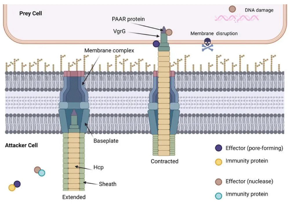
Structurally, the T6SS resembles an inverted contractile bacteriophage tail composed of three major components: a membrane complex, a baseplate complex, and a contractile sheath surrounding an inner tube. The membrane complex anchors the secretion apparatus within the bacterial envelope, while the contractile sheath rapidly contracts to propel the inner Hcp tube and the VgrG–PAAR spike complex into target cells. This mechanical injection process delivers numerous antibacterial and anti-eukaryotic effector proteins directly into recipient cells without requiring extracellular diffusion. The highly conserved core proteins, designated TssA through TssM, coordinate assembly, contraction, recycling, and regulation of the secretion apparatus, ensuring efficient delivery of virulence factors during infection.
Genomic analyses have demonstrated that K. pneumoniae contains multiple T6SS gene clusters exhibiting considerable structural diversity among clinical isolates. Comparative genomic studies indicate that hypervirulent and multidrug-resistant strains frequently harbor complete T6SS loci together with accessory effector and immunity genes that increase bacterial competitiveness and pathogenic potential. Expression of the T6SS is tightly regulated by environmental signals including iron limitation, osmotic stress, oxygen availability, quorum sensing, nutrient deprivation, and host-derived inflammatory mediators. Multiple transcriptional regulators coordinate activation of T6SS genes, allowing bacteria to conserve metabolic energy while rapidly responding to changing environmental conditions encountered during infection.
One of the principal biological functions of the T6SS is interbacterial competition. By injecting antibacterial toxins that degrade peptidoglycan, nucleic acids, phospholipids, or cellular membranes, K. pneumoniae eliminates competing microorganisms occupying the same ecological niche. This competitive advantage facilitates intestinal colonization, respiratory tract persistence, and establishment of infection within polymicrobial environments. Simultaneously, bacteria producing corresponding immunity proteins remain protected against their own effectors, ensuring selective survival of genetically related populations. The ability to dominate microbial communities contributes substantially to successful colonization before clinical infection becomes established.
Beyond bacterial competition, the T6SS directly modulates interactions with host cells. Experimental studies demonstrate that disruption of essential T6SS structural genes significantly reduces bacterial adhesion, invasion, biofilm formation, epithelial colonization, and virulence in both cell culture and animal infection models. Several T6SS effectors influence mitochondrial function, intracellular calcium signaling, inflammatory mediator production, cytoskeletal organization, and innate immune activation. These alterations impair normal cellular homeostasis while creating conditions favorable for bacterial survival and dissemination throughout infected tissues.
Recent mechanistic investigations have revealed an unexpected role for the T6SS in regulating vascular biology. Vascular endothelial cells are responsible for maintaining vascular tone, nitric oxide production, barrier integrity, leukocyte trafficking, coagulation, and microvascular perfusion. During K. pneumoniae infection, the T6SS effector protein VgrG4 activates the mitochondrial receptor NLRX1, leading to excessive mitochondrial reactive oxygen species generation. This oxidative stress activates protein kinase Cβ (PKCβ), which phosphorylates endothelial nitric oxide synthase (eNOS) at its inhibitory regulatory site. Consequently, endothelial nitric oxide production declines, resulting in impaired endothelium-dependent vasodilation, increased vascular stiffness, endothelial activation, and reduced tissue perfusion. These findings establish a direct molecular connection between bacterial virulence and vascular dysfunction.
In addition to suppressing nitric oxide signaling, T6SS-mediated mitochondrial dysfunction disrupts endothelium-dependent hyperpolarization by impairing calcium-activated potassium channel signaling. Reduced endothelial responsiveness promotes leukocyte adhesion, platelet activation, oxidative injury, vascular leakage, and microvascular thrombosis. Collectively, these pathological events contribute to tissue hypoxia, systemic inflammation, septic organ dysfunction, and adverse clinical outcomes frequently observed in severe K. pneumoniae infections. Emerging evidence therefore suggests that vascular injury represents an active consequence of bacterial virulence rather than merely a secondary manifestation of overwhelming inflammation.
Recognition of the T6SS as a major virulence determinant has stimulated the development of anti-virulence therapeutic strategies. Current approaches include inhibition of T6SS assembly, suppression of effector secretion, blockade of VgrG-associated signaling pathways, neutralization of oxidative stress, preservation of endothelial nitric oxide signaling, monoclonal antibody therapy, bacteriophage-derived interventions, and nanoparticle-mediated delivery of anti-virulence compounds. Unlike conventional antibiotics, these approaches aim to reduce bacterial pathogenicity without directly inhibiting bacterial growth, thereby decreasing selective pressure for antimicrobial resistance. Continued investigation of T6SS biology and host–pathogen interactions is expected to accelerate development of innovative therapeutics capable of restoring vascular homeostasis while improving outcomes in infections caused by multidrug-resistant and hypervirulent K. pneumoniae.
Mechanisms regulating vascular homeostasis
Vascular homeostasis is maintained through a complex network of endothelial, vascular smooth muscle, immune, and neurohumoral signaling pathways that collectively regulate vascular tone, permeability, coagulation, inflammation, and tissue perfusion. The vascular endothelium forms a highly specialized monolayer lining the interior surface of blood vessels and serves as a dynamic biological interface between circulating blood and peripheral tissues. Under physiological conditions, endothelial cells continuously monitor mechanical forces, circulating metabolites, inflammatory mediators, and microbial products, responding through tightly regulated signaling pathways that preserve vascular integrity and organ function. Disruption of these regulatory mechanisms during bacterial infection results in endothelial dysfunction, impaired vasodilation, vascular leakage, thrombosis, and multiple-organ injury, all of which contribute significantly to the morbidity and mortality associated with severe Klebsiella pneumoniae infections. (nature.com).
Nitric oxide (NO) is the principal mediator of vascular homeostasis and is synthesized predominantly by endothelial nitric oxide synthase (eNOS). Following stimulation by shear stress, acetylcholine, bradykinin, vascular endothelial growth factor, and insulin, eNOS converts L-arginine into nitric oxide and L-citrulline in the presence of tetrahydrobiopterin and other essential cofactors. Nitric oxide diffuses rapidly into adjacent vascular smooth muscle cells, where it activates soluble guanylate cyclase, increases cyclic guanosine monophosphate (cGMP) production, and promotes smooth muscle relaxation, thereby maintaining physiological vasodilation. Beyond its vasodilatory effects, nitric oxide inhibits platelet aggregation, suppresses leukocyte adhesion, reduces oxidative stress, limits vascular smooth muscle proliferation, and preserves endothelial barrier function. Consequently, diminished nitric oxide bioavailability is widely recognized as one of the earliest indicators of endothelial dysfunction during infectious and inflammatory diseases.
Endothelial nitric oxide synthase activity is tightly controlled through phosphorylation, intracellular calcium signaling, protein–protein interactions, and subcellular localization. Phosphorylation of activating residues enhances nitric oxide production, whereas phosphorylation at inhibitory sites suppresses enzymatic activity. Multiple intracellular kinases, phosphatases, and adaptor proteins coordinate these regulatory events to ensure precise control of vascular tone. During bacterial infection, excessive inflammatory signaling and oxidative stress disturb these regulatory pathways, leading to impaired eNOS activation, reduced nitric oxide synthesis, and diminished endothelium-dependent vasodilation. Recent experimental studies have demonstrated that K. pneumoniae virulence factors interfere directly with these signaling mechanisms through coordinated actions of capsular polysaccharides and Type VI secretion system effectors, providing a molecular explanation for infection-associated vascular dysfunction. (nature.com).
Reactive oxygen species (ROS) are another major determinant of vascular homeostasis. Under normal physiological conditions, low concentrations of superoxide, hydrogen peroxide, and related oxidants participate in cellular signaling and adaptive responses. However, excessive ROS production during bacterial infection overwhelms endogenous antioxidant defenses, resulting in oxidative stress and endothelial injury. Mitochondria, nicotinamide adenine dinucleotide phosphate (NADPH) oxidases, xanthine oxidase, and activated inflammatory leukocytes represent major intracellular sources of reactive oxygen species. Elevated ROS not only damage lipids, proteins, and nucleic acids but also react rapidly with nitric oxide to generate peroxynitrite, thereby further reducing nitric oxide bioavailability and amplifying endothelial dysfunction.
The endothelial glycocalyx also plays a critical role in maintaining vascular homeostasis. This carbohydrate-rich extracellular layer, composed of proteoglycans, glycosaminoglycans, glycoproteins, and plasma proteins, protects endothelial cells from mechanical injury while regulating vascular permeability, mechanotransduction, leukocyte adhesion, and coagulation. During severe bacterial infection, inflammatory cytokines, bacterial toxins, oxidative stress, and matrix-degrading enzymes promote glycocalyx degradation, exposing the endothelial surface to circulating inflammatory cells and coagulation factors. Loss of glycocalyx integrity contributes to increased vascular permeability, tissue edema, microvascular thrombosis, and impaired organ perfusion, features commonly observed during septic complications associated with K. pneumoniae infection.
Inflammatory signaling pathways further influence vascular function through coordinated activation of pattern-recognition receptors, including Toll-like receptors and nucleotide-binding oligomerization domain-like receptors. Recognition of bacterial lipopolysaccharide and other pathogen-associated molecular patterns activates nuclear factor-κB (NF-κB), mitogen-activated protein kinases (MAPKs), and inflammasome pathways, resulting in the production of tumor necrosis factor-α, interleukin-1β, interleukin-6, interferons, and chemokines. Although these responses contribute to bacterial clearance, sustained activation promotes endothelial activation, leukocyte recruitment, oxidative stress, vascular leakage, and coagulation abnormalities that ultimately compromise vascular homeostasis.
Maintenance of vascular integrity also depends upon balanced regulation of coagulation and fibrinolysis. Healthy endothelial cells express anticoagulant molecules including thrombomodulin, tissue factor pathway inhibitor, and heparan sulfate while simultaneously suppressing platelet activation. During systemic bacterial infection, endothelial injury shifts this balance toward a procoagulant phenotype characterized by tissue factor expression, platelet activation, fibrin deposition, and impaired fibrinolysis. These changes facilitate formation of microvascular thrombi, restrict tissue perfusion, and contribute to disseminated intravascular coagulation and multiple-organ dysfunction frequently observed in severe sepsis.
Emerging experimental evidence indicates that K. pneumoniae can disrupt vascular homeostasis through interactions between bacterial virulence factors and host endothelial signaling. CPS has been associated with phosphatase-dependent changes in eNOS activation, whereas selected T6SS-associated mechanisms have been linked to mitochondrial oxidative stress and inhibitory regulation of eNOS. These observations provide a plausible mechanistic framework, but the relative contribution of each pathway in patients remains to be established.
Understanding the mechanisms regulating vascular homeostasis has important therapeutic implications. Preservation of endothelial nitric oxide signaling, reduction of oxidative stress, stabilization of the endothelial glycocalyx, modulation of inflammatory pathways, and inhibition of bacterial virulence factors represent promising strategies for preventing vascular injury during K. pneumoniae infection. Combining host-directed therapies with conventional antimicrobial treatment may provide superior protection against endothelial dysfunction, improve tissue perfusion, reduce organ failure, and ultimately enhance survival in patients with multidrug-resistant and hypervirulent K. pneumoniae infection.
Capsule- and type VI secretion system-mediated endothelial dysfunction
Endothelial dysfunction has emerged as a central pathogenic event during severe Klebsiella pneumoniae infection, linking bacterial virulence with vascular injury, impaired tissue perfusion, and organ failure. Traditionally, endothelial damage associated with Gram-negative infections was attributed primarily to excessive systemic inflammation and lipopolysaccharide-induced cytokine release. However, recent evidence indicates that K. pneumoniae actively manipulates endothelial signaling through its major virulence determinants, particularly capsular polysaccharides (CPS) and the Type VI secretion system (T6SS). These virulence factors directly interfere with intracellular pathways that regulate nitric oxide (NO) production, endothelial barrier integrity, oxidative balance, and inflammatory responses, thereby disrupting vascular homeostasis independently of generalized inflammatory injury (Figure 2).
Source: Created by Haider et al 2026
Capsular polysaccharides and endothelial signaling
Capsular polysaccharides form a thick extracellular matrix that enables K. pneumoniae to evade complement activation, inhibit phagocytosis, and resist antimicrobial peptides. Beyond these protective functions, CPS interacts directly with endothelial cells and alters intracellular signaling pathways involved in vascular regulation. Experimental studies have demonstrated that CPS suppresses endothelial nitric oxide synthase (eNOS) activation by promoting phosphatase-dependent dephosphorylation of activating eNOS residues. Reduced eNOS activity leads to diminished NO production, impaired vascular relaxation, and decreased microvascular perfusion.
The decline in nitric oxide bioavailability has several downstream consequences. NO normally suppresses platelet aggregation, limits leukocyte adhesion, and inhibits vascular smooth muscle proliferation. Consequently, CPS-mediated reduction in NO promotes endothelial activation, enhances expression of vascular adhesion molecules, facilitates inflammatory cell recruitment, and increases vascular resistance. Persistent endothelial dysfunction further accelerates tissue hypoxia and inflammatory injury, creating conditions that favor bacterial survival and dissemination (Table 1).
Type VI secretion system and mitochondrial dysfunction
The Type VI secretion system represents another critical mechanism through which K. pneumoniae alters endothelial physiology. This contractile secretion apparatus injects effector proteins directly into host cells, allowing bacteria to manipulate intracellular signaling with remarkable precision. Recent investigations have identified VgrG-associated effectors capable of inducing mitochondrial dysfunction within endothelial cells.
Mitochondrial injury results in excessive production of reactive oxygen species (ROS), activation of protein kinase Cβ (PKCβ), and inhibitory phosphorylation of eNOS. These molecular events substantially reduce nitric oxide synthesis while simultaneously increasing oxidative stress. ROS further react with available nitric oxide to generate peroxynitrite, thereby amplifying endothelial dysfunction through oxidative modification of proteins, lipids, and DNA. The combined effects of reduced NO production and excessive oxidative stress markedly impair endothelium-dependent vasodilation (Table 2).
Potential interactions between CPS and T6SS
CPS and T6SS should be viewed as distinct virulence systems that may contribute to a common pathogenic environment rather than as a single, proven regulatory pathway. CPS primarily provides a surface barrier and modifies interactions with complement, phagocytes, antimicrobial peptides, and host cells. T6SS, in contrast, is a contact-dependent secretion apparatus that delivers effectors and is strongly linked to bacterial competition and selected host-cell phenotypes. Experimental evidence supports independent effects of both systems on host responses, but direct evidence demonstrating that CPS controls T6SS expression, or that T6SS directly controls CPS production during vascular infection, remains limited. The proposed interaction is therefore best presented as a working model requiring further experimental validation.
In severe infection, the effects of multiple virulence factors may converge on endothelial activation. Capsule-mediated immune evasion can increase bacterial persistence, while T6SS activity and other bacterial products may alter host-cell signaling. LPS-driven inflammation, siderophore-dependent bacterial survival, and biofilm-associated persistence can further sustain the inflammatory burden. These relationships are biologically plausible, but their relative contribution to vascular injury is likely to vary with strain, infection site, bacterial burden, and host condition.
Inflammatory amplification
Endothelial dysfunction induced by CPS and T6SS is further intensified through activation of inflammatory signaling pathways. Recognition of bacterial components by pattern-recognition receptors stimulates activation of nuclear factor-κB (NF-κB), mitogen-activated protein kinases (MAPKs), and inflammasome complexes, leading to increased production of tumor necrosis factor-α (TNF-α), interleukin-1β (IL-1β), interleukin-6 (IL-6), and other pro-inflammatory mediators. These cytokines enhance endothelial permeability, reduce barrier integrity, increase oxidative stress, and perpetuate vascular inflammation.
Inflammatory cytokines also promote expression of endothelial adhesion molecules, including intercellular adhesion molecule-1 (ICAM-1), vascular cell adhesion molecule-1 (VCAM-1), and E-selectin, facilitating recruitment of neutrophils and monocytes to sites of infection. Although these responses contribute to bacterial clearance, excessive activation frequently results in collateral vascular injury and tissue destruction.
Endothelial barrier disruption
Maintenance of endothelial barrier integrity is essential for preventing plasma leakage and maintaining adequate tissue perfusion. During K. pneumoniae infection, CPS- and T6SS-mediated signaling disrupts intercellular junction proteins such as vascular endothelial cadherin (VE-cadherin), occludin, and claudins. Loss of these junctional complexes increases endothelial permeability, allowing plasma proteins, inflammatory mediators, and immune cells to enter surrounding tissues.
Excessive vascular leakage contributes to tissue edema, impaired oxygen diffusion, reduced organ perfusion, and progression toward acute respiratory distress syndrome (ARDS), septic shock, and multiple-organ failure. These pathological changes are particularly important in pulmonary and systemic K. pneumoniae infections, where endothelial barrier failure is closely associated with adverse clinical outcomes.
Clinical implications
Recognition that endothelial dysfunction results from direct bacterial virulence rather than solely from systemic inflammation has important therapeutic implications. Strategies designed to preserve endothelial nitric oxide signaling, inhibit oxidative stress, stabilize endothelial junctions, and neutralize CPS or T6SS activity may significantly reduce vascular injury without exerting selective pressure for antibiotic resistance.
Future therapeutic approaches may combine conventional antimicrobial therapy with anti-virulence agents, endothelial-protective drugs, monoclonal antibodies, bacteriophage-derived enzymes, and nanoparticle-based drug delivery systems. Such combination strategies have the potential to restore vascular homeostasis, improve tissue perfusion, reduce organ dysfunction, and enhance survival in patients with multidrug-resistant and hypervirulent K. pneumoniae infections.
Inflammation, oxidative stress, and immune responses
Inflammation represents one of the earliest host defense mechanisms activated following Klebsiella pneumoniae infection. Recognition of bacterial pathogen-associated molecular patterns (PAMPs), including lipopolysaccharide (LPS), capsular polysaccharides, outer membrane proteins, fimbriae, and secreted virulence factors, by pattern-recognition receptors (PRRs) expressed on macrophages, dendritic cells, neutrophils, and endothelial cells initiates a cascade of innate immune responses. Toll-like receptors (TLRs), nucleotide-binding oligomerization domain (NOD)-like receptors, and C-type lectin receptors activate intracellular signaling pathways that culminate in the production of inflammatory cytokines, chemokines, and antimicrobial molecules. Although these responses are essential for restricting bacterial growth, excessive or sustained immune activation contributes significantly to endothelial dysfunction, vascular injury, and tissue damage.
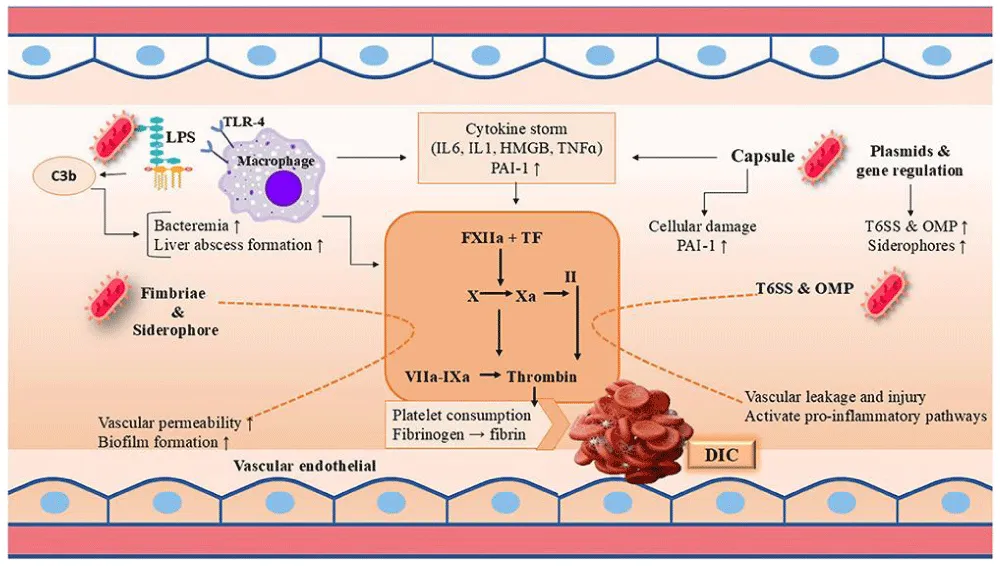
Among the pattern-recognition receptors, Toll-like receptor 4 (TLR4) plays a pivotal role in detecting the lipid A component of bacterial lipopolysaccharide. Binding of LPS to TLR4 activates both myeloid differentiation primary response protein 88 (MyD88)-dependent and Toll/interleukin-1 receptor domain-containing adaptor-inducing interferon-β (TRIF)-dependent signaling pathways. These pathways stimulate nuclear factor-κB (NF-κB), interferon regulatory factors, and mitogen-activated protein kinases (MAPKs), resulting in transcription of numerous pro-inflammatory genes. Elevated concentrations of tumor necrosis factor-α (TNF-α), interleukin-1β (IL-1β), interleukin-6 (IL-6), interleukin-8 (CXCL8), interferon-γ (IFN-γ), and granulocyte colony-stimulating factor amplify local and systemic inflammatory responses. While these mediators promote recruitment and activation of neutrophils and macrophages, uncontrolled cytokine production may culminate in cytokine storm, endothelial injury, septic shock, and multiple-organ dysfunction.
Neutrophils constitute the first line of cellular defense against K. pneumoniae. Rapid migration of neutrophils into infected tissues facilitates bacterial clearance through phagocytosis, degranulation, reactive oxygen species generation, and formation of neutrophil extracellular traps (NETs). However, hypervirulent strains possessing thick capsular polysaccharides exhibit increased resistance to neutrophil-mediated killing by reducing phagocytic uptake and limiting complement deposition. Persistent neutrophil activation also promotes collateral tissue injury through excessive release of proteases, elastase, myeloperoxidase, and oxidizing molecules that damage endothelial cells and surrounding tissues.
Macrophages are equally important in orchestrating antibacterial immunity. Resident alveolar macrophages, Kupffer cells within the liver, and circulating monocyte-derived macrophages recognize K. pneumoniae through multiple pattern-recognition receptors and initiate inflammatory signaling while coordinating adaptive immune responses. Activated macrophages produce nitric oxide, reactive oxygen species, inflammatory cytokines, and chemokines that recruit additional immune cells to sites of infection. Nevertheless, K. pneumoniae employs multiple immune-evasion mechanisms, including capsule-mediated inhibition of phagocytosis, suppression of complement activation, and modulation of intracellular signaling pathways, allowing bacterial persistence despite robust inflammatory responses.
Oxidative stress has emerged as a major determinant of vascular injury during K. pneumoniae infection. Physiologically, low concentrations of reactive oxygen species (ROS) participate in antimicrobial defense and intracellular signaling. During severe infection, however, excessive ROS generated by activated neutrophils, macrophages, endothelial cells, mitochondria, and NADPH oxidases overwhelm endogenous antioxidant systems. Superoxide anions, hydrogen peroxide, hydroxyl radicals, and peroxynitrite oxidize proteins, lipids, and nucleic acids, resulting in cellular dysfunction and apoptosis. Oxidative stress also decreases nitric oxide bioavailability through rapid formation of peroxynitrite, thereby impairing endothelial-dependent vasodilation and exacerbating vascular dysfunction.
Recent mechanistic studies suggest that Type VI secretion system (T6SS) effector proteins directly enhance mitochondrial ROS production within endothelial cells. Increased oxidative stress activates protein kinase Cβ, inhibits endothelial nitric oxide synthase (eNOS), disrupts mitochondrial respiration, and impairs ATP production. Simultaneously, capsular polysaccharides contribute to endothelial dysfunction by suppressing phosphatase-regulated eNOS activation. Together, these complementary mechanisms create a pro-oxidative vascular environment characterized by reduced nitric oxide synthesis, impaired vasodilation, endothelial activation, and increased vascular permeability.
Adaptive immune responses also influence the outcome of K. pneumoniae infection. Dendritic cells process bacterial antigens and present them to naïve T lymphocytes, promoting differentiation of T helper (Th1, Th17) cells and activation of cytotoxic T lymphocytes. B lymphocytes generate antibodies directed against capsular polysaccharides, lipopolysaccharide, outer membrane proteins, and other bacterial antigens. Protective antibodies facilitate complement activation, opsonophagocytosis, and bacterial clearance, providing the rationale for development of capsule-based vaccines and monoclonal antibody therapies. Nevertheless, extensive antigenic diversity among capsular serotypes remains a significant obstacle to broad-spectrum vaccine development.
Accumulating evidence indicates that inflammatory injury associated with K. pneumoniae infection is not solely the consequence of host immune activation but also results from active manipulation of host signaling pathways by bacterial virulence factors. Crosstalk between inflammation, oxidative stress, endothelial dysfunction, mitochondrial injury, and coagulation abnormalities establishes a self-amplifying cycle that promotes bacterial dissemination and organ damage. Understanding these interconnected mechanisms is essential for identifying therapeutic strategies capable of simultaneously suppressing excessive inflammation, preserving vascular homeostasis, and enhancing antibacterial immunity.
Emerging therapeutic approaches aim to modulate host inflammatory responses while avoiding excessive immunosuppression. Antioxidants, mitochondrial protective agents, cytokine inhibitors, endothelial-protective compounds, monoclonal antibodies targeting bacterial virulence factors, bacteriophage-derived enzymes, immunomodulatory peptides, and nanoparticle-based drug delivery systems are currently being investigated as adjunctive therapies. Combining these host-directed interventions with effective antimicrobial treatment may improve bacterial clearance, reduce endothelial injury, preserve organ function, and enhance survival among patients with severe multidrug-resistant K. pneumoniae infections (Figure 3).
Source: Created by Haider et al 2026
Experimental models investigating vascular injury
Experimental models have substantially advanced understanding of how Klebsiella pneumoniae disrupts vascular homeostasis and contributes to endothelial dysfunction. These systems provide valuable platforms for investigating host–pathogen interactions, identifying bacterial virulence mechanisms, evaluating immune responses, and assessing the therapeutic potential of emerging interventions. Because vascular injury during K. pneumoniae infection results from complex interactions among endothelial cells, immune cells, vascular smooth muscle, and circulating inflammatory mediators, the integration of In vitro, Ex vivo, and in vivo models is essential for elucidating disease mechanisms and facilitating translational research.
In vitro endothelial models
Cultured endothelial cells remain the cornerstone of mechanistic studies examining vascular injury caused by K. pneumoniae. Human umbilical vein endothelial cells (HUVECs), human pulmonary microvascular endothelial cells (HPMECs), human aortic endothelial cells (HAECs), and other primary endothelial cell cultures are commonly used to investigate bacterial adhesion, inflammatory signaling, endothelial barrier integrity, nitric oxide production, oxidative stress, apoptosis, and mitochondrial dysfunction.
These models enable detailed analysis of endothelial nitric oxide synthase (eNOS) regulation, intracellular calcium signaling, reactive oxygen species (ROS) generation, and cytokine secretion following exposure to capsular polysaccharides or Type VI secretion system (T6SS) effectors. Modern molecular techniques, including CRISPR-Cas9 genome editing, RNA interference, transcriptomic profiling, and proteomic analysis, have further strengthened these experimental platforms by allowing precise identification of host signaling pathways involved in endothelial dysfunction.
Three-dimensional endothelial cultures and microfluidic vascular organ-on-chip systems have further improved physiological relevance by reproducing blood flow, shear stress, and endothelial barrier architecture. These advanced models more closely mimic the human microvascular environment and provide greater insight into bacterial-induced vascular injury than conventional monolayer cultures.
Ex vivo vascular function studies
Isolated blood vessel preparations provide direct functional assessment of vascular responses following exposure to bacterial virulence factors. Segments of the aorta, mesenteric arteries, pulmonary arteries, and resistance vessels are commonly examined using wire or pressure myography to measure endothelial-dependent and endothelial-independent vasodilatory responses.
Acetylcholine-induced relaxation serves as an indicator of endothelial function, whereas sodium nitroprusside-mediated relaxation evaluates vascular smooth muscle responsiveness. Selective impairment of acetylcholine-induced vasodilation after exposure to K. pneumoniae virulence factors indicates direct endothelial injury rather than generalized smooth muscle dysfunction. Additional measurements of nitric oxide bioavailability, vascular stiffness, oxidative stress, and endothelial calcium signaling further characterize alterations in vascular physiology.
Animal models
Animal models remain indispensable for investigating the systemic effects of K. pneumoniae infection. Murine models are widely employed because of their well-characterized immune systems, availability of genetically modified strains, and reproducibility. Depending on the experimental objective, infection may be established through intranasal, intratracheal, intravenous, or intraperitoneal inoculation.
Pulmonary infection models reproduce bacterial pneumonia and facilitate evaluation of endothelial barrier disruption, inflammatory cell recruitment, pulmonary edema, and acute respiratory distress syndrome. Intravenous infection models simulate bacteremia and sepsis, allowing investigation of systemic vascular dysfunction, coagulation abnormalities, endothelial activation, and multiple-organ injury. Liver abscess models are particularly valuable for studying hypervirulent strains, which frequently disseminate beyond the liver to distant organs.
Genetically engineered mice deficient in inflammatory mediators, endothelial signaling proteins, oxidative stress regulators, or innate immune receptors have provided important insights into the molecular pathways responsible for bacterial virulence and host susceptibility.
Advanced molecular technologies
Recent technological advances have transformed investigations of K. pneumoniae pathogenesis. High-throughput transcriptomics, proteomics, metabolomics, lipidomics, and single-cell RNA sequencing enable comprehensive characterization of host and bacterial responses throughout infection.
Similarly, confocal microscopy, intravital microscopy, electron microscopy, fluorescence imaging, and multiphoton microscopy permit direct visualization of bacterial dissemination, endothelial junction integrity, leukocyte trafficking, vascular permeability, and microvascular blood flow. These complementary techniques provide spatial and temporal information regarding vascular injury that cannot be obtained through conventional histological methods.
Translational experimental models
Bridging the gap between laboratory research and clinical application remains a major objective in infectious disease research. Humanized mouse models, patient-derived endothelial cells, organoids, precision-cut tissue slices, and vascular organ-on-chip platforms increasingly provide clinically relevant systems for evaluating bacterial virulence and therapeutic efficacy.
Integration of clinical isolates obtained from patients with multidrug-resistant and hypervirulent K. pneumoniae infections further enhances translational relevance by reflecting the genetic diversity encountered in healthcare settings. These models support evaluation of individualized host responses and facilitate development of precision medicine approaches for severe bacterial infections.
Limitations and future perspectives
Although each experimental model provides valuable information, none fully reproduces the complexity of human infection. Cell culture systems offer exceptional mechanistic resolution but lack systemic immune interactions. Animal models replicate physiological responses but may not accurately reflect all aspects of human vascular biology because of species-specific differences. Organ-on-chip technologies more closely mimic the human microenvironment; however, their widespread application remains limited by technical complexity and cost.
Future research should integrate multi-omics technologies, artificial intelligence, computational modeling, advanced imaging, and patient-derived experimental systems to achieve a more comprehensive understanding of host–pathogen interactions. Such multidisciplinary approaches are expected to accelerate the identification of biomarkers, therapeutic targets, and anti-virulence strategies capable of preserving vascular homeostasis while improving clinical outcomes in infections caused by multidrug-resistant and hypervirulent Klebsiella pneumoniae.
Emerging therapeutic strategies
The rapid global emergence of multidrug-resistant (MDR), extensively drug-resistant (XDR), carbapenem-resistant (CRKP), and hypervirulent Klebsiella pneumoniae has significantly reduced the effectiveness of conventional antimicrobial therapy. Although antibiotics remain the cornerstone of treatment, increasing resistance, biofilm formation, and immune evasion have highlighted the need for innovative therapeutic strategies that extend beyond direct bacterial killing. Recent research has shifted toward anti-virulence approaches designed to attenuate bacterial pathogenicity while preserving the host microbiota and minimizing the selective pressure responsible for antimicrobial resistance. Because capsular polysaccharides (CPS) and the Type VI secretion system (T6SS) are central mediators of endothelial dysfunction and vascular injury, they have emerged as attractive therapeutic targets (Figure 4).
Source: Created by Haider et al 2026
Anti-capsular therapies
Capsular polysaccharides represent one of the most promising targets for next-generation therapeutics. The capsule protects K. pneumoniae from complement-mediated lysis, phagocytosis, antimicrobial peptides, and oxidative injury while facilitating bacterial persistence within host tissues. Consequently, disruption of capsule integrity can substantially reduce bacterial virulence.
Monoclonal antibodies directed against capsular polysaccharides have demonstrated encouraging results in preclinical studies by enhancing complement activation, promoting opsonophagocytic killing, and improving bacterial clearance. Similarly, glycoconjugate vaccines targeting conserved capsular antigens are being investigated as preventive strategies for individuals at high risk of healthcare-associated infections. Bacteriophage-derived capsule depolymerases provide another promising approach by enzymatically degrading the capsule, thereby exposing bacteria to host immune defenses and improving antibiotic susceptibility.
Small-molecule inhibitors targeting enzymes involved in capsule biosynthesis, polymerization, and export have also gained increasing attention. Inhibition of proteins such as Wza, Wzb, Wzc, Wzx, and Wzy may reduce capsule production, attenuate bacterial virulence, and restore host immune recognition without directly affecting bacterial viability.
Targeting the type VI secretion system
The Type VI secretion system has emerged as an attractive anti-virulence target because of its essential role in bacterial competition, host colonization, immune modulation, and endothelial dysfunction. Pharmacological inhibition of T6SS assembly or effector secretion may significantly reduce bacterial pathogenicity while exerting minimal selective pressure for antimicrobial resistance.
Current experimental approaches include inhibition of ATP-dependent assembly proteins, disruption of sheath contraction, blockade of VgrG–PAAR spike formation, and suppression of T6SS gene expression through transcriptional regulators. Neutralization of individual effector proteins capable of inducing mitochondrial dysfunction, oxidative stress, and endothelial injury represents another promising therapeutic strategy. Although no T6SS-specific inhibitors have yet reached routine clinical use, continued structural and molecular characterization of this secretion apparatus is expected to facilitate rational drug development.
Host-directed therapies
Preservation of endothelial function has become an important therapeutic objective in severe K. pneumoniae infection. Host-directed therapies seek to reduce tissue injury by protecting vascular integrity rather than directly eliminating bacteria.
Potential approaches include enhancement of endothelial nitric oxide synthase (eNOS) activity, restoration of nitric oxide bioavailability, stabilization of the endothelial glycocalyx, suppression of excessive oxidative stress, and modulation of inflammatory signaling pathways. Pharmacological agents targeting mitochondrial dysfunction, reactive oxygen species production, protein kinase activation, and endothelial barrier disruption may reduce vascular injury while preserving organ perfusion.
Selective immunomodulation also offers therapeutic potential by limiting excessive inflammatory responses without impairing bacterial clearance. Balanced regulation of cytokine production may reduce endothelial activation, vascular leakage, and multiple-organ dysfunction during severe infection.
Clinical Evidence and Adjunctive Pharmacological Therapy
Current clinical management of severe K. pneumoniae infection remains centered on appropriate antimicrobial therapy, source control, and supportive critical-care measures. For carbapenem-resistant Enterobacterales, contemporary guidance includes newer β-lactam/β-lactamase inhibitor combinations and cefiderocol for selected infections according to the resistance mechanism and susceptibility profile. These therapies are clinically established options in appropriate settings, whereas CPS-targeting antibodies, T6SS inhibitors, endothelial-protective agents, and most phage-based strategies remain investigational. Experimental anti-virulence approaches should therefore be presented as potential adjuncts rather than substitutes for effective antimicrobial treatment.
Host-directed treatment is an attractive adjunctive concept because endothelial injury is an important component of severe sepsis. However, evidence for routine use of specific eNOS activators, antioxidants, glycocalyx stabilizers, or PKC-directed agents in K. pneumoniae infection is not yet sufficient to support clinical recommendations. Future trials should define patient selection, timing, pharmacokinetics, safety, and interaction with antimicrobial therapy before these strategies can be incorporated into practice.
Bacteriophage therapy
The renewed interest in bacteriophage therapy has generated considerable enthusiasm for treatment of multidrug-resistant K. pneumoniae. Lytic bacteriophages selectively infect bacterial cells while sparing the normal microbiota. In addition to direct bacterial killing, many bacteriophages produce capsule depolymerases that degrade capsular polysaccharides, thereby enhancing bacterial susceptibility to host immune responses and antimicrobial agents.
Phage cocktails containing multiple complementary bacteriophages may reduce the likelihood of bacterial resistance while expanding antimicrobial spectrum. Combination therapy involving bacteriophages and conventional antibiotics has demonstrated synergistic activity in several experimental infection models.
Antimicrobial peptides and immunotherapy
Host-derived antimicrobial peptides constitute an important component of innate immunity and have attracted increasing interest as potential therapeutic agents. These peptides disrupt bacterial membranes, inhibit biofilm formation, neutralize endotoxins, and modulate inflammatory responses.
Immunotherapeutic approaches include monoclonal antibodies targeting bacterial virulence factors, passive immunization, engineered antibodies with enhanced complement activation, and therapeutic vaccines designed to stimulate protective humoral and cellular immune responses. Such interventions may provide valuable adjunctive treatment for immunocompromised individuals at high risk of invasive K. pneumoniae infection.
Nanotechnology-based drug delivery
Nanomedicine offers new opportunities for improving treatment efficacy while reducing systemic toxicity. Liposomes, polymeric nanoparticles, lipid nanoparticles, dendrimers, exosomes, and inorganic nanocarriers can enhance targeted delivery of antibiotics, anti-virulence compounds, antioxidants, and immunomodulatory agents directly to infected tissues.
Nanoparticle-based formulations improve drug stability, prolong circulation time, facilitate intracellular delivery, and overcome barriers associated with bacterial biofilms. Functionalization of nanoparticles with antibodies, peptides, or receptor-specific ligands may further increase targeting specificity while minimizing off-target effects.
Combination therapeutic strategies
Given the complexity of K. pneumoniae pathogenesis, combination therapy is likely to provide superior clinical outcomes compared with single-agent treatment. Simultaneous targeting of bacterial viability, virulence factors, endothelial dysfunction, oxidative stress, and dysregulated immune responses may interrupt multiple pathogenic pathways responsible for disease progression.
Future treatment protocols may combine conventional antibiotics with anti-capsular antibodies, T6SS inhibitors, bacteriophages, endothelial-protective agents, antioxidants, and nanoparticle-mediated drug delivery systems. Such multimodal approaches have the potential to improve bacterial clearance, restore vascular homeostasis, reduce antimicrobial resistance, and enhance patient survival.
Translational Comparison of Therapeutic Approaches
Conventional antimicrobial therapy remains the clinical standard and directly targets bacterial viability; its effectiveness depends on susceptibility and adequate source control. Anti-capsular antibodies, capsule depolymerases, and capsule-biosynthesis inhibitors are pathogen-directed approaches that may restore immune recognition but face challenges related to capsule diversity and strain specificity. T6SS inhibitors are mechanistically attractive but are at an earlier developmental stage, with no T6SS-specific agent established for routine clinical use. Bacteriophages and phage-derived depolymerases offer strain-specific options and may be useful against selected resistant isolates, although manufacturing, regulatory, pharmacokinetic, and resistance issues remain. Host-directed strategies aim to preserve endothelial function rather than directly kill bacteria and therefore are best considered adjunctive until clinical efficacy is demonstrated. Nanotechnology may serve as a delivery platform across several of these approaches, but its clinical value depends on reproducible manufacturing, biodistribution, safety, and pharmacological validation.
Future clinical translation
Successful translation of these emerging therapeutic strategies into clinical practice will require multidisciplinary collaboration among microbiologists, immunologists, vascular biologists, pharmacologists, and clinicians. Future clinical trials should prioritize evaluation of safety, pharmacokinetics, optimal therapeutic combinations, biomarker-guided patient selection, and long-term outcomes. Advances in precision medicine, artificial intelligence, multi-omics technologies, and systems biology are expected to accelerate development of personalized therapeutic approaches capable of simultaneously targeting bacterial virulence and preserving vascular function. These innovations may ultimately redefine the management of multidrug-resistant and hypervirulent Klebsiella pneumoniae infections while improving clinical outcomes worldwide.
Future perspectives
Despite substantial advances in understanding the molecular pathogenesis of Klebsiella pneumoniae, significant knowledge gaps remain regarding the mechanisms by which bacterial virulence factors disrupt vascular homeostasis and contribute to disease severity. Most current evidence has been generated using experimental cell culture systems and animal models, whereas validation in human tissues and clinical cohorts remains limited. Future investigations should therefore prioritize translational studies that integrate molecular microbiology, vascular biology, immunology, and clinical medicine to establish a comprehensive understanding of host–pathogen interactions during K. pneumoniae infection.
One of the highest priorities is the identification of bacterial and host biomarkers that accurately predict endothelial dysfunction, vascular injury, and clinical outcomes. Circulating markers of endothelial activation, inflammatory cytokines, oxidative stress mediators, glycocalyx degradation products, and endothelial nitric oxide synthase (eNOS) activity may facilitate early diagnosis and risk stratification in patients with severe infection. Simultaneously, bacterial genomic profiling could identify highly virulent strains possessing enhanced capsular polysaccharide synthesis, Type VI secretion system (T6SS) activity, or additional virulence determinants associated with poor prognosis.
Advances in high-throughput sequencing and multi-omics technologies are expected to transform future investigations of K. pneumoniae pathogenesis. Integrated analyses combining genomics, transcriptomics, proteomics, metabolomics, lipidomics, and single-cell RNA sequencing will provide comprehensive characterization of bacterial adaptation, host immune responses, and vascular signaling pathways throughout the course of infection. These approaches will facilitate identification of previously unrecognized molecular interactions that may serve as diagnostic biomarkers or therapeutic targets.
Artificial intelligence (AI), machine learning, and computational systems biology are likely to become increasingly valuable tools for infectious disease research. Predictive computational models integrating clinical data, imaging findings, bacterial genomic information, and host molecular signatures may improve early diagnosis, predict disease progression, and optimize individualized therapeutic strategies. AI-assisted drug discovery may also accelerate identification of novel inhibitors targeting capsular polysaccharide biosynthesis, T6SS assembly, bacterial metabolism, and endothelial signaling pathways.
Future therapeutic development should increasingly emphasize anti-virulence strategies rather than relying exclusively on conventional antibiotics. Selective inhibition of capsule biosynthesis, blockade of T6SS-mediated effector secretion, disruption of bacterial communication systems, and preservation of endothelial function may substantially reduce bacterial pathogenicity while minimizing the selective pressure responsible for antimicrobial resistance. Such host-directed and pathogen-directed therapies could be combined with existing antimicrobial agents to enhance bacterial clearance while limiting tissue injury and vascular dysfunction.
Nanotechnology is expected to play a transformative role in the next generation of antimicrobial therapies. Nanoparticle-based drug delivery systems offer opportunities for targeted delivery of antibiotics, antioxidants, immunomodulators, monoclonal antibodies, nucleic acid therapeutics, and anti-virulence compounds directly to infected tissues. Functionalized nanoparticles capable of recognizing bacterial surface antigens or inflamed endothelial cells may improve therapeutic specificity while reducing systemic toxicity. Continued optimization of biodegradable nanocarriers, controlled drug-release systems, and stimulus-responsive nanoparticles will further enhance clinical applicability.
The development of effective vaccines remains another important research priority. Although considerable diversity among capsular serotypes has complicated vaccine design, advances in structural glycobiology, reverse vaccinology, and protein engineering provide new opportunities for generating broad-spectrum vaccines targeting conserved bacterial antigens. Combined vaccine formulations incorporating capsular polysaccharides, outer membrane proteins, siderophore receptors, and T6SS-associated proteins may provide broader and more durable protection against multidrug-resistant and hypervirulent K. pneumoniae strains.
Precision medicine approaches are also expected to influence future management of K. pneumoniae infections. Integration of bacterial genomic sequencing, host genetic susceptibility, immune profiling, and circulating biomarkers may facilitate personalized treatment strategies based on the characteristics of both the pathogen and the patient. Such individualized approaches could optimize antimicrobial selection, identify patients most likely to benefit from adjunctive immunomodulatory therapy, and improve overall clinical outcomes.
From a translational perspective, future research should incorporate advanced experimental platforms that more accurately reproduce human vascular physiology. Human organoids, vascular organ-on-chip systems, three-dimensional bioprinted tissues, and patient-derived endothelial cell models provide physiologically relevant environments for investigating host–pathogen interactions and evaluating novel therapeutics. These models may reduce dependence on animal experimentation while improving the predictive value of preclinical studies.
Finally, successful translation of laboratory discoveries into clinical practice will require close collaboration among microbiologists, vascular biologists, immunologists, pharmacologists, biomedical engineers, infectious disease specialists, and critical care physicians. Multicenter clinical studies, standardized experimental protocols, and international data-sharing initiatives will be essential for validating emerging biomarkers and therapeutic strategies. Continued interdisciplinary research is expected to accelerate the development of innovative interventions that preserve vascular homeostasis, reduce bacterial virulence, and improve survival among patients with severe Klebsiella pneumoniae infection [1-30].
Conclusion
Klebsiella pneumoniae remains one of the most formidable bacterial pathogens responsible for severe healthcare-associated and community-acquired infections worldwide. The continued emergence of multidrug-resistant, carbapenem-resistant, and hypervirulent strains has intensified the global need for innovative therapeutic strategies that extend beyond conventional antimicrobial treatment. While capsular polysaccharides and the Type VI secretion system (T6SS) have long been recognized as essential virulence determinants that facilitate immune evasion, bacterial competition, and host colonization, recent evidence has expanded their significance by demonstrating their direct involvement in the disruption of vascular homeostasis.
The evidence reviewed here supports a role for K. pneumoniae virulence factors in endothelial injury, but the strength of evidence differs substantially across mechanisms. CPS and T6SS can affect host responses in experimental systems, including pathways involving eNOS, oxidative stress, inflammation, and barrier integrity. These findings provide a useful mechanistic framework for understanding vascular complications of severe infection, but they should not be interpreted as proof that a unified CPS–T6SS vascular pathway has been established in patients.
From a therapeutic perspective, anti-capsular and T6SS-directed strategies are promising research areas, while appropriate antimicrobial therapy remains the foundation of clinical management. Host-directed interventions may eventually complement antimicrobial treatment by limiting endothelial injury, but their efficacy and safety require clinical validation. The most realistic near-term strategy is therefore a combined framework in which established antimicrobial treatment is supported by carefully selected adjunctive interventions when evidence becomes available.
Future work should focus on translating mechanistic findings into human evidence. Patient-derived endothelial models, vascular organ-on-chip systems, clinical isolates, multi-omics analyses, and biomarker-guided studies may help determine which bacterial and host pathways are most relevant to disease severity. Well-designed clinical studies will also be needed to establish whether targeting virulence or endothelial injury improves outcomes beyond effective antimicrobial therapy alone.
Important uncertainties remain, particularly regarding the contribution of individual virulence factors in patients, the timing of endothelial injury, and the extent to which experimental pathways are conserved across diverse K. pneumoniae lineages. Future research should therefore prioritize clinically anchored studies that link bacterial genotype and phenotype with endothelial biomarkers, organ dysfunction, treatment response, and patient outcomes.
Overall, CPS and T6SS are important components of K. pneumoniae pathogenicity and may contribute to vascular dysfunction through different, partially overlapping mechanisms. The current evidence supports further investigation of these pathways, but it also argues for careful separation of established clinical evidence from promising preclinical hypotheses. Such an evidence-based approach will improve the translational value of future research and help identify interventions that can reduce severe K. pneumoniae disease while preserving vascular function.
Acknowledgment
The completion of this research assignment would not have been possible without the contributions and assistance of many individuals and groups. We’re. Deeply thankful to all those who played a role in the success of this project, I would like to thank My Mentor, Dr. Naweed Imam Syed, Professor in the Department of Cell Biology at the University of Calgary, for their useful input and guidance for the duration of the research project. Their insights and understanding have been instrumental in shaping the path of this undertaking.
Authors' contribution
I would like to express our sincere gratitude to all the members of our take a look at who generously shared their time, studies, and insights with us. Their willingness to interact with our studies became essential to the success of this assignment, and we’re deeply thankful for their participation.
Funding and financial support
The authors received no financial support for the research, authorship, and/or publication of this article
- Liu W, Li M, Cao S, Ishaq HM, Zhao H, Yang F, et al. The biological and regulatory role of Type VI secretion system of Klebsiella pneumoniae. Infect Drug Resist. 2023;16:6911-6922. Available from: https://doi.org/10.2147/idr.s426657
- Lery LMS, Batista PR. Piecing together the puzzle: current knowledge and open questions about the Klebsiella pneumoniae Type VI secretion system (T6SS). Mem Inst Oswaldo Cruz. 2026;121:e260010. Available from: https://doi.org/10.1590/0074-02760260010
- Li Y, Zhang Y, Wang X, et al. Klebsiella pneumoniae capsular polysaccharide: mechanism in regulation of synthesis, virulence, and pathogenicity. Virulence. 2025;16(1):2439509. Available from: https://doi.org/10.1080/21505594.2024.2439509
- Bray AS, Broberg CA, Hudson AW, Wu W, Nagpal R, Islam M, et al. Klebsiella pneumoniae employs a Type VI secretion system to overcome microbiota-mediated colonization resistance. Nat Commun. 2025;16: Available from: https://www.nature.com/articles/s41467-025-56309-8
- Russo TA, Marr CM. Hypervirulent Klebsiella pneumoniae. Clin Microbiol Rev. 2019;32(3):e00001-19. Available from: https://doi.org/10.1128/cmr.00001-19
- Wyres KL, Holt KE. Klebsiella pneumoniae as a key trafficker of drug resistance genes from environmental to clinically important bacteria. Curr Opin Microbiol. 2018;45:131-139. Available from: https://doi.org/10.1016/j.mib.2018.04.004
- Wyres KL, Lam MMC, Holt KE. Population genomics of Klebsiella pneumoniae. Nat Rev Microbiol. 2020;18:344-359. Available from: https://doi.org/10.1038/s41579-019-0315-1
- Paczosa MK, Mecsas J. Klebsiella pneumoniae: going on the offense with a strong defense. Microbiol Mol Biol Rev. 2016;80(3):629-661. Available from: https://doi.org/10.1128/MMBR.00078-15
- Martin RM, Bachman MA. Colonization, infection, and the accessory genome of Klebsiella pneumoniae. Front Cell Infect Microbiol. 2018;8:4. Available from: https://www.frontiersin.org/journals/cellular-and-infection-microbiology/articles/10.3389/fcimb.2018.00004/full
- Navon-Venezia S, Kondratyeva K, Carattoli A. Klebsiella pneumoniae: a major worldwide source and shuttle for antibiotic resistance. FEMS Microbiol Rev. 2017;41(3):252-275. Available from: https://doi.org/10.1093/femsre/fux013
- Shon AS, Bajwa RPS, Russo TA. Hypervirulent (hypermucoviscous) Klebsiella pneumoniae: a new and dangerous breed. Virulence. 2013;4(2):107-118. Available from: https://doi.org/10.4161/viru.22718
- Clegg S, Murphy CN. Epidemiology and virulence of Klebsiella pneumoniae. Microbiol Spectr. 2016;4(1). Available from: https://doi.org/10.1128/microbiolspec.uti-0005-2012
- Podschun R, Ullmann U. Klebsiella spp. as nosocomial pathogens: epidemiology, taxonomy, typing methods, and pathogenicity factors. Clin Microbiol Rev. 1998;11(4):589-603. Available from: https://doi.org/10.1128/cmr.11.4.589
- Rojas LJ, Salim M, Cober E, et al. Colistin resistance in carbapenem-resistant Klebsiella pneumoniae: laboratory detection and clinical implications. J Clin Microbiol. 2017;55:1884-1890.
- Pitout JDD, Peirano G, Kock MM, Strydom KA, Matsumura Y. The global ascendancy of OXA-48-type carbapenemases. Clin Microbiol Rev. 2019;33:e00102-19.
- Rendueles O. Deciphering the role of the capsule of Klebsiella pneumoniae during pathogenesis. Curr Opin Microbiol. 2020;54:31-36. Available from: https://doi.org/10.1016/j.mib.2020.01.002
- Pan YJ, Lin TL, Chen CT, Chen YY, Hsieh PF, Hsu CR, et al. Capsular types of Klebsiella pneumoniae revisited by whole-genome sequencing and their relationship to virulence. Virulence. 2015;6(6):591-603. Available from: doi:10.1080/21505594.2015.1058477.
- Lam MMC, Wyres KL, Duchêne S, Wick RR, Judd LM, Gan YH, et al. Population genomics of hypervirulent and multidrug-resistant Klebsiella pneumoniae. Nat Microbiol. 2018;3(9):986-998. Available from: doi:10.1038/s41564-018-0207-4.
- Follador R, Heinz E, Wyres KL, Ellington MJ, Kowarik M, Holt KE, et al. The diversity of Klebsiella pneumoniae surface polysaccharides. Microb Genom. 2016;2(8):e000073. Available from: https://doi.org/10.1099/mgen.0.000073
- Struve C, Roe CC, Stegger M, Stahlhut SG, Hansen DS, Engelthaler DM, et al. Mapping the evolution of hypervirulent Klebsiella pneumoniae. mBio. 2015;6(4):e00630-15. Available from: https://doi.org/10.1128/mbio.00630-15
- Yang FL, Yang AS, Chang YC, Tsai YJ, Lin TL, Wang JT. Capsule-deficient Klebsiella pneumoniae exhibits reduced virulence and increased susceptibility to host immune responses. Infect Immun. 2011;79(10):4106-4115. Available from: doi:10.1128/IAI.05115-11.
- Monjarás Feria J, Valvano MA. An overview of anti-virulence strategies against Gram-negative bacterial pathogens. Front Cell Infect Microbiol. 2020;10:157. Available from: https://www.frontiersin.org/journals/cellular-and-infection-microbiology/articles/10.3389/fcimb.2020.00157/full
- Costa TRD, Felisberto-Rodrigues C, Meir A, Prevost MS, Redzej A, Trokter M, et al. Secretion systems in Gram-negative bacteria: structural and mechanistic insights. Nat Rev Microbiol. 2015;13(6):343-359. Available from: https://www.nature.com/articles/nrmicro3456
- Ho BT, Dong TG, Mekalanos JJ. A view to a kill: the bacterial Type VI secretion system. Cell Host Microbe. 2014;15(1):9-21. Available from: https://doi.org/10.1016/j.chom.2013.11.008
- Basler M. Type VI secretion system: secretion by a contractile nanomachine. Philos Trans R Soc Lond B Biol Sci. 2015;370(1679):20150021. Available from: https://doi.org/10.1098/rstb.2015.0021
- Durand E, Cambillau C, Cascales E, Journet L. VgrG, Tae, PAAR and beyond: the versatile arsenal of Type VI secretion effectors. Trends Microbiol. 2014;22(9):498-507. Available from: https://doi.org/10.1016/j.tim.2014.06.004
- Cianfanelli FR, Monlezun L, Coulthurst SJ. Aim, load, fire: the Type VI secretion system, a bacterial nanoweapon. Trends Microbiol. 2016;24(1):51-62. Available from: https://doi.org/10.1016/j.tim.2015.10.005
- Hernandez RE, Gallegos-Monterrosa R, Coulthurst SJ. Type VI secretion system regulation and its role in bacterial competition and pathogenesis. Curr Opin Microbiol. 2020;54:55-62. Available from: doi:10.1016/j.mib.2020.01.007.
- Förstner KU, Ringel MT, Costa TRD. Structure, assembly and regulation of bacterial Type VI secretion systems. Nat Rev Microbiol. 2022;20(11):637-651. Available from: doi:10.1038/s41579-022-00725-4.
- Silverman JM, Brunet YR, Cascales E, Mougous JD. Structure and regulation of the Type VI secretion system. Annu Rev Microbiol. 2012;66:453-472. Available from: https://doi.org/10.1146/annurev-micro-121809-151619

 Save to Mendeley
Article Alerts
Subscribe to our articles alerts and stay tuned.
 This work is licensed under a Creative Commons Attribution 4.0 International License.