Archive of Urological Research
Biomechanical Interpretation of Clinical Cystometrogram
Retired from the Department of Medical Physics and Technology, Delft Technical University and Erasmus University Rotterdam, Netherlands
Author and article information
Cite this as
Van Duyl WA. Biomechanical Interpretation of Clinical Cystometrogram. Arch Urol Res. 2026; 10(2): 6-11. Available from: 10.17352/aur.000057
Copyright License
© 2026 Van Duyl WA. This is an open-access article distributed under the terms of the Creative Commons Attribution License, which permits unrestricted use, distribution, and reproduction in any medium, provided the original author and source are credited.After a short history of clinical consideration of biomechanical properties of bladder function and incontinence, standard clinical mechanical interpretation of cystometrograms is discussed. The interpretation is reconsidered according to a modified biomechanical model and published experimental results related to the model. The objective is to promote clinical evaluation of cystometrograms according to the presented model. A crucial aspect of the model is that actual total bladder volume V is separated into an elastic part VE related to actual detrusor pressure pd and a contractile part VR with zero detrusor pressure. An argument to apply this model is that VE/VR depends on the history of elongations and stimulated or spontaneous contractions, and makes the pressure-volume relation not characteristic of a particular bladder.
Urine, continuously secreted by the two kidneys, is propelled into the bladder as boluses via two ureters by peristaltic contraction waves. The bladder functions as temporary storage, in order to expel the gathered urine occasionally under neurological control by muscular contraction of the bladder wall and preferably under voluntary control. Hence, this cyclic functioning of the bladder has two different phases, the collection or storage phase and the expulsion or voiding phase. During the collection phase, the bladder wall contains a slowly increasing volume of urine. To accommodate the increasing volume of urine, the bladder wall is elongated by stress in the wall. The increasing stress in the wall is related to pressure in the bladder. The energy that is needed for the elongation of the bladder wall is delivered by the mechanics of the peristaltic propulsion of the ureters. Distensibility of a material under stress is a well-known feature. So, in principle, the storage function of the bladder can be realised by passive properties of bladder tissue like elasticity and plasticity, i.e. without any other type of active property or neurological intervention. In particular, the pressure-volume relation of the bladder can be compared with that of an elastic balloon. Bladder pressure during the collection phase is low, so that resistance for ureteral outflow into the bladder is low. In the collection phase, the urethra normally is closed to prevent involuntary loss of urine. To start the voiding phase, bladder pressure needs to be raised to a higher level to open the urethra. Then reflux from the bladder into the ureters is prevented by the local anatomic structure of the bladder wall. To continue urethral outflow in the expulsion phase, pressure needs to be maintained while the bladder wall is contracting and bladder volume is decreasing. At the end of the expulsion phase, a new collection phase starts. It is relevant to note that in the cyclic function of the bladder, contraction of the bladder wall in the expulsion phase normally implies complete recovery of the large elongated state of bladder attained during the collection phase. In contrast to distensibility, is contraction or recovery of the elongated state not a basic mechanical feature of materials? In bladder wall contraction, tissue is realised by a specific delicate structure that can be stimulated to contract, hence stimulated to recover the passive elongated state of bladder wall. While elongation of the bladder wall in the collection phase can be considered a manifestation of a passive property energised by the external source of the ureteral propulsion, is contraction a manifestation of bladder tissue that needs energy to be delivered by the specific internal structure? Contraction is an active property of bladder tissue. Nevertheless, in the cyclic function of the bladder, the different properties of passive elongations and active contractions obviously have reciprocal effects on the elongated state of the bladder wall. During the cyclic elongations and contractions of the bladder wall, the total amount of tissue Vt is constant. This implies that elongation of the wall is accompanied by thinning of the wall and contraction by thickening. The property of expulsion by stimulated contraction is the comparison of the pressure-volume relation of the bladder with a balloon inflated.
Normally, bladder contraction is under neurological control of the central nervous system via efferent nerves. To occasionally empty the bladder, the central nervous system needs to be informed about the filled state of the bladder via afferent nerves. Neurological control of contraction changes from voluntary to involuntary if bladder volume has passed a certain level. So, eventually afferent information will pass a threshold, followed by efferent stimuli to contract the bladder wall. The volume that can be gathered in the bladder just before stimuli are generated to initiate the start of voiding determines what is called the capacity of the bladder. Loss of urine out of normal voluntary control is related to human complaints of urinary incontinence.
In 1971, Caldwell of the Royal Devon and Exeter Hospital, investigating the mechanism of closing and opening of the urethra, organised in Exeter a meeting announced as the “Continent Club” to discuss basic and clinical aspects of incontinence. At that meeting, physicist D.J. Griffiths introduced the collapsible tube as a mechanical model for the urethra [1]. While that meeting was attended by about 70 researchers, it was the start of the “International Continence Society” ICS that nowadays has more than 3000 members and organises annual meetings around the world to promote multidisciplinary discussions about causes and interventions for different types of incontinence. Since then, in particular, biomechanical aspects of bladder function also are recognised as relevant for clinical diagnosis. Following standardised clinical measurement procedures referred to as urodynamics, defined parameters are evaluated to compare them to normal values. In 1977 Griffiths [2] declared that bladder expulsion function may be studied clinically, without interference from the urethra, through just two mechanical parameters: the maximum bladder pressure (normal 50-100 cm H 2O) and the maximum flow (normal 35-120 ml/s), which may be determined during voluntary interruption of voiding [2,3]. An extra parameter for contractility has been proposed to evaluate the contraction function of the bladder in the expulsion phase [4]. The collection function of the bladder is evaluated through a set of parameters derived from filling cystometry with a continuously closed urethra. The normal values of the mechanical parameters vary within large ranges.
The biomechanical aspects of the complete bladder cyclic function are still challenged in physiological and pathophysiological studies. In the present article, a more detailed mechanical interpretation of the results of slow filling cystometry is reviewed. A mechanical perspective on total cyclic bladder function based on the model presented here for the collection function is given in [5].
Standard clinical interpretation of a cystometrogram
Pressure within the bladder in-situ is composed of three components: 1-a main component caused by stress in the wall related to the elongated state, 2-a hydrostatic component caused by the weight of urine, 3-a component transferred from pressure outside the environment of the bladder. The pressure component related to stress in the bladder wall is named detrusor pressure pd. Variation of the weight of urine and change of the position of the body cause variation of the distribution of stress in the wall and may be accompanied by variation in the distribution of elongated states of bladder wall. Because detrusor pressure is low, small variations of the disturbing pressure components are often significant. Such disturbing variations will become larger if bladder volume increases. For a study of the properties of the tissue of the bladder wall from recorded pressure-volume relations, the hydrostatic effects need to be excluded. This has been realised in a particular experimental setup to study pig bladder in-vitro [6]. Such a setup is not possible in clinical cystometry. In clinical cystometry, hydrostatic effects on bladder pressure, measured at one fixed point within bladder volume, are taken relative to a constant calibration level, and the patient is kept in a constant position. Furthermore, to correct for the disturbing contribution from the environment of the bladder to the measured bladder pressure, simultaneously abdominal pressure is measured. Subtraction of recorded abdominal pressure from recorded bladder pressure is supposed to yield the recording of detrusor pressure. In slow-filling cystometry, bladder pressure and abdominal pressure are recorded while the bladder is filled from outside via a catheter in the urethra at a filling rate of approximately 1ml/s. Figure 1 is an example of a clinical cystometrogram. The ratio of a change in volume ΔV and the associated change in detrusor pressure Δpd is taken as a measure of distensibility of the bladder wall, and this ratio is named by ICS bladder compliance: C= ΔV/ Δpd. Compliance along bladder volume is a constant. If the rise of detrusor pressure pd is linear with increasing volume V, then compliance C is constant and is a measure of the slope of the cystometrogram. Because of thinning of the bladder wall with increasing volume, stiffness of bladder tissue needs to increase within a large range of elongation to yield an almost constant compliance [7]. But, in cystometrograms, C normally varies more or less along volume. Variation of C can be derived from the ratio of small variation of detrusor pressure Δpd associated with small values ΔV taken along the range of V in the cystometrogram. Normal values of compliance obtained from slow-filling cystometry may vary between 10-40 ml/cmH2O. An abnormally low compliance of a hypertonic bladder reflects low distensibility of the bladder wall. Low compliance may hinder ureteral efflux or may cause involuntary loss of urine, and may cause nephritis. Chronic obstruction of the urethra may cause gradual thickening of the bladder wall, hence an increase of bladder tissue Vt, which is accompanied by reduction of bladder compliance. Low compliance reduces the capacity of the bladder. Obviously, there are several clinical reasons to derive compliance C from a slow-filling cystometrogram. To obtain comparable results, ICS promotes good practice of clinical cystometry [8]. Nevertheless, there is doubt about the reliability and clinical value of the standard way of evaluation of compliance [9]. The passive properties of the bladder in terms of elasticity, viscosity and plasticity have been determined in detail on tissue strips of pig bladder [10]. The interpretation of cystometrograms in the next section throws light on a source of large variations of standardised measures of compliance.
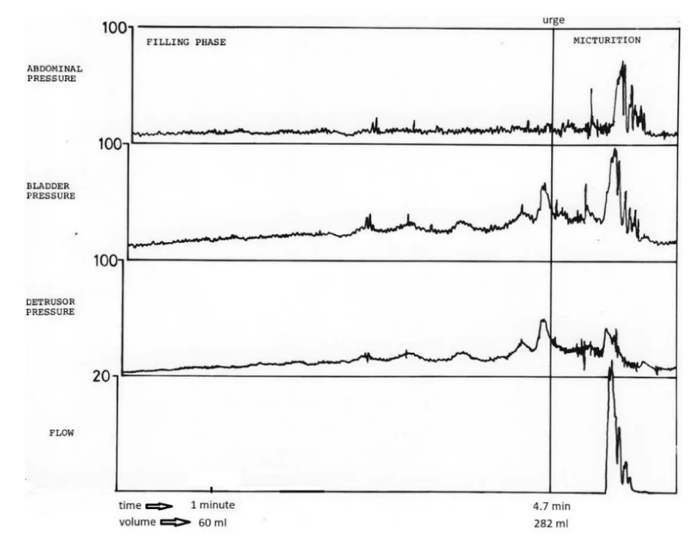
Instead of a smooth increase of detrusor pressure with increasing volume, detrusor pressure usually follows wavelike courses as shown in the cystometrogram of Figure 1. Normally, these pressure waves are transient and return to the previous pressure level. Probably because of this transient character of the waves, they are considered to be superimposed on a smooth tonic pressure component. This implies that tonic pressure and the pressure waves are considered as independent components of detrusor pressure. While the tonic component can be ascribed to passive properties of distensibility of the bladder wall, the pressure waves are obviously caused by active contractions of the bladder wall. Because no stimuli for these contractions are identified, they are supposed to be generated spontaneously.
Spontaneous pressure waves can be accompanied by sensations of urge to void or even can cause leakage. In cases where complaints of urge coincide with spontaneous pressure waves, the complaints are diagnosed as motor urge. Sensations of urge are generated by afferent nervous activity from the bladder. Complaints of urge not associated with detected pressure waves may be diagnosed as sensory urge. Nowadays, spontaneous activity of the bladder related to complaints is ascribed to bladder overactivity.
A modified concept of bladder compliance
If an amount ΔV is added fast or almost stepwise to bladder volume V, then detrusor pressure pd also increases almost stepwise. The step in detrusor pressure is caused by the almost stepwise increase of elastic elongation of the bladder wall. If after the stepwise increase in pressure bladder volume V is constant, then detrusor pressure pd gradually decreases at a decreasing rate, eventually ending at a constant level a little higher than before the addition of ΔV. During this isovolumetric pressure decay, the stepwise increase of the elastic elongated state is obviously gradually compensated by elongation of non-elastic structures of the bladder wall. This compensating elongation can be identified as the passive mechanical property of plasticity of bladder tissue [11]. The mechanical properties of elasticity and plasticity are distributed in the bladder wall. Plastic elongation is a type of elongation caused by stress only if stress is above a threshold level, and then elongation takes place with a decaying velocity, ending at a certain elongated state. The attained plastic elongated state remains as long as stress is below the newly set threshold level. The attained plastic elongated state can be lengthened more only if the previously set threshold is passed. So, the threshold level is higher for a more plastic elongated state of tissue. The observed gradual isovolumetric pressure decay after a stepwise increase of volume now can be ascribed to gradual plastic elongation of bladder tissue while the elastic elongation of the bladder wall is releasing. This phenomenon of isovolumetric pressure decay is named pressure relaxation. In a simple mechanical model for pressure relaxation the two different properties elasticity and plasticity, which both are distributed properties in bladder wall, can be lumped together in overall properties of elasticity and plasticity of bladder wall and schematically represented by two discrete elements set in series as shown in the model of Figure 2, The model is an arrangement in series of two elements named “series elastic element (SEE)” and “plasto/contractile element (PCE)”. Here, the elements represent, respectively, the overall elasticity and overall plasticity of the bladder wall in an artificially separated way.
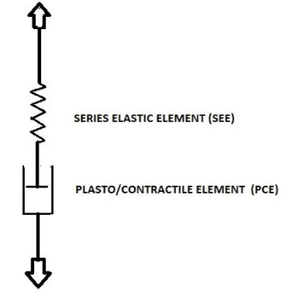
By this model, we explain the initial stepwise increase of pressure as a stepwise elongation of SEE, followed by a gradual decrease of isovolumetric pressure or relaxation as the gradual elongation of PCE is accompanied by equal release of the elongated state of SEE.
If, instead of adding ΔV, a small amount ΔV is withdrawn almost stepwise out of bladder volume V, then detrusor pressure pd decreases almost stepwise. This stepwise pressure decrease is caused by the release of part of the elastic elongated states distributed in the bladder wall. If, after the almost stepwise decrease of the elastic elongated state, bladder volume is constant, then detrusor pressure pd gradually increases, ending at a constant level a little lower than before withdrawing ΔV. The observed gradual pressure increase after the stepwise decrease of volume is named pressure recovery. In terms of the mechanical model of Figure 2, the isovolumetric pressure increase during pressure recovery can be explained by a gradual increase of the elongated state of SEE, which is compensating for an equal gradual contraction of PCE, represented also by this element. The different properties of plastic (P) elongation and contractile (C) shortening are expressed by the length of the same element PCE. In this way, relaxation and recovery are expressed as reverse phenomena. The expression of these reverse phenomena in the length of just one element PCE is in harmony with the evidence of complete inversion of the elongated state of bladder wall during each functional cycle of the bladder. In the model, the length of PCE is expressed as the resultant balance of two different distributions of passive elongations and active contractions within the bladder wall. Because passive plastic elongation and active contractions are basically different properties in different distributions in the structure of bladder tissue, the way PCE is elongated during relaxation and is contracted during recovery are determined by different parameters.
Also, SEE in Figure 2 represents the resultant sum of distributed completely invertible elastic elongated states within bladder tissue. In contrast to PCE, stress is needed to maintain the elongated state of SEE. So far, the model of Figure 2 has been used to explain the different phenomena of pressure relaxation and recovery.
The model of Figure 2 can also be used to describe the variable elongated state of the bladder wall during filling of the bladder [11]. During the collection phase, the plastic elongated state of PCE is gradually increasing because of increasing stress generated by the elongation of SEE. In the voiding phase, the elongated state of PCE is reset by stimulated contraction. The elongation of the bladder wall during filling is almost equal in two dimensions and is proportional to the radius of the bladder approximated as a sphere. The Figure 2 can represent the circumferential length of the spherical bladder.
Now the model of Figure 2 suggests dividing the virtually total volume V of the bladder into two parts: one part determined by the length of SEE and the other part determined by the length of PCE. The amount of volume ΔV that needs to be withdrawn stepwise out of V just to attain zero detrusor pressure is related to the total release of the elongated state of SEE. The volume ΔV needed to be withdrawn for pd=0 is named the Elastic Volume VE. The Elastic Volume VE is defined as that part of total volume V that causes a certain detrusor pressure pd and is determined by elastic properties ascribed to SEE. The bladder volume remaining after withdrawal of the Elastic Volume, encompassed with detrusor pressure pd=0, is named Rest Volume VR so that: V=VE+VR. According to the model of Figure 2, Rest Volume VR is determined by the elongated state of PCE. In connection with the concept of Elastic Volume VE, a parameter Elastic Compliance CE is introduced, defined by: CE= VE/pd. In the parameter of compliance C, as defined by ICS and derived from the steepness of a cystometrogram, a significant part ΔV of V in C= ΔV/ Δpd concerns Rest Volume and does not contribute to detrusor pressure Δpd. Consequently, the standard measure of compliance C is considerably larger than the Elastic Compliance CE: C>>CE. The difference between C and CE depends on the elongated state of PCE, which is a variable balance between passive elongation and active contraction and may be affected by relaxations and recoveries after disturbing variation in volume V. In standard cystometry, the variable parts Elastic Volume VE and Rest Volume VR are not distinguished in V so that their different effects on variations of compliance C are masked.
The definition of the parts VE and VR of bladder volume V suggests deriving in practice their approximated values from the determination of CE= VE/pd by measuring the step in pd after a fast withdrawal or addition of a known part ΔV. But, of course, it is easier to derive the parameters VE, VR and CE on bladders in vitro than on bladders in-situ. Experiments on pig bladders in-vitro have shown that Rest Volume VR is a significant part of total volume V and may even be larger than Elastic Volume VE [12]. Furthermore, it turned out that the ratio VE/VR of a particular bladder is variable depending on the history of passive elongation and active contraction of the bladder wall. Spontaneous contractions, relaxations and recoveries are part of that history. Although in a slow-filling cystometrogram the increase of V can be accompanied by a small increase in pd, it is concluded here that detrusor pressure is related only to part VE of total bladder volume V, so that according to the new interpretation of a cystometrogram detrusor pressure is not a measure of the filled state of the bladder.
Spontaneous contractions detected as pressure waves are distributed in the bladder wall within a wide spectrum of amplitudes and have different effects on bladder function
In terms of the model of Figure 2, the transient pressure waves, superimposed on tonic pressure as shown in Figure 1, can be ascribed to transient variation of the length of PCE, which under isovolumetric conditions is transferred to variation of the length of SEE and consequently to variation in detrusor pressure. Each transient pressure wave is generated by an active contraction of PCE followed by a passive plastic elongation of PCE. It has been shown, originally in strips of pig bladder [13] and later on in human bladder in-situ [14], that spontaneous contractions are distributed in bladder tissue. The extent of regional spontaneous activity varies and migrates. The rhythmic nature of the distributed activity may become synchronised. This character of distributed contractions has been confirmed in many studies.
The smallest possible transient contraction is generated by a single smooth muscle cell. Because the smallest contractions are generated by smooth muscle cells, contraction activity at small scale is named MicroMotions MM [13,14]. The distribution of regional transient contractions in the bladder wall can be described by a network of connected elements, each represented by the model of Figure 2, all with different elongated states. The size or number of simultaneously contracting cells in the bladder wall determines the amplitude of a detrusor pressure wave. Consequently, pressure waves will be spontaneously generated with amplitudes in a wide range. In a region with a completely transient contraction, the elongated state of PCE of the involved elements is supposed to return to the previous elongated states. If the regional contraction is not completely transient, then the plastic elongated state of PCE’s in that region is shorter than before the contraction. Not-completely-transient contractions also can be spontaneously generated and distributed in the bladder wall. Under isovolumetric conditions, not-completely-transient regional contractions in the bladder wall are compensated by elastic elongations or plastic elongations probably also elsewhere in the wall. Hence, not-completely-transient contractions may increase the elongated states of the SEE’s in the network and consequently increase stress and pressure. So, spontaneously generated not-completely-transient regional contractions may increase tonic detrusor pressure. This means that tonic pressure and spontaneous contractions are not independent as previously supposed. Spontaneous increase of tonic pressure caused by distributed spontaneous not-completely-transient regional contractions can make the bladder unstable.
The potential role of micromotions has been discussed by Drake et al [15]. In an experimental pilot study of MM in human bladder, it has been concluded that not only detected spontaneous pressure waves can be associated with sensations of urge but that also regional MM-activity, not detected as pressure waves, can be associated with sensations [14]. But also that not all MM-activity is associated with sensations. Such not sensed MM-activity still may be the source of neural activity and be the source of activity that has been described as neural noise [16]. Regional contraction activity or MM, sensed or not sensed, whether or not detected as pressure waves, can be the source of afferent nervous activity that informs CNS about the actual filling state of the bladder. Then, after the conclusion that detrusor pressure cannot be taken as a measure of total volume, a question arises as to which mechanical parameter of the bladder is coded in the afferent activity to represent actual volume.
Taking account of the variable thickness of the bladder wall with variable volume, it can be shown for a spherical bladder that stress σ in the wall is related to pressure p and volume V approximately according to the simple formula: σ ≈ (3/2Vt) pdV. [17]. So, contrary to detrusor pressure pd, stress σ in the wall is proportional to volume V and hence stress σ can be coded as a measure of actual volume V, provided that detrusor pressure pd has a reproducible value in relation to variable V and varies spontaneously only within a small range. In a study of the relation between afferent nerve activity and bladder volume during the cyclic bladder function of a rat, a correlation has been determined between afferent nerve activity and bladder volume V, detrusor pressure pd, circumferential wall tension T and stress σ in the wall. It turned out that nervous activity correlates the best with stress in the wall [16]. Afferent nerve activity depended linearly on stress within 6%, and both slope and offset were independent of the filling rate of the bladder. These experimental results confirm the conclusion drawn from the equation and are compatible with the conclusion drawn from the model of Figure 2 that detrusor pressure is determined only by the elastic part VE of total volume V and support the importance that the increase of detrusor pressure with increasing volume V is reproducible and almost constant. Spontaneous increase in stress, reflected in an increase in pressure, can become coded in afferent nerve activity that normally codes for a larger volume. Then passage of the threshold can initiate inadequate sensations of urge. For adequate coding of afferent nerve activity during a normal collection function of the bladder with physiological filling up to normal capacity, tonic and phasic detrusor pressure needs to be maintained within a small reproducible range.
Variations of the surrounding of the bladder and variations of the position of the body may change the distribution of plastic elongated states and reduce tonic detrusor pressure. Such reductions in pressure can be restored by regional spontaneous not-completely-transient-contractions or MM. From this point of view, spontaneous not-completely-transient-contractions are functional in the maintenance of a tonic detrusor pressure within an interval compatible with normal coding of nerve activity for actual volume. The process of recovery of disturbing elongations by regional contractions and MM has been ascribed a physiological meaning for accommodation of a certain detrusor pressure to actual bladder volume [18]. In particular, this process of accommodation of detrusor pressure to actual volume can be ascribed to distributed, not-sensed low-level activity or MM. To prevent bladder instability, these functional low-level, not-completely transient phasic contractions need to be limited. Hence, the conditions for the transition of completely-transient-spontaneous- contractions or MM to not-completely-transient-contractions are both physiological and clinically relevant, but still a mystery [19]. An accommodated state of the bladder is also reached in tetraplegic patients and in bladders in-vitro. So, accommodation of tonic pressure to volume seems to be an autonomic property of the bladder, i.e. without intervention of the central nervous system.
Conclusion
In deviation from routine clinical cystometry, the slowly filled bladder volume V is divided into elastic volume VE and rest volume VR, respectively, based on distributed elasticity and plasticity/contractility in the bladder wall and is represented by a simple discrete mechanical model consisting of SEE in series with PCE. This approach enables differential assessment of elasticity as a source of pressure separated from plasticity/contractility as a measure of the elongated state of the bladder wall that depends on the history or balance between passive plastic elongations and active muscular contractions. A measure of elastic compliance CE relates pressure to VE. Because the ratio VE/VR is variable, also for a certain volume V, the steepness of a cystometrogram as a measure of bladder compliance C is not an adequate measure of the pressure-volume relation of a bladder. Contractions are generated spontaneously and distributed in the bladder wall within a wide range of amplitude from micro- to macro-motions, but are not always observed in pressure as transient waves. Non-transient contractions cause an increase in tonic pressure, may make a bladder unstable, but also have a physiological meaning for the autonomic process of accommodation of pressure to volume in the collection phase. A reproducible accommodated state of tonic detrusor pressure is important for adequate sensory coding of stress in the bladder wall as a measure of actual bladder volume. Conditions for the transition of spontaneous contraction from completely to not-completely transient are still a mystery.
- Griffiths DJ. The mechanics of the urethra and of micturition. Br J Urol. 1973;45:497-507. Available from: https://doi.org/10.1111/j.1464-410x.1973.tb06812.x
- Griffiths DJ. Urodynamic assessment of bladder function. Br J Urol. 1977;49:29-36. Availa-ble from: https://doi.org/10.1111/j.1464-410x.1977.tb04518.x
- Griffiths DJ. Urodynamics: the mechanics and hydrodynamics of the lower urinary tract. Medical Physics Handbooks. Vol. 4. Bristol: Adam Hilger Ltd; 1980. ISBN: 0-85274-507-9.
- Van Duyl WA, Coolsaet BLRA, Van Mastrigt R. A new clinical parameter for the assess-ment of the contractility of the urinary bladder. Urol Int. 1978;33(1-3):31-39. Available from: https://doi.org/10.1159/000280173
- Van Duyl WA. A consistent mechanical perspective on bladder function: connecting storage and voiding phases. Bladder. 2026;13(2):e212000092. Available from: https://doi.org/10.14440/bladder.0271
- Van Duyl WA. Biomechanics of urinary bladder: slow-filling and slow-emptying cystometry and accommodation. Bladder. 2021;8(1):e451-12. Available from: https://doi.org/10.14440/bladder.2021.826
- Coolsaet BLRA, Van Duyl WA, Van Mastrigt R, Van der Zwart A. Visco-elastic properties of the bladder wall. Urol Int. 1975;30(1):16-26. Available from: https://doi.org/10.1159/000279953
- Rosier PFWM, Schaefer W, Lose G, Goldman HB, Guralnick M, Eustice S, Dickinson T, Hashim H. International Continence Society Good Urodynamic Practices and Terms 2016: urodynamics, uroflowmetry, cystometry, and pressure-flow study. Neurourol Uro-dyn. 2017;36(5):1243-1260. Available from: https://doi.org/10.1002/nau.23124
- Wyndaele JJ, Gammie A, Bruschini H, De Wachter S, Fry CH, Jabr RI, Kirschner-Hermanns R, Madersbacher H. Bladder compliance: what does it represent? Can we measure it, and is it clinically relevant? Neurourol Urodyn. 2011;30(5):714-722. Available from: https://doi.org/10.1002/nau.21129
- Van Mastrigt R, Coolsaet BLRA, Van Duyl WA. Passive properties of the urinary bladder in the collection phase. Med Biol Eng Comput. 1978;16(5):471-482. Available from: https://doi.org/10.1007/BF02457796
- Van Duyl WA. Simplified analysis of cystometrograms of urinary bladder in terms of elastici-ty and plasticity. J Urol Nephrol Stud. 2023;4(3):461-470. Available from: https://doi.org/10.32474/JUNS.2023.04.000187
- Van Duyl WA. Accommodated tonic detrusor pressure determined by the combination of variable elastic volume and variable elastic compliance explained by means of sliding fil-aments model. J Urol Nephrol Stud. 2022;4(1). Available from: https://doi.org/10.32474/JUNS.2022.04.000176
- Van Duyl WA. Spontaneous contractions in urinary bladder smooth muscle: preliminary re-sults. Neurourol Urodyn. 1985;4(4):301-307.Available from: https://doi.org/10.1002/nau.1930040406
- Van Os-Bossagh P, Kosterman LM, Hop WC, et al. Micromotions of bladder wall in chronic pelvic pain: a pilot study. Int Urogynecol J. 2001;12(2):89-96. Available from: https://doi.org/10.1007/s001920170071
- Drake MJ, Kanai A, Bijos DA, Ikeda Y, Zabbarova I, Vahabi B, et al. The potential role of unregulated autonomous bladder micromotions in urinary storage and voiding dysfunc-tion, overactive bladder and detrusor underactivity. BJU Int. 2017;119(1):22-29. Availa-ble from: https://doi.org/10.1111/bju.13598
- Biallosterski BT, Van Koeveringe GA, Van Kerrebroeck PE, Gillespie JI, De Wachter SG. Nonvoiding activity of the guinea pig bladder. J Urol. 2011;186(2):721-727. Available from: https://doi.org/10.1016/j.juro.2011.03.123
- Le Feber J, Van Asselt E, Van Mastrigt R. Afferent bladder nerve activity in the rat: a mech-anism for starting and stopping voiding contractions. Urol Res. 2004;32:395-405. Availa-ble from: https://doi.org/10.1007/s00240-004-0416-8
- Van Duyl WA, Coolsaet BLRA. Biomechanics of the urinary bladder: spontaneous contrac-tion activity and micromotions related to accommodation. Int Urol Nephrol. 2021;53(7):1345-1353. Available from: https://doi.org/10.1007/s11255-021-02814-w
- Van Duyl WA. History and mystery of micromotions in the wall of urinary bladder: a review of chain of coupled investigations of spontaneous contractions on pig bladder and human bladder. J Urol Nephrol Stud. 2024;4(4). Available from: https://doi.org/10.32474/JUNS.2024.04.00020



 Save to Mendeley
Article Alerts
Subscribe to our articles alerts and stay tuned.
 This work is licensed under a Creative Commons Attribution 4.0 International License.