Archive of Urological Research
Giant Vesical Calculus with Vesico-Vaginal Fistula and Multiple Sewing Needles in the Peritoneal Cavity in a 13 year old Female
Madras University and the Royal Colleges of Surgeons of England and Edinburgh, Urologist, St Philomena’s Hospital, Bangalore, India
Author and article information
Cite this as
Vijayan P. Giant Vesical Calculus with Vesico-Vaginal Fistula and Multiple Sewing Needles in the Peritoneal Cavity in a 13 year old Female. Arch Urol Res. 2025; 9(1): 001-003. Available from: 10.17352/aur.000055
Copyright License
© 2025 Vijayan P. This is an open-access article distributed under the terms of the Creative Commons Attribution License, which permits unrestricted use, distribution, and reproduction in any medium, provided the original author and source are credited.A 13 year old post pubertal nomadic girl came to us with a history of recurrent lower abdominal pain and urinary incontinence of 3 years duration. She also had intermittent hematuria. She also stated that her late (mentally deranged) mother had inserted multiple needles into her (patient’s) vagina when she was 5 years of age. She underwent a successful 2 stage surgical procedure for removal of the needles, calculus and repair of the fistula.
Vesico-Vaginal Fistula (VVF) is an abnormal communication between the urinary bladder and the vagina causing urinary incontinence. It usually occurs in women in the child bearing age or the elderly but rare in the younger age group. VVF resulting from a large bladder stone causing pressure necrosis of the bladder wall is very uncommon. Foreign bodies in the bladder can also cause stone formation and fistulous communication with the vagina. The present case had a large bladder stone and VVF. She also had multiple metallic foreign bodies (sewing needles) in the peritoneal cavity.
Case5 report
- A 13 year old post pubertal nomadic girl hailing from an extremely poor economic background, moving from place to place selling glass beads came to us with a history of recurrent lower abdominal pain and urinary incontinence of 3 years duration. She also had intermittent hematuria. She attained menarche at the age of 12 years and stated that her late mother who suffered from mental illness had inserted multiple needles into her (patient’s) vagina when she was 5 years old. On examination she was poorly nourished (Ht 4ft 6 in) wt (25 kg) anemic with Peri-orbital edema. She had suprapubic tenderness and her Undergarments were soaked with urine.
Abdominal examination did not reveal any distension of the urinary bladder.
Her virginal orifice showed continuous leakage of urine. The rest of the abdomen revealed no abnormal findings
Lab investigations:
- Lab: Hb 9 g
- RBS 120 mg
- Urea 62 mg
- Creatinine 2.3 mg
The urine was heavily infected (E. Coli).)
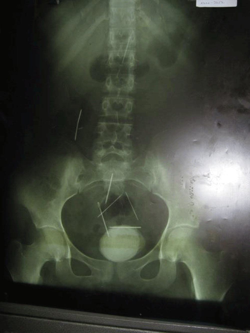
- X-Ray (Figure 1)
- Large vesical calculus
- Multiple radio-opaque linear objects in the abdominal cavity
- CT Scan Confirmed metallic objects in peritoneal cavity
- One such object located in the under surface of liver
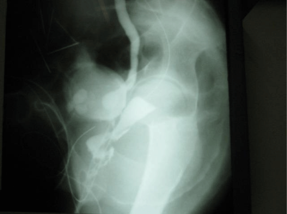
Cystography showed (Figure 2)
- Vesical calculus almost filling up the bladder
- Marked Lt vesico- ureteric reflux
- Lt hydro-ureteronephrosis
- Fistula between vagina and bladder close to bladder neck
Under the cover of antibiotics she was taken up for surgical treatment.
Under general anesthesia
- Cystoscopy confirmed vesical calculus
- Vesico vaginal fistula close to bladder neck
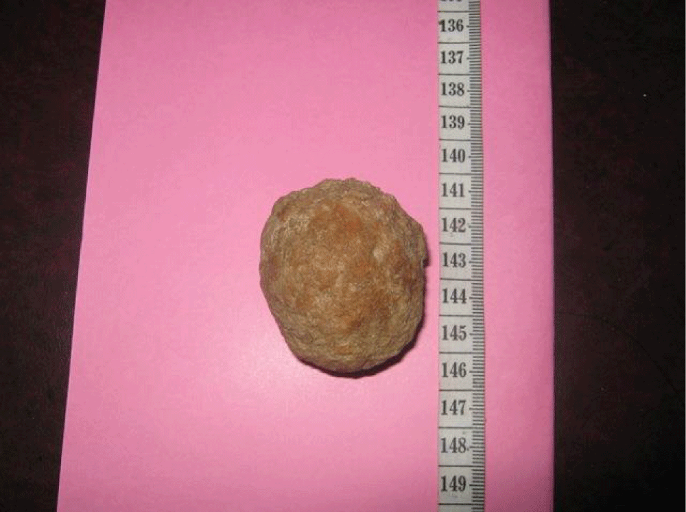
Through the abdominal route, the bladder was opened. The stone was occupying the entire lumen of the bladder and adherent to the grossly thickened inflamed wall. The stone measuring 6.5 cms X 6 cms was separated from the bladder wall and removed (Figure 3). There was a 1 cm fistulous communication between the bladder wall (close to the bladder neck) and the anterior vaginal wall.
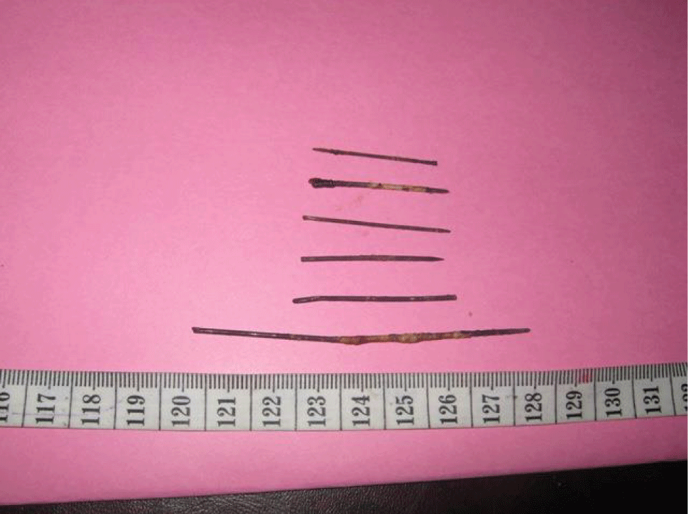
The peritoneal cavity was opened and 5 sewing needles were identified and removed (Figure 4) One needle was found to be densely adherent to the common bile duct area it was felt that attempts to remove might be hazardous and hence abandoned.
In view of the heavily infected bladder wall, it was decided to repair the fistula after 3 months. A supra-pubic catheter was kept and the bladder closed.
3 months later she was readmitted
- General condition much better
- Wt 35 Kg
- Hb 12 g
- Urea 37 mg
- Creatinine 1.4
- Leakage of urine per vagina continues as before
Again under general anesthesia, the vesico-vaginal fistula was approached through the vaginal route and successfully closed. Martius fat pad flap from thr Rt. Labium Majus was interposed between the vaginal wall and the bladder to strengthen the repair. The suprapubic and urethral catheters placed at the time of surgery were removed after 2 weeks. She found herself to be dry and voiding urine normally with good control.
She was followed up for 5 years and she was leading a normal life. She got married after 3 years and delivered a male child by Caesarean section 2 years later.
The lone sewing needle was still at the same site and causing no problems.
Discussion
VVF may be very rarely congenital but usually acquired. In the developed countries it may follow radiation, pelvic malignancy or surgical procedure (Hysterectomy). Obstructed labour is a common cause in the developing countries [1]. Foreign bodies can also cause stone formation in about 10% of cases [2]. Vesical calculus causing pressure necrosis of the bladder is rare [3].
The symptoms are varied, recurring UTI, pyuria, suprapubic pain and predominantly urinary incontinence.
Apart from routine investigations i.e., blood chemistry, urine analysis, culture and antibiotic sensitivity are mandatory.
Specific investigations include ultra-sonography, KUB (X-ray) initially and occasionally contrast study.
The management of VVF will depend on the presence of infection and the state of the bladder wall.
If it is severely inflamed, a 2-stage procedure is recommended [4].
In the present case, a 2 stage abdominal approach was adopted i.e., removal of the stone initially and a delayed repair of the fistula 3 months later in view of the bladder being grossly inflamed as a result of infection.
The presence of sewing needles in the peritoneal cavity was probably incidental and had no influence on the causation of stone or fistula. All but one needle were removed. The lone needle was left in situ as it was feared that attempts to remove it might cause unexpected complications involving the common bile duct.
Treatments cured her
Conclusion
This is a unique case for 2 reasons.
1. The patient had undergone child abuse by her mother who had inserted sewing needles into her vagina.
2. She being from a poor economic background developed a large bladder stone which caused a urinary fistula between bladder and vagina.
Surgical treatment in 2 stages cured her.
- Cheng MH, Choo HT, Horng HC, Hung MJ, Huang BS, Wong PH. Vesical calculus associated with vesico-vaginal fistula. Gynecol Minim Invasive Ther. 2014;3:23-25. Available from: https://doi.org/10.1016/j.gmit.2013.07.009
- Dalela D, Goel A, Shakhwar SN, Singh KM. Vesical calculi with unrepaired vesicovaginal fistula: a clinical appraisal of an uncommon association. J Urol. 2003;170(6 Pt 1):2206-2208. Available from: https://doi.org/10.1097/01.ju.0000095503.76155.30
- Palinrungi MA, Syahrir S, Kholis K, Syarif M, Faruk M. Giant bladder stone formed around sewing-needle in childhood: a case report and literature review. Urol Case Rep. 2020;29:101101. Available from: https://doi.org/10.1016/j.eucr.2019.101101
- Shephard SN, Langmang SJ, Kirshner CV. Bladder stones in vesico-vaginal fistula: Is concurrent repair an option? Experience with 87 patients. Int Urogynecol J. 2017;28:569-574. Available from: https://doi.org/10.1007/s00192-016-3142-1
Article Alerts
Subscribe to our articles alerts and stay tuned.
 This work is licensed under a Creative Commons Attribution 4.0 International License.


 Save to Mendeley
