International Journal of Spine Research
Multi-Level forestier syndrome in the cervical vertebra with an unusual radiographic appearance: Case report
Deniz Sirinoglu, Mehmet Volkan Aydin*, Idris Avci, Gizem Meral Atiş, Buse Sarıgül and Mehmet Volkan Aydın
Cite this as
Sirinoglu D, Aydin MV, Avci I, Atiş GM, Sarıgül B, et al. (2020) Multi-Level forestier syndrome in the cervical vertebra with an unusual radiographic appearance: Case report. Int J Spine Res 2(1): 001-003. DOI: 10.17352/ijsr.000006We present a rare case of Forestier disease with multi-level vertebra involvement from the upper cervical to the thoracic area which has not been reported in the literature before.
A 65- year old male patient was admitted to our outpatient clinic with neck pain, dysphagia and sleep apnea for over 5 months. On his cervical CT scan revealed broad ossification of the anterior longitudinal ligament from C2 to T1 with anteriorly beaking osteophytes surrounding the vertebral bodies causing compression of the trachea and esophagus on C2 and C3. With the diagnosis of Diffuse Idiopathic Skeletal Hyperostosis (DISH), the patient underwent surgery. With an anterolateral approach the ossified pathological segment was removed with a high-speed drill and the patient’s symptoms revealed immediately after the surgery.
Forestier disease or DISH is defined as a non-inflammatory ossification of spinal and peripheral elements. It is mostly seen in the thoracic region. In the cervical region it is mostly seen in the subaxial segment between C4-C7. Dysphagia, being the most prominent symptom in patients with cervical DISH. The fact that makes our case unique is the multiple cervical vertebra involvement of DISH starting in the upper cervical vertebra which has not been reported in the literature in this variety before.
In our patient the ossification mimicking a bird’s beak had vertical growing characteristics despite being in the upper cervical area. Also, multi-level involvement in the cervical spine is rare because the cervical vertebrae are more mobile than the ones in the thoracic area. In our patient ossification from C2 to T1 was present which has not been presented in the literature before. Rapid improvement of the patient’s symptoms confirmed with the radiological images strenghten the hypothesis that patients with dysphagia, especially in the ones with weight loss due to it, have better outcomes when approached surgically than conservatively.
Background
Diffuse Idiopathic Skeletal Hyperostosis (DISH) or Forestier disease is defined as a non-inflammatory ossification of ligaments of the spinal colon [1]. It is mostly seen in the upper thoracic vertebra but can also occur anywhere on the vertebral colon [2]. Estimated prevalence is suggested between 2.9%-28% in general [3]. Mostly identified as an incidental finding on plain radiographs or computerized tomography most cases are asymptomatic. But based on location and size symptoms may vary from neck and back pain, to dysphagia and eating disabilities. The incidence rises with age and several metabolic disorders like diabetes mellitus and hypercholesterolemia are risk factors for DISH [2,4].
We present a rare case of Forestier disease with multi-level vertebra involvement from the upper cervical to the thoracic area which has not been reported in the literature before.
Clinical presentation
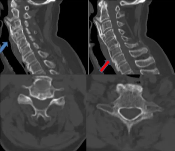
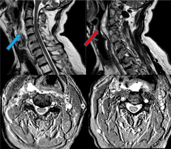
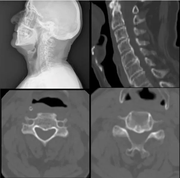
A 65- year old male patient with history of diabetes mellitus was admitted to our outpatient clinic with neck pain, dysphagia and sleep apnea for over 5 months. Previously evaluated by the otolaryngology department a esophagography a slight compression of the upper cervical esophagus segment was observed. He stated that he lost 6kg in 6months due to the dysphagia. On his cervical CT scan revealed broad ossification of the anterior longitudinal ligament from C2 to T1 with anteriorly beaking osteophytes surrounding the vertebral bodies causing compression of the trachea and esophagus on C2 and C3 (Figure 1). Magnetic Resonance Imaging (MRI) showed that there were no disc pathologies; vertebral heights and spinal alimentation were normal. The spinal canal and spinal cord were intact. (Figure 2). With the diagnosis of DISH, the patient underwent surgery. With an anterolateral approach the ossified pathological segment was removed with a high-speed drill and no compression of the surrounding soft tissue fractures were confirmed. The patient’s symptoms revealed immediately after the surgery and on the postoperative control CT scan, total clearance of the ossified structures were observed (Figure 3).
Discussion
Forestier disease is defined as a non-inflammatory ossification of spinal and peripheral elements [5,6] and was first reported by Forestier and Rotes-Querol in 1950 [5] and later named as DISH by Resnick, et al., in 1975 [7]. Three criteria for diagnosis is crucial for this condition: ossification of the anterior longitudinal ligament in at least four vertebral bodies successively, preservation of intervertebral disc height and intra-articular osseous fusion or sclerosis with absence of apophyseal joint bone ankylosis and sacroiliac joint erosion [5,8]. It is mostly seen in the thoracic region [5,9], but cases of the other vertebra segments are reported as well. In the cervical region it is mostly seen in the subaxial segment between C4-C7 [10]. As the thoracic vertebrae are immobile ossification of the structures tend to grow more vertically and can involve multiple levels, whereas cervical vertebra being more mobile they mostly show a horizontal growing pattern [7]. The etiology of DISH still remains unclear. Incidences vary between 2.9-28% in general [3]. Obesity, diabetes mellitus, hypertension, acromegaly, hypervitaminosis A are some risk factors for Forestier disease [8,11]. Most of the patients are asymptomatic are only diagnosed incidentally on radiographic findings but may vary depending on the size and localization of the ossifications [9]. Dysphagia, being the most prominent symptom is seen in 17- 28% of patients with cervical DISH due to compression of the esophagus by the osteophytes [5,8,9]. Some patients may present with pain and restriction of range of motion [12] or suffer from dyspnea caused by tracheal obstruction, sleep apnea and stridor [13]. Also in rare cases, osteophytes compressing the jugular foramen, sympathetic chain, vertebral artery were reported [9]. Treatment may be either conservative or surgical however conservative treatment is adequate for most of the patients [5]. For patients with dysphagia, airway compromise and rapid weight loss, surgery should be the initial choice of treatment [8]. For the surgical treatment, anterior approach with special caution to the adhesions between the osteophytes and pharyngo-esophageal segment, may be preferred [9].
Conclusion
The fact that makes our case unique is the multiple cervical vertebra involvement of DISH starting in the upper cervical vertebra which has not been reported in the literature in this variety before. Forestier disease, being more common in the thoracic and lower cervical area, show a vertical growing pattern in the thoracic and horizontal pattern in the cervical area. In our patient the ossification mimicking a bird’s beak had vertical growing characteristics despite being in the upper cervical area. Also, multi-level involvement in the cervical spine is rare because the cervical vertebrae are more mobile than the ones in the thoracic area. In our patient ossification from C2 to T1 was present which has not been presented in the literature before. Rapid improvement of the patient’s symptoms confirmed with the radiological images strenghten the hypothesis that patients with dysphagia, especially in the ones with weight loss due to it, have better outcomes when approached surgically than conservatively. In our opinion, it is not necessary to remove all the ossificated levels. In our case, we just drilled the segment in contact with the esophagus and trachea and had excellent results.
- Toyoda H, Terai H, Yamada K, Suzuki A, Dohzono S, et al. (2017) Prevalence of diffuse idiopathic skeletal hyperostosis in patients with spinal disorders. Asian Spine J 11: 63-70. Link: http://bit.ly/36NLDGt
- Lecerf P, Malard O (2010) How to diagnose and treat symptomatic anterior cervical osteophytes? Eur Ann Otorhinolaryngol Head Neck Dis 127: 111-116. Link: http://bit.ly/37QUIOM
- Egerter AC, Kim ES, Lee DJ (2015) Dysphagia Secondary to Anterior Osteophytes of the Cervical Spine. Global Spine J 5: e78-e83. Link: http://bit.ly/39TeZVH
- Ramírez-Villaescusa J, Restrepo-Pérez M, Ruiz-Picazo D (2016) Spinal Epidural Hematoma Related to Vertebral Fracture in an Atypical Rigid Diffuse Idiopathic Skeletal Hyperostosis: A Case Report. Geriatr Orthop Surg Rehabil 8: 18-22. Link: http://bit.ly/37R49he
- Najib J, Goutagny S, Peyre M, Faillot T, Kalamarides M (2014) Forestier's disease presenting with dysphagia and disphonia. Pan Afr Med J 17: 168. Link: http://bit.ly/2FvkWKq
- Omar A, Mesfin A (2017) Odontoid Fracture in a Patient With Diffuse Idiopathic Skeletal Hyperostosis. Geriatr Orthop Surg Rehabil 8: 14-17. Link: http://bit.ly/2T6Rvq5
- Resnick D, Shaul SR, Robins JM (1975) Diffuse idiopathic skeletal hyperostosis (DISH): Forestier's disease with extraspinal manifestations. Radiology 115: 513-524. Link: http://bit.ly/37N4LnU
- Goico-Alburquerque A, Zulfiqar B, Antoine R, Samee M (2017) Diffuse Idiopathic Skeletal Hyperostosis: Persistent Sore Throat and Dysphagia in an Elderly Smoker Male. Case Rep Med 2017: 2567672. Link: http://bit.ly/301J4Om
- Srivastava SK, Bhosale SK, Lohiya TA, Aggarwal RA (2016) Giant Cervical Osteophyte: An Unusual Cause of Dysphagia. J Clin Diagn Res 10: MD01–MD02. Link: http://bit.ly/2t2LfF5
- Özkırış M, Okur A, Kapusuz Z (2014) Forestier's syndrome: a rare cause of dysphagia. Kulak Burun Bogaz Ihtis Derg 24: 54-57. Link: http://bit.ly/2uzxFJZ
- Mader R (2008) Diffuse idiopathic skeletal hyperostosis: time for a change. J Rheumatol 35: 377-379. Link: http://bit.ly/35xYN8N
- Bakker JT, Kuperus JS, Kujif HJ, Oner FC, de Jong PA, et al. (2017) Morphological characteristics of diffuse idiopathic skeletal hyperostosis in the cervical spine. PLoS One 12: e0188414. Link: http://bit.ly/2sRVrjZ
- McCafferty RR, Harrison MJ, Tamas LB, Larkins MV (1995) Ossification of the anterior longitudinal ligament and Forestier's disease: an analysis of seven cases. J Neurosurg 83: 13-17. Link: http://bit.ly/2QXFzEx
Article Alerts
Subscribe to our articles alerts and stay tuned.
 This work is licensed under a Creative Commons Attribution 4.0 International License.


 Save to Mendeley
