International Journal of Spine Research
Comparative study of the bone graft area and fusion rate in unilateral transforaminal lumbar interbody fusion between Endoscopic and Miniopen procedures. A technical note and preliminary report
Kang Taek Lim1*, Hyung-Suk Kim1, Elmer Jose Arevalo Meceda3,4, Han Gawi Nam1 and Chun-Kun Park1,2
2Professor Emeritus, Department of Neurosurgery, The Catholic University of Korea, Seoul, St. Mary’s Hospital, 222 Banpo-daero, Seocho-gu, Seoul 06591, Korea
3Department of Neurosciences, University of the East Ramon Magsaysay Memorial Medical Center, Aurora Boulevard, Quezon City, Metro Manila, Philippines
4Department of Surgery, Section of Neurosurgery, Bicol Medical Center, Naga City, Camarines Sur, Philippines
Cite this as
Lim KT, Kim HS, Arevalo Meceda EJ, Nam HG, Park CK (2020) Comparative study of the bone graft area and fusion rate in unilateral transforaminal lumbar interbody fusion between Endoscopic and Mini-open procedures. A technical note and preliminary report. Int J Spine Res 2(1): 029-036. DOI: 10.17352/ijsr.000011Spinal fusion surgery can now be performed through the endoscopic approach. Adequate endplate preparation and sufficient contact between bone graft or bone graft substitutes with the surfaces of the vertebral endplates are main factors to achieve successful arthrodesis. The purpose of this study are to compare the bone graft area, ratio of allograft-bonegraft (allo-bone) to total disc area, fusion rate, functional and radiographic outcomes between Endoscopic and Mini-open TLIF and to introduce the endoscopic technique of endplate preparation and implantation.
Methods: Hospital records of 59 patients who underwent TLIF (Endoscopic fusion, 23; Mini-open TLIF, 36) for the period of January 2017 to June 2018, were reviewed. Immediate postoperative CT scans were used to measure the bone graft area of an index segment by getting a mid-disc level slice on CT. The bone graft area and the ratio of the area occupied by allo-bone against the total disc area were computed. Perioperative outcomes such as operation time, length of hospital stay, and incidence of surgical complications were also recorded and analyzed. The Visual Analog Scale (VAS) were assessed 1week, 1month, and 12 months after surgery. Oswestry Disability Index (ODI) were evaluated preoperative, and 12months after surgery in both groups. The restoration of disc height (DH), segmental lumbar lordosis (SLL), fusion rate were also reviewed at 12months postoperatively.
The endoscopic technique of endplate preparation and implantation were described in detail.
Results: The bone graft area ratio were significantly higher in endoscopic group (42.4±20.3%) than in the Mini-open group (32.3±2.8%), (p<0.01) in immediate postoperative CT scan.
The VAS were significant lower in endoscopic TLIF at postoperative 1week than Mini-open TLIF (p < 0.05), and both were identically improved at 12months with no significant difference between the two groups as was the ODI.
DH, SLL were significantly increased in both group at 12 months (p<0.01).
Fusion rate for both procedures were considered to be satisfactory at 12months postoperatively, but there was not significantly different between the two groups (Endoscopic TLIF, 95.6%, Mini-open TLIF, 94.4%, p= 0.977).
Conclusion: The bone graft area and area ratio were significantly higher in Endoscopic group than Mini-open TLIF. Both techniques however, provided excellent clinical results with low complication rates. More effective instruments should be developed for Endoscopic TLIF to reduce the operation time.
Introduction
The TLIF procedure was first introduced by Harms and Rollinger as an alternative to PLIF in 1982 [1,2], Foley, et al, first described the minimally invasive TLIF procedure in 2003 [3] and It has since become an increasingly popular method of lumbar arthrodesis in spine fusion [4,5].
Mummaneni and Rodts [6], introduced the Mini-open TLIF technique, in which an expandable tubular retractor is used in a Wiltse-type approach. This procedure provides direct simultaneous visualization of both pedicles and allows placement of pedicle screws through the expandable retractor.
Mini-open TLIF was recently proposed as an alternative treatment for spondylolisthesis with the benefit of providing that unilateral access via the facet joint to decompress the exiting and traversing nerve root directly and provides excellent clinical results such as high fusion rates, restoring disc space height, lumbar lordosis, and coronal-sagittal balance of the spine with smaller operative wounds, reduced trauma to adjacent tissue, and a more rapid postoperative recovery [7-9].
Despite the many advantages, the main setback was the difficulty encountered while preparing proper fusion bed and an increased risk of dural injury was also reported [10], due to the narrow operating space.
A proper preparation of the endplate is important to effect a solid intradiscal fusion by maximizing the surface area available for implant and allo-bone graft seating. The endoscopic technique may have the potential advantage of good visualization during this stage. Direct endoscopic guidance during preparation of adequate fusion bed and during insertion of the cage may result to lesser risk of neurological injury and better fusion rates.
As spinal endoscopic science, techniques, and instruments developed, the endoscopic approach now became a new option for the surgeon to perform spinal fusion surgery [11,12]. It is thought that insertion of endoscopy system into the disc space allows very clear visualization inside the disc which facilitates safer discectomy resulting to creation of a bigger fusion space than Mini-open technique.
In this study, we compared the interbody bone graft area and area ratio after Endoscopic TLIF and Mini-open TLIF to assess their relationship to fusion rates and clinical, radiological outcomes.
Materials and methods
Patients and clinical assessment
The study was conducted at Good Doctor Teun Teun Hospital, Anyang, South Korea. The hospital review board approved the study protocol. The study was done through a retrospective review of hospital records of 59 consecutive patients who had been diagnosed with one level lumbar stenosis with unstable Grade I-II spondylolisthesis. Twenty three (23) patients had undergone Endoscopic TLIF and thirty six (36) patients had undergone Mini-open TLIF for the period of January 2017 to June 2018. All endoscopic fusion surgeries were performed by one surgeon and another surgeon performed all the Mini-open procedures, both using a single cage filled with allo-bone and stabilized by percutaneous pedicle screws-rods. The operators are both senior minimal invasive spine surgeons of our institution with more than 20 years of experience and each of them are known masters of their surgical technique of choice.
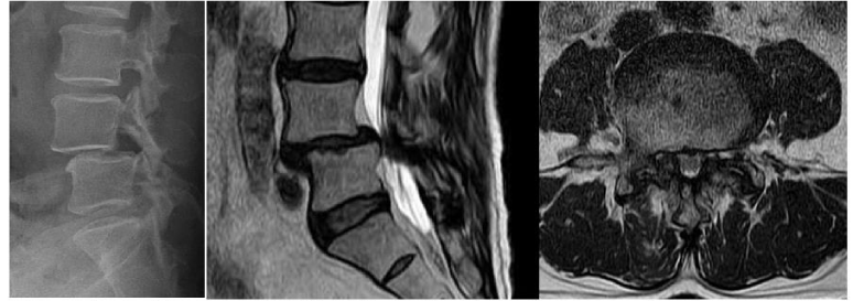
The inclusion criteria were single level lumbar stenosis with unstable grade 1or 2 spondylolisthesis causing low back pain and radiculopathy which correlated well with radiological features on MRI, CT scan and plain radiography that were refractory to conservative treatment for a period of at least six weeks (Figure 1). Patients with stable spondylolisthesis, multiple level stenosis, those with a history of previous lumbar spine surgery and those with degenerative spondylolisthesis grade 3 and above were excluded.
The demographics, mean age, gender, operation time, length of hospital stay, and complications were recorded through review of medical records. Magnetic Resonance Imaging (MRI) were done within 24 hours postoperative for all patients to demonstrate any complications.
All 59 patients were clinically assessed based on the VAS score (visual analog scale score for the back), preoperatively and at postoperative 1week, 1 months and 12 months. Also, the patients were assessed based on Oswestry Disability Index (ODI) preoperatively and at postoperative 12 months.
Radiographic assessment
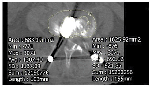
In order to objectively determine whether endoscopic procedures augment TLIF and enhance fusion bed preparation, the radiological assessment included immediate postoperative CT of the level of interest. The bone graft area was measured by the PACS INFINITE system software (S. Korea) on axial view of the CT scan.
The ratio of the area of the fusion bed occupied by the morselized allo-bone graft and the cage at the mid-disc level versus the area of the whole endplate were calculated at the same axial level as bone graft area (Figure 2).
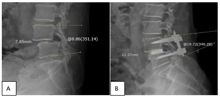
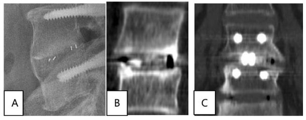
Disc Height (DH), Segmental Lumbar Lordosis (SLL), and fusion rate were assessed to compare radiologic difference between two groups at the 12months postoperatively. The DH was defined as the interbody height measured by a line through the mid-point perpendicular to the endplate line of the vertebral body at the fusion disc level. The SLL was measured as the Cobb angle of the superior and inferior endplates at the fused level pre and postoperatively (Figure 3). The fusion rates were assessed based on Brantigen and Steffee criteria [13], standing anterior-posterior and lateral with dynamic radiographs of the lumbar spine were performed at the 12months postoperatively. Radiographic fusion was confirmed by the presence of continuous bridging bone between the both adjacent vertebral bodies with the cage (classification D), a sclerotic line between the graft and vertebral bone (Classification E), no cage migration or collapse and no screw loosening on dynamic x-ray (Figure 4). If plain x-ray did not show a clear fusion, patients were evaluated with CT. The coronal CT images was essential radiological study to confirm the vertical band-like bone bridge on both sides of titanium case at the graft sites (Figure 4).
Surgical technique
The Endoscopic TLIF and Mini-open TLIF involves positioning the patient prone on a Wilson frame after the patient was put under epidural anesthesia with mask sedation. A unilateral paramedian 8mm skin incision, 3cm from midline of the level of interest was made for Endoscopic TLIF and placement of working sleeve in the middle of facet joint on the symptomatic side, a 25mm skin incision was made for Mini-open approach.
Unilateral total facetectomy and laminotomy was performed using Kerrison punch and high-speed drill to be able to clearly visualize the underlying traversing and exiting root in both procedures.
The ipsilateral traversing and exiting nerve roots were carefully identified after total facetectomy. The contralateral traversing nerve root was decompressed by medial facetectomy. In the Mini-open approach, after ipsilateral decompression of the lateral recess, the table, the microscope, and the tubular retractor can be tilted to access and decompress the central, contralateral lateral recess and foramen if necessary.
Double sealing technique [14], using Tachosil® (a sealant agent) was used to repair observed incidental durotomy that happens during the operation and conversion to open surgery was no longer necessary to close the tear.
In the endoscopic TLIF technique, the working sleeve bevel was used to medially retract the traversing root and thecal sac into the center of spinal canal to protect the nerve before annulotomy/ discectomy. A nerve root retractor was used for the same purpose in Mini-open technique.
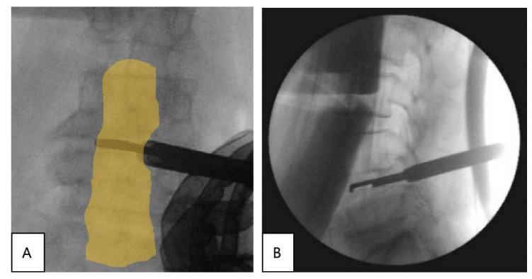
The annulus was incised between the thecal sac or traversing nerve root and exiting nerve root and a wide window was made on the ipsilateral paramedian posterior annulus. After annulotomy, proper endplate preparation was done under direct endoscopic visualization. Discectomy for adequate fusion bed was performed using disc shaver, forceps and curettes (Figure 5).
Under the endoscopic view, disc material and cartilaginous endplate were thoroughly removed, leaving the bony endplates intact. In both procedures, care should be taken not to injure the bony endplates to reduce the chance of vertebral subsidence against the interbody cage.
After preparing the endplates, operator use shaver to measure the appropriate size and length of the interbody cage in both groups. Then, allo-bone graft was packed inside the fusion space created (Figure 6). Allo-bone graft delivery device is inserted into the disc space, and the anterior and contralateral portions of the disc space was tightly packed with Demineralized Bone Matrix (DBM) combined with available autologous bone graft to promote interbody fusion. Graft impactors were used to maximize the amount of DBM that can be placed into the interbody space (Figure 6).
A single cage (Medusa®, 3D printed alloy porous cage, Meddysey, S. Korea) was packed with DBM and was inserted into the interbody space through the soft tissue retractor under the endoscopic guidance and placed anteriorly and as centrally as possible to correct lordosis and for reduction slippage (Figure 7). A PEEK, polyetheretherketone cage (AnyPlus® lumbar Interbody cage, GS medical, S. Korea ) filled with DBM graft was inserted into the central part of the disc space in Mini-open TLIF. Cage position was confirmed with C arm fluoroscopy during implantation.
The percutaneous pedicle screw fixation used in Endoscopic TLIF (MISS® Pedicle screw, Meddyssey, S. Korea) and those used in Mini open TLIF (Anyfix® GS Medical, S. Korea ) were identical percutaneous systems.
After checking the proper placement of interbody cage and screws, the surgeon tightened the screws and rods in compression to restore the normal lordosis and prevent cage migration. Placement of surgical drains were done in both groups for minimizing hematoma formation. All patients are mobilized 6hrs postoperatively as comfort allowed them to improve general outcomes and reduce adverse events. Drain is removed 1day postoperatively in both groups.
Results
Demographic data
The study involved 59 patients who had been diagnosed with lumbar stenosis with one level unstable spondylolisthesis. 23 patients (mean age ± SD: 62.7 ± 4.1) underwent Endoscopic TLIF all performed by one operator and 36 patients (mean age ± SD: 58.2 ± 8.2) underwent Mini-open TLIF, all by another operator. There were no significant difference in the ages between the 2 groups. The operative levels ranged from L2–3 to L5–S1: L2–3 in 3 patients, L3-4 in 7 patients, L4–5 in 32 patients, and L5–S1 in 17 patients (Table 1). The most common operated spinal level was L4-L5 in both groups followed by L5-S1.
Clinical outcomes
The mean operative time was longer in Endoscopic TLIF (163.6±52.1 minutes) than in Mini-open TLIF group (152.5±53.4 minutes) which failed to show significance (P=0.142). The average hospital stay was shorter for Endoscopic TLIF than for Mini-open TLIF group which was statistically significant. (2.80±4.23 vs 5.38 ± 0.52, p < 0.01)
The perioperative complications recorded were dural tear in 3 patients and postoperative epidural hematoma in 1 patient, no deep infection, no root injury were reported in both groups (Table 2). The incidental durotomies happened once in Endoscopic TLIF and twice in Mini-open TLIF. Tachosil sealing technique were done in all instances. Revision surgery was not required in the immediate postoperative period in both groups. One case of symptomatic epidural hematoma happened in the Endoscopic TLIF group but resolved without surgical intervention.
The results of VAS for back pain showed a statistically significant (p<0.01) improvement in both groups postoperatively. The 1 week VAS for back pain was significantly lower in Endoscopic TLIF compared to Mini-open TLIF patients (Table 3). This may be related to less damage of muscle.
The VAS for back pain at 1month and 12months were both improved and there was no statistically significant difference in both groups (p>0.05) (Table 3).
The mean ODI value for the two groups both significantly improved from 52.6 (Endoscopic TLIF) and 57.2 (Mini-open TLIF) preoperatively to 22.0 (Endoscopic TLIF), 21.6 (Mini-open TLIF) postoperatively at 12 months (p<0.01) (Table 3, Figure 8).
Radiographic outcomes
The Disc height and segmental lumbar lordosis showed a statistically significant improvement between preoperative and postoperative 12months in both groups (Table 4).
Intervertebral DH changed from 8.5±5.2 preoperatively to 12±3.1 postoperatively in Endoscopic TLIF group and from 8.9±3.2 to 11.3±3.1 in Mini-open TLIF at 12months follow-up. SLL improved from 8.7±4.2 preoperatively to 10.7±7.2 at 12months postoperatively in Endoscopic TLIF group and from 7.2±2.7 at pre-operation to 10.3±4.6 at 12months in Mini-open TLIF. There were statistically significant differences in the change of disc height, segmental lumbar lordosis pre and postoperatively in two groups (p<0.01).
The bone graft area were assessed by computed tomography (CT) at immediate postoperative period. The bone graft area ratio was defined as the bone graft area, occupied allo-bone and interbody cage divided by the total endplate area of the mid portion of the axial disc space. The bone graft area ratio was significantly higher in the Endoscopic TLIF group (42.4±20.3%) than in the Mini-open TLIF group (32.3±2.8%, p<0.01) (Table 4).
The 12months antero-posterior, lateral radiographs and dynamic conventional radiography views were used to assess the solid fusion status. The presence of bone bridge between cage and vertebral body, the absence of lucency around the cages and no adverse dynamic changes such as subsidence, loss of correction, cage dislodgment, and loosening of instrumentation were assessed. As it is hard to confirm the bone bridge through the simple x-ray in the titanium cage, additional CT scan was needed to evaluate the solid fusion (Figure 4). The fusion rate showed no difference between the two groups, reaching 95.6% Grade E in Endoscopic TLIF and 94.4% in Mini-open TLIF using Brantigan and Steffe classification. Failure of fusion were noted in 1case in Endoscopic TLIF and in 2 cases in Mini-open TLIF which had cage migration of >3 mm. These 3 cases required subsequent revision.
Discussion
Tatsumi, et al. [15], reported that the average area of endplate preparation in MIS-TLIF (39.2%) was lower than that in MIS-PLIF (46.7%), but the fusion rate and clinical outcomes shows similar results [15-17]. With just unilateral total facetectomy, unilateral approach TLIF can provide wide surface areas of vertebral body-to-implant contact due to the adequacy of disc space preparation.
The Mini open-TLIF that utilizes expandable tubular retractors and a single cage for interbody support have gained in popularity due to its many advantages such as decreased trauma to back muscles, less intraoperative blood loss, and less damage to bony structures and shorter duration of hospital stay compared to traditional Open-PLIF over recent years.
Khan, et al. [18], reported a meta-analysis of Mini open-TLIF versus open TLIF and found that the Mini open-TLIF can significantly reduce the blood loss, length of hospital stay and complications of fusion surgery, however, the fusion rate and operative time were similar.
From a biologic and structural standpoint, widening of the contact area between the vertebral body and cage and the presence of osteo-inductive activity from fusion material in the fusion site was key to promote increased interbody fusion.
In terms of fusion rates, biomechanical analysis showed that the stiffness of lumbar interbody fusion with a single cage is almost equivalent to that with two standard cages [19]. The fusion rates were related to the bone graft area ratio as much as to proper endplate preparation. To increase fusion rates, it was important to adequately prepare the fusion bed without damage to the bony endplates. The interbody graft area should be significantly greater than 30% of the total endplate area to provide enough contact area between the vertebral body and cages [20,21].
In the last 6 years, interbody fusion using full endoscopic approach has been introduced in spine operation.[11-12] One of the advantages of the endoscopic approach include further reduction of approach injury to the posterior spinal musculature and this may account for the decreased postoperative pain and disability leading to shorter recovery. This was apparent in the significantly shorter duration of hospital stay in our study (Table 2). It was relatively easy to decompress the posterior structures including the ipsilateral facet, lamina, ligamentum flavum and contralateral facet joints due to its steerable function and high resolution-magnification visualization afforded by the endoscope.
For endplate preparation, the endoscopic approach may be superior to Mini-open approach, because it can facilitate better access beyond the midline of posterior annulus, enabling the surgeon to reach the contralateral disc space more easily (Figure 9). Endoscopy allows direct view inside the disc space with a wider contralateral exposure of the fusion segment, which permits efficient disc space clearance and thus wider fusion bed, increasing the contact surface area for fusion.
The significant greater bone graft area ratio in immediate post-operative scans with the endoscopic TLIF compared to Mini-open TLIF (Table 4) may be a direct consequence of the endoscopic technique effecting greater surface area bed for fusion to occur. This however did not result to a statistically significant higher fusion rate in endoscopic TLIF.
The operation time, though not significant, is higher in Endoscopic TLIF (Table 2). The authors agree that the endplate preparation was still a time-consuming process using conventional instruments. Developing new instruments for endoscopic system may be necessary to lessen duration of the procedure.
Endoscopic TLIF, conducted through the unilateral approach can also demonstrate clearly direct spinal decompression as well as contralateral indirect decompression. Postoperative MRI shows marked increase the cross-sectional area and foraminal height (Figure 10).
Limitations of study
A longer follow-up time with a larger patient size are needed to compare the fusion rate and clinical results between two groups objectively. Second, the bone graft area was measured only at the mid portion of the disc space and may be a source of observational bias, a volumetric measurement of the whole bone graft area may have revealed more accurate results. Furthermore, a randomized controlled trial looking at the same endpoints using a larger patient size for both groups with a longer follow up period may be more conclusive.
Conclusion
The bone graft area was significantly higher in endoscopic TLIF compared Mini-open TLIF however the fusion rate were equivalent at 12 months post-operative. Endoscopic TLIF may be superior in creating increased bone graft surface of fusion bed, with shorter hospital stays but with a longer operating time compared to Mini-open TLIF. Endoscopic TLIF and Mini-open TLIF were equally efficacious in managing back pain, with improved ODI, and improved disc height and segmental lumbar lordosis. The endoscopic approach was able to provide sufficient disc clearance of fusion bed to achieve a solid arthrodesis while minimizing neural retraction and shows similar fusion rate with Mini-open TLIF at 12month after surgery. Good visualization of anatomical structure with less bleeding during endoscopic procedures were several of the advantages noted for more safe and effective fusion.
- Harms JG, Jeszenszky D (1998) The unilateral transforaminal approach for posteriorlumbar interbody fusion. Orthop Traumatol 6: 88-99. Link: https://bit.ly/2VT8uNw
- Harms JG, Rollinger H (1982) Die operative Behandlung der Spondylolisthese durch dorsale Aufrichtung und ventrale Verblockung. Z Orthop 120: 343-347. Link: https://bit.ly/35vcRBF
- Foley KT, Holly LT, Schwender JD (2003) Minimally invasive lumbar fusion. Spine (Phila Pa 1976) 28: S26-S35. Link: https://bit.ly/2WhK9Aa
- Isaacs RE, Podichetty VK, Santiago P, Sandhu FA, Spears J, et al. (2005) Minimally invasive microendoscopy-assisted transforaminal interbody fusion with instrumentation. J Neurosurg Spine 3: 98-105. Link: https://bit.ly/2YrQNX2
- Jang JS, Lee SH (2005) Minimally invasive transforaminal lumbar interbody fusion with ipsilateral pedicle screw and contralateral facet screw fixation. J Neurosurg Spine 3: 218-223. Link: https://bit.ly/2StIZ3d
- Mummaneni PV, Rodts GE (2005) The mini-open transforaminal lumbar interbody fusion. Neurosurgery 57: 256-261. Link: https://bit.ly/2Ysm8sW
- Virdee JS, Nadig A, Anagnostopoulos G, George KJ (2017) Comparison of peri-operative and 12-month lifestyle outcomes in minimally invasive transforaminal lumbar interbody fusion versus conventional lumbar fusion. Br J Neurosurg 31: 167-171. Link: https://bit.ly/2WclooI
- Djurasovic M, Rouben DP, Glassman SD, Casnellie MT, Carreon LY, et al. (2016) Clinical outcomes of minimally invasive versus open TLIF: a propensity-matched cohort study. Am J Orthop 45: E77- E82. Link: https://bit.ly/2Svsa8h
- Goldstein CL, Phillips FM, Rampersaud YR (2016) Comparative effectiveness and economic evaluations of open versus minimally invasive posterior or transforaminal lumbar interbody fusion: a systematic review. Spine 41: S74- S89. Link: https://bit.ly/3aYbPPA
- Foley KT, Lefkowitz MA (2002) Advances in minimally invasive spine surgery. Clin Neurosurg 49: 499-517. Link: https://bit.ly/3d939aE
- Butler A, Alam M, Wiley K, Ghasem A, Rush A, et al. (2019) Endoscopic Lumbar Surgery: The State of the Art in 2019. Neurospine 16: 15-23. Link: https://bit.ly/3d3vXRO
- Kolcun JPG, Brusko GD, Basil GW, Epstein R, Wang MY (2019) Endoscopic transforaminal lumbar interbody fusion without general anesthesia: operative and clinical outcomes in 100 consecutive patients with a minimum 1-year follow-up. Neurosurgical Focus 46: E14. Link: https://bit.ly/2ycgyQG
- Brantigan JW, Steffee AD, Geiger JM (1991) A carbon fiber implant to aid interbody lumbar fusion. Mechanical testing. Spine 16: S277-S282. Link: https://bit.ly/3f6n9N7
- Nam HGW, Kim HS, Park JS, Lee DK, Park CK, et al. (2018) Double-Layer TachoSil Packing for Management of Incidental Durotomy During Percutaneous Stenoscopic Lumbar Decompression. World Neurosurg 120: 448-456. Link: https://bit.ly/3aSS1NP
- Tatsumi R, Lee YP, Khajavi K, Taylor W, Chen F, Bae H (2015) In vitro comparison of endplate preparation between four mini-open interbody fusion approaches. Eur Spine J 24. Link: https://bit.ly/3f6nsaJ
- JinTao Q, Yu T, Mei W, Xu-Dong T, Tian-Jian Z, et al. (2015) Comparison of MIS vs. open PLIF/TLIF with regard to clinical improvement, fusion rate, and incidence of major complication: a meta-analysis. Eur Spine J 24: 1058-1065. Link: https://bit.ly/2KTAbzK
- Seng C, Siddiqui MA, Wong KP, Zhang K, Yeo W, et al. (2013) Five-year outcomes of minimally invasive versus open transforaminal lumbar interbody fusion: a matched-pair comparison study. Spine 38: 2049-2055. Link: https://bit.ly/35q38g7
- Khan NR, Clark AJ, Lee SL, Venable GT, Rossi NB, et al. (2015) Surgical outcomes for minimally invasive vs open transforaminal lumbar interbody fusion: an updated systematic review and meta-analysis. Neurosurgery 77: 847-874. Link: https://bit.ly/3dkjoSD
- Ames CP, Acosta FL, Chi J, Iyengar J, Muiru W, et al. (2005) Biomechanical comparison of posterior lumbar interbody fusion and transforaminal lumbar interbody fusion performed at 1 and 2 levels. Spine (Phila Pa 1976) 30: E562- E566. Link: https://bit.ly/2WxFkTx
- Closkey RF, Parsons JR, Lee CK, Blacksin MF, Zimmerman MC (1993) Mechanics of interbody spinal fusion. Analysis of critical bone graft area. Spine (Phila Pa 1976) 18: 1011-1015. Link: https://bit.ly/2WhNcbn
- Potter BK, Freedman BA, Verwiebe EG, Hall JM, Polly DW, et al. (2005) Transforaminal lumbar interbody fusion: clinical and radiographic results and complications in 100 consecutive patients. J Spinal Disord Tech 18: 337-346. Link: https://bit.ly/2WgU0pW
Article Alerts
Subscribe to our articles alerts and stay tuned.
 This work is licensed under a Creative Commons Attribution 4.0 International License.



 Save to Mendeley
