International Journal of Spine Research
Lumbar Facet Joint Cysts: Evaluating Clinical Trends and Treatment Outcomes in a Single-Centre Retrospective Case Series
Spinal Department, University Hospital of Wales, UK
Author and article information
Cite this as
Sherif R, Carter J, McCarthy MJH. Lumbar Facet Joint Cysts: Evaluating Clinical Trends and Treatment Outcomes in a Single-Centre Retrospective Case Series. Int J Spine Res. 2024; 6(1): 001-007. Available from: 10.17352/ijsr.000025
Copyright License
© 2024 Sherif R, et al. This is an open-access article distributed under the terms of the Creative Commons Attribution License, which permits unrestricted use, distribution, and reproduction in any medium, provided the original author and source are credited.Introduction: Lumbar facet joint cysts are a rare but significant cause of back and radicular pain. Their infrequency leaves literature sparse and optimal management debated. This study analyzed the characteristics and treatment outcomes of facet joint cysts in patients within our Health Board, evaluating adherence to current management guidelines and treatment efficacy.
Materials and methods: A retrospective review included 87 patients diagnosed with lumbar facet joint cysts via MRI over 5 years. MRI findings were analyzed for cyst characteristics and associated spinal pathologies, while clinical records were reviewed for symptomatology and treatments received.
Results: Lumbar facet joint cysts were identified in 0.39% (87/22,292) of lumbar spine MRIs. Patients had a mean age of 61.2 years and a male-to-female ratio of 35:52. Cysts most commonly occurred at L4-L5 (50%) and L3-L4 (26.2%), with 62.8% causing neural compromise, predominantly affecting the left L5 nerve root. Symptoms were present in 70.1% of patients, with 43.7% undergoing interventions. Among treated patients, 89.5% received facet joint injections, but only 45.2% experienced short-term relief. Surgical outcomes were universally positive at 6 weeks, although 21.4% required further interventions.
Conclusion: While injections offer limited short-term relief, surgical treatment consistently provides superior outcomes. However, considering the risks of surgery, facet joint injections should remain the first-line approach, reserving surgery for refractory cases. This pragmatic strategy balances symptom control with patient safety, optimizing outcomes for this challenging condition.
Facet joint cysts, also known as juxtafacet cysts, are fluid-filled sacs arising from the synovial membrane of spinal facet joints, typically linked to degenerative changes. Distinguished by their synovial lining and continuity with the joint capsule, they may cause symptoms like radiculopathy or neural compression, though many are asymptomatic. Lumbar facet joint cysts are a rare but significant cause of lower back pain and radiculopathy, arising from degenerative arthritis of the zygapophyseal joint [1]. These cysts form when synovial fluid herniates through a defect in the joint capsule and are characterized by a continuation of the synovium on imaging [2]. Although facet cysts, like osteoarthritis, can occur at any spinal level, they are most frequently observed in the lumbar spine, particularly at L4-L5, the most mobile vertebral segment [3]. Additionally, facet cysts are associated with a higher incidence of spondylosis and spondylolisthesis at the affected vertebral level [4].
In many cases, facet joint cysts are asymptomatic and incidentally detected on imaging. However, when these cysts cause neural compression, they can present with various symptoms, including severe radiculopathy, and in rare instances, cauda equina syndrome [5,6]. Management of symptomatic cysts typically begins with conservative approaches, such as analgesia, physiotherapy, and corticosteroid injections. These injections aim to aspirate, rupture, or reduce the cyst, but their success is variable. If conservative methods fail, surgical decompression is often pursued. The optimal management approach remains debated: while some studies advocate for a conservative-first strategy, others recommend surgery as a first-line option, citing the inconsistent outcomes of non-invasive methods [1,7].
This study aims to evaluate the demographics, imaging characteristics, and treatment outcomes of lumbar facet joint cysts, focusing on adherence to current literature recommendations and treatment efficacy. It seeks to determine whether these cysts were managed in line with established guidelines and to assess patient outcomes post-treatment. Additionally, the research aims to document the clinical and demographic features of lumbar facet joint cysts in a cohort from our Health Board, offering valuable insights into this rare condition.
Materials and methods
Selection criteria and parameters
Patients were identified through a radiological database search for reports containing the term “facet joint cyst” in MRI Spine Lumbar and Sacrum (MRI SLS) scans conducted within our Health Board over 5 years. The inclusion criteria were patients aged 18 years or older who had undergone MRI Lumbar Spine during the specified period, resulting in a cohort of 87 patients. Each MRI was reviewed using Impax imaging software, and clinical correspondence was accessed through the Clinical Portal.
Imaging evaluation
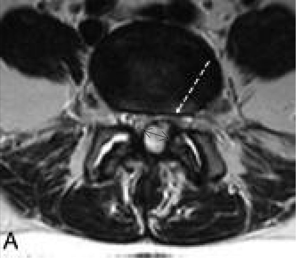
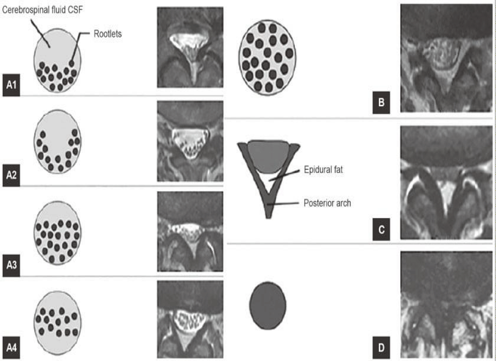
The spinal level of each cyst was recorded. The largest axial diameter of the cyst (in mm) was measured on imaging using Impax software, and the total surface area of the cyst (in mm²) was calculated from the same axial slice (Figure 1). Spinal canal compromise was determined by measuring the free canal area (excluding the cyst) and dividing it by the total canal area (including the cyst), expressed as a percentage. Spinal canal stenosis at the level of the cyst was graded according to the criteria proposed by Schizas, et al. (Figure 2). Radiology reports were reviewed for evidence of nerve compression, documenting which nerves were affected if compression was present. Additional findings, such as coexistent spondylolisthesis, spondylosis, and facet joint osteoarthritis, were logged along with their corresponding spinal levels. Information about any prior spinal surgeries and the levels involved was also recorded.
Patient characteristics and professional correspondence
Patient records were accessed through the Clinical Portal, which included correspondence between the Orthopaedic Surgical Department, Spinal Physiotherapy Team, Chronic Pain Management Team, and General Practitioners. Patients were categorized as symptomatic or asymptomatic (incidental findings). For symptomatic cases, details of symptomatology, such as back pain and/or radicular pain, were recorded. For radicular symptoms, the affected limbs were noted. The initial non-invasive management prescribed or self-administered for symptom control was documented.
Treatment and outcomes
Hospital interventions, including facet joint injections ± nerve root blocks and surgical treatments, were evaluated. For each patient, the time between MRI and the first intervention was recorded. Post-treatment outcomes were assessed through clinical correspondence, with 3-month follow-ups for injections and 6-week follow-ups for surgery. Subsequent follow-ups were also reviewed to identify further rounds of injections or progression to surgery. For patients requiring surgery after injections, the time between the first injection and the surgery was noted.
For surgical cases, outcomes were assessed in terms of symptom resolution and complications. Patients receiving surgery as a first-line treatment were evaluated for post-surgical outcomes and any additional treatments required. Surgical complications, such as infections or recurrence, were documented. This methodology allowed for a comprehensive evaluation of the imaging, management, and outcomes of lumbar facet joint cysts in the cohort, providing insights into their clinical course and treatment effectiveness.
Data collection and statical analysis
All collected data were securely stored in an encrypted Microsoft Office Excel 2011 database for analysis and interpretation. Statistical analyses were performed using IBM SPSS Software Version 23. A T-test was conducted to evaluate the relationship between the percentage of canal compromise caused by the cyst and the likelihood of requiring spinal injections or surgery. Additionally, a T-test was used to compare the percentage of canal compromise between symptomatic and asymptomatic patient groups, providing insights into the impact of canal compromise on symptomatology and treatment trends.
Results
In this study, a total of 22,292 Magnetic Resonance Imaging (MRI) scans were performed for the evaluation of the lumbar spine, revealing 87 patients diagnosed with lumbar facet joint cysts, resulting in an incidence rate of 0.39% across this patient population. The mean age of the cohort was calculated at 61.2 years, with a range spanning from 27 to 94 years and a standard deviation of 14.3 years. The demographic distribution included 35 males and 52 females, indicating a slight female predominance in the presentation of this condition.
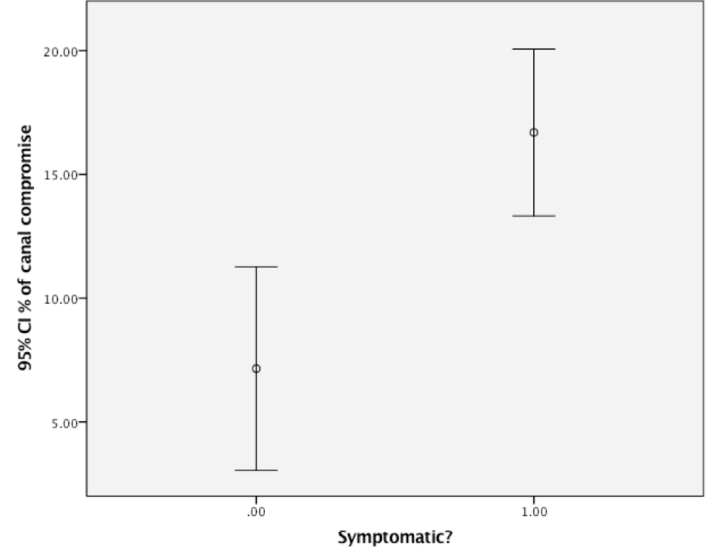
Among the identified patients, a total of 94 cysts were documented, which included 7 patients presenting with additional cysts. The side distribution of cysts showed that 46 cysts were located on the right side, whereas 48 cysts were on the left. A noteworthy observation was that 59 cysts resulted in nerve root compression, impacting 57 patients in the process (Figure 3). The mean maximum diameter of the cysts was determined to be 7.39 mm with a standard deviation of 3.23 mm, highlighting the variability in cyst size. Further assessments revealed the mean percentage of spinal canal compromise, which was calculated to be 13.84% (standard deviation of 13.04%). This percentage was notably higher in those patients who reported symptoms, averaging 16.70% (standard deviation of 13.15%), suggesting a correlation between the severity of symptoms and the extent of canal compromise (Figure 4). The localization of the cysts by spinal level demonstrated the following distribution: 5 cysts at the L2-L3 level, 22 cysts at L3-L4, 47 cysts at L4-L5, and 20 cysts at L5-S1 (Figure 5).
The grading of spinal canal stenosis, based on Schizas, et al.’s criteria (Table 1), revealed significant narrowing in symptomatic patients. Among the 87 cases, 46 (52.9%) exhibited spondylolisthesis, with 28 cysts (29.8%) located at the same level, supporting the link between spinal instability and cyst formation. Table 2 shows L4-L5 as the most affected level for spondylolisthesis, spondylosis, and facet joint osteoarthritis. Notably, 15 patients had prior spinal surgeries, with 11 symptomatic and 6 cysts occurring at the previous surgical level, emphasizing the role of degenerative changes and surgical history in cyst development.
When evaluating the symptomatic profiles of the patients, 61 patients were identified as symptomatic. Of these, 55 reported symptoms that were directly correlated with underlying nerve root compression, while 6 patients exhibited symptoms in the absence of evident compression on imaging. Specifically, back pain was documented by 45 patients, while 44 patients reported radicular symptoms. It is noteworthy that 36 patients experienced a combination of both back pain and radiculopathy. This symptomatology underscores the complexity and variability of patient presentations associated with lumbar facet cysts (Table 3).
Regarding treatment approaches, all 52 symptomatic patients received an initial management protocol that included analgesic therapy. In addition, 26 patients were referred for physiotherapy as an adjunctive treatment. Other treatment modalities included 2 patients who sought chiropractic care, 1 who pursued hydrotherapy, 1 who utilized Transcutaneous Electrical Nerve Stimulation (TENS), and 2 patients who explored acupuncture. These varying approaches illustrate the multi-faceted nature of managing symptoms associated with lumbar facet joint cysts.
A total of 38 patients proceeded to undergo hospital interventions, with a mean waiting interval of 9.68 months (standard deviation of 12.3 months) from the MRI date to the actual intervention. Among these patients, 34 underwent facet joint injections, with nine patients requiring multiple sets of injections due to recurring symptoms. Some patients expressed a preference for surgical intervention over conservative measures and opted for immediate surgery rather than undergoing injections. In total, 19 of the 52 patients needed surgical interventions; importantly, 5 patients remained on the waiting list at the time of data analysis.
The injections used in this study were outpatient procedures performed under local anesthetic and guided by X-ray fluoroscopy, involving a combination of corticosteroids and local anesthetic agents chosen at the discretion of the surgeon or radiologist. Injections were indicated for symptomatic lumbar facet joint cysts causing back pain or radiculopathy refractory to conservative management, such as analgesia or physiotherapy. Surgery was reserved for patients with persistent or worsening symptoms. Post-intervention care varied, with no specific standardized treatments, but patients often received additional conservative measures, such as analgesics or physiotherapy, as needed.
Complications resulting from the intervention were minimal but noteworthy; among the 14 patients who underwent surgical procedures, 2 complications were reported. One patient experienced a 2:1 heart block intraoperatively, an important consideration in anesthetic management, while another patient developed an epidural hematoma, highlighting the potential risks associated with surgical management of this condition.
At the three-month follow-up after receiving injection treatments, 14 out of 34 patients (approximately 41%) reported an improvement in their symptoms, demonstrating some initial efficacy of the conservative treatment approach. Conversely, 16 patients (47%) indicated that they felt no benefit from the injections, and 1 patient reported an exacerbation of their symptoms following the procedure. Additionally, 3 patients had not attended their follow-up appointments, leaving their outcomes undetermined.
For the cohort that underwent surgical interventions, the six-week post-operative follow-up revealed that all patients reported a subjective decrease in their symptoms. Among these surgical patients, 2 individuals indicated a complete resolution of their pain, which speaks to the effectiveness of the surgical approach for certain patients. However, it is important to note that subsequent follow-ups indicated that 3 patients who had undergone surgery required additional surgical interventions due to continued symptomatic persistence. Among these, 2 cases required further intervention at the original cyst level, while 1 case necessitated a procedure at a different spinal level. This data highlights the ongoing challenges and complexities in the management of lumbar facet joint cysts and the potential for ongoing symptomatology despite initial surgical intervention.
In summary, this comprehensive review of outcomes associated with lumbar facet joint cysts provides valuable insights into the demographics, clinical characteristics, treatment modalities, and patient outcomes. The findings underscore the need for a tailored approach to treatment, taking into account the variability in symptom presentation and patient responses to both conservative and surgical interventions.
Discussion
Facet joint cysts are believed to develop as a consequence of hypermobility and instability within the spinal column, often secondary to degenerative changes. This hypothesis is reinforced by the frequent association of facet joint cysts with intervertebral disc degeneration and degenerative spondylolisthesis, particularly at the L4-L5 segment, which is recognized as the most mobile area of the spine [10,11]. The incidence of degenerative spondylolisthesis is reported to be 21.2% in MRI and CT scans within the general population [12]. In our study, 52.9% of patients had degenerative spondylolisthesis, with 60.9% of these cases occurring at the level of the cysts. The L4-L5 segment was also identified as the most frequently affected level for spondylosis, facet joint osteoarthritis, and degenerative spondylolisthesis in our cohort, further supporting the notion that increased spinal mobility accelerates the degenerative cascade, which includes the formation of cysts.
Facet joint cysts are relatively rare, with an incidence ranging from 0.1% to 1% in radiological assessments (CT and MRI) [13,15]. In this study, we observed a crude incidence of 0.39%, which aligns with existing literature. The mean age of patients in our cohort was 61.2 years, corroborating findings that suggest facet joint cysts most commonly affect individuals in their seventh decade of life [14]. Additionally, a slight female predominance was noted, with a male-to-female ratio of 40:60, consistent with previous studies [15].
In our cohort, 50% of cysts were located at the L4-L5 level, followed by 26.2% at L3-L4, which reflects findings reported in other research [8]. The mean cyst diameter of 7.39 mm indicates that the cysts in this study were relatively small compared to those documented in other case reports [11]. Utilizing the Schizas grading system, the most prevalent spinal canal stenosis grade was A. It is noteworthy that symptomatic patients exhibited greater canal compromise than the overall cohort; however, statistical analysis revealed no significant correlation between canal compromise and the likelihood of requiring injections or surgical intervention.
Both back pain and radiculopathy were nearly equally prevalent among patients, with one additional patient reporting back pain compared to those experiencing radicular pain. The average time from MRI to the first hospital intervention was 9.68 months, which may be considered suboptimal given the symptom burden experienced by some patients. Notably, four patients opted for surgical treatment before receiving injections, deviating from current guidelines that advocate for conservative management as the first-line approach, while the remaining 34 patients adhered to established protocols.
Percutaneous spinal injections are generally regarded as the first-line treatment for facet joint cysts, yet their efficacy can be limited. A meta-analysis by Shuang, et al. [16], which included 544 patients, reported a post-injection success rate of 55.8%, one of the highest rates documented in the literature. Other studies have echoed similar findings. For instance, Hsu et al. [17] reported positive outcomes in 8 out of 8 surgical patients compared to only 2 out of 4 following corticosteroid injections. In a follow-up study by Parlier, et al. [14], of 30 patients who underwent facet joint injections, 18 experienced poor outcomes, and 14 required surgical intervention within six months.
In our study, 14 out of 31 patients who attended their three-month follow-up reported symptom improvement following injections. However, 15 of these 31 patients ultimately required surgery due to persistent symptoms. This aligns with existing literature trends, where injection success rates hover around 50%. Despite these modest results, the significant risks associated with surgical treatment make conservative management a rational first-line approach.
Studies investigating the role of obesity and other comorbidities in the management of lumbar facet joint cysts emphasize their significant influence on treatment outcomes. Hatgis, et al. highlighted that obesity contributes to the development and progression of lumbar facet cysts due to increased mechanical stress, impacting treatment efficacy and long-term outcomes [18]. Bagley, et al. discussed how obesity exacerbates spinal conditions like lumbar stenosis and facet joint cysts, complicating both conservative and surgical treatments [19]. Similarly, Santellán-Hernández, et al. noted a higher prevalence of obesity in patients with facet joint syndromes and emphasized its potential to alter therapeutic responses, particularly in minimally invasive treatments [20].
Xu, et al. analyzed recurrent back pain and cyst recurrence post-surgery, finding obesity to be a contributing factor influencing recurrence rates and overall recovery [21]. Lastly, Ambrosio, et al. demonstrated that comorbidities, including obesity, often affect patient selection and outcomes in minimally invasive interventions for lumbar facet joint syndromes, underscoring the need for personalized treatment approaches [22]. These findings collectively underscore the importance of considering obesity and related demographic factors when evaluating and managing lumbar facet joint cysts. These insights could guide tailored treatment strategies to improve patient outcomes.
Limitations of the Study
This retrospective study was dependent on the accuracy of clinical documentation within the Clinical Portal. Some patients had accessible imaging but lacked corresponding clinical records, which limited our ability to analyze symptomatology and treatment outcomes comprehensively. Furthermore, some patients included in treatment statistics had not yet attended their follow-up appointments to assess outcomes. The presence of concomitant spinal pathologies, both before and following cyst diagnosis, posed another challenge, as these comorbidities likely influenced treatment outcomes, introducing confounding variables that could skew our results.
Future research should focus on expanding demographic analyses to include comorbidities and their impact on both conservative and surgical treatment outcomes. Prospective, multi-center studies with larger sample sizes could provide more robust data on the long-term efficacy of injections versus surgery. Additionally, advancements in imaging and predictive modeling could help identify patients most likely to benefit from specific interventions, improving treatment selection and timing.
The key takeaway from this study is that percutaneous injections for lumbar facet joint cysts provide symptomatic relief in approximately half of the cases and should remain the preferred first-line treatment due to their minimally invasive nature and lower risk compared to surgical options. From the authors’ perspective, this finding reinforces the need for a cautious and patient-centered approach, prioritizing conservative management whenever feasible. While injections offer a safe and effective option for many, their limited success rate highlights the importance of ongoing assessment and potential progression to surgery for refractory cases. Additionally, the authors believe that understanding patient-specific factors, such as comorbidities like obesity, can further refine treatment strategies and enhance outcomes. This perspective underscores the necessity for future research to develop predictive models and innovative treatment methods to address the variability in patient responses and improve care for individuals with lumbar facet joint cysts.
Conclusion
While percutaneous injections for facet joint cysts yield positive outcomes in approximately 50% of cases, the associated risks of surgical management render it an impractical first-line choice for many patients. Our study highlights the importance of individualized treatment approaches, emphasizing that injections should remain the preferred initial strategy due to their lower risk profile. The findings underscore a critical need for further research to refine treatment protocols and improve patient outcomes, particularly given the complex interplay of degenerative changes and symptomatology associated with facet joint cysts. By focusing on a more nuanced understanding of these dynamics, clinicians can better tailor interventions to enhance patient care and quality of life.
Declarations
Availability of data and material: All data and materials referenced in this study are derived from previously published sources and are publicly available. Specific references are provided in the manuscript.
Authors’ contributions: All authors contributed to the conception, design, and drafting of the manuscript.
Data availability statement: No new data were generated or analyzed in this study. This review is based solely on previously published studies, which are cited within the manuscript. All data supporting the findings of this review are available in the referenced articles.
Author contributions statement: All authors made substantial contributions to the conception and design of the study, data collection, and analysis.
All authors have read and approved the final version of the manuscript and agree to be accountable for all aspects of the work, including its accuracy and integrity.
- Boviatsis EJ, Stavrinou LC, Kouyialis AT, Gavra MM, Stavrinou PC, Themistokleous M, et al. Spinal synovial cysts: pathogenesis, diagnosis and surgical treatment in a series of seven cases and literature review. Eur Spine J. 2008;17(6):831-7. Available from: https://doi.org/10.1007/s00586-007-0563-z
- Alicioglu B, Sut N. Synovial cysts of the lumbar facet joints: a retrospective magnetic resonance imaging study investigating their relation with degenerative spondylolisthesis. Prague Med Rep. 2009;110(4):301–9. Available from: https://pubmed.ncbi.nlm.nih.gov/20059882/
- Atkinson JLD, Lyons MK, Wharen RE, Deen HG, Zimmerman RS, Lemens SM. Surgical evaluation and management of lumbar synovial cysts: The Mayo Clinic experience. J Neurosurg. 2000;93(1 Suppl):53–7. Available from: https://doi.org/10.3171/spi.2000.93.1.0053
- Ustuner E, Tanju S, Dusunceli E, Deda H, Erden I. Synovial cyst: an uncommon cause of back pain. Curr Probl Diagn Radiol. 2007;36(1):48-50. Available from: https://doi.org/10.1067/j.cpradiol.2006.08.002
- Baum JA, Hanley EN. Intraspinal synovial cyst simulating spinal stenosis. Spine. 1986;11(5):487–9. Available from: https://doi.org/10.1097/00007632-198606000-00018
- Shaw M, Birch N. Facet joint cysts causing cauda equina compression. J Spinal Disord Tech. 2004;17(6):442–5. Available from: https://doi.org/10.1097/01.bsd.0000112086.85112.cf
- Yurt A, Seçer M, Aydın M, Akçay E, Ertürk AR, Akkol İ, et al. Surgical management of juxtafacet cysts in the lumbar spine. Int J Surg. 2016;29:9-11. Available from: https://doi.org/10.1016/j.ijsu.2016.03.003
- Cambron SC, McIntyre JJ, Guerin SJ, Li Z, Pastel DA. Lumbar facet joint synovial cysts: does T2 signal intensity predict outcomes after percutaneous rupture? AJNR Am J Neuroradiol. 2013;34(8):1661-4. Available from: https://doi.org/10.3174/ajnr.a3441
- Schizas C, Theumann N, Burn A, Tansey R, Wardlaw D, Smith FW, et al. Qualitative grading of severity of lumbar spinal stenosis based on the morphology of the dural sac on magnetic resonance images. Spine (Phila Pa 1976). 2010;35(21):1919-24. Available from: https://doi.org/10.1097/brs.0b013e3181d359bd
- Pait G, Raja A, Guererro C. Assessment and characteristics of intraspinal cystic lesions. Semin Spine Surg. 2006;18(3):139-47. Available from: http://dx.doi.org/10.1053%2Fj.semss.2006.06.005
- Trummer M, Flaschka G, Tillich M, Homann CN, Unger F, Eustacchio S. Diagnosis and surgical management of intraspinal synovial cysts: report of 19 cases. J Neurol Neurosurg Psychiatry. 2001;70(1):74-7. Available from: https://doi.org/10.1136/jnnp.70.1.74
- Kalichman L, Kim DH, Li L, Guermazi A, Berkin V, Hunter DJ. Spondylolysis and spondylolisthesis: prevalence and association with low back pain in the adult community-based population. Spine (Phila Pa 1976). 2009;34(2):199-205.
- Mercader J, Gomez J, Cardenal C. Intraspinal synovial cyst: Diagnosis by CT. Neuroradiology. 1985;27(4):346–348. Available from: https://doi.org/10.1007/bf00339570
- Parlier-Cuau C, Wybier M, Nizard R, Champsaur P, Le Hir P, Laredo JD. Symptomatic lumbar facet joint synovial cysts: clinical assessment of facet joint steroid injection after 1 and 6 months and long-term follow-up in 30 patients. Radiology. 1999;210(2):509-13.
- Shah R, Lutz G. Lumbar intraspinal synovial cysts: Conservative management and review of the world’s literature. Spine J. 2003;3(6):489–488. Available from: https://doi.org/10.1016/s1529-9430(03)00148-7
- Shuang F, Hou S, Zhu J, Ren DF, Cao Z, Tang JG. Percutaneous resolution of lumbar facet joint cysts as an alternative treatment to surgery: A meta-analysis. PLOS One. 2014;9(1):e84198. Available from: https://journals.plos.org/plosone/article?id=10.1371/journal.pone.0084198
- Hsu KY, Zucherman JF, Shea WJ, Jeffrey RA. Lumbar intraspinal synovial and ganglion cysts (facet cysts). Ten-year experience in evaluation and treatment. Spine (Phila Pa 1976). 1995;20(1):80-9. Available from: https://doi.org/10.1097/00007632-199501000-00015
- Hatgis J, Granville M, Berti A, Jacobson RE. Targeted radiofrequency ablation as an adjunct in treatment of lumbar facet cysts. Cureus. 2017;9(6):e1355. Available from: https://doi.org/10.7759/cureus.1355
- Bagley C, MacAllister M, Dosselman L, Moreno J, MacAllister L. Current concepts and recent advances in understanding and managing lumbar spine stenosis. F1000Research. 2019;8:137. Available from: https://doi.org/10.12688/f1000research.16082.1
- Santellán-Hernández JO, Mendoza-Gómez RF, Gómez-Ruiz A. Postsurgical lumbar facet joint syndrome: Therapeutic results of facet infiltration. Medicina Clínica Práctica. 2021;4:100164.
- Xu R, McGirt MJ, Parker SL, Bydon M, Olivi A. Factors associated with recurrent back pain and cyst recurrence after surgical resection of spinal synovial cysts. Spine J. 2010;10(1):55–61. Available from: https://doi.org/10.1016/j.spinee.2009.10.002
- Ambrosio L, Vadalà G, Russo F, Papalia R, Denaro V, Iacono F. Interventional minimally invasive treatments for chronic low back pain caused by lumbar facet joint syndrome: A systematic review. Global Spine J. 2023;13(4):1163-1179. Available from: https://doi.org/10.1177/21925682221142264
Article Alerts
Subscribe to our articles alerts and stay tuned.
 This work is licensed under a Creative Commons Attribution 4.0 International License.


 Save to Mendeley
