Open Journal of Orthopedics and Rheumatology
Bridging and Repair of First Metatarsal Fracture with Chronic Pseudoarthrosis Following Multiple Surgical Interventions Using a Minimally Invasive Approach
Michael A Scarpone1 and Charles C Lee2*
1Department of Orthopedics, Trinity Health System, Catholic Health Initiatives, 15655 St Rt 170 Suite B, Calcutta, OH 43920, USA
2Department of Cell Biology and Human Anatomy, School of Medicine, University of California, Davis 95616, USA
Cite this as
Scarpone MA, Lee CC. Bridging and Repair of First Metatarsal Fracture with Chronic Pseudoarthrosis Following Multiple Surgical Interventions Using a Minimally Invasive Approach. Open J Orthop Rheumatol. 2025;10(1):001-004. Available from: 10.17352/ojor.000050Copyright License
© 2025 Scarpone MA,et al. This is an open-access article distributed under the terms of the Creative Commons Attribution License, which permits unrestricted use, distribution, and reproduction in any medium, provided the original author and source are credited.Metatarsal fractures are a common type of injury to the foot and can result in chronic pain and disability if the fracture site fails to fuse after a series of bone fusion procedures with or without instrumentation, resulting in pseudarthrosis, which may ultimately require amputation. A 58-year-old male patient without any known risk factors presented with pseudarthrosis and chronic pain in the right foot after 4 failed bone fusion procedures on the first metatarsal bone where amputation was considered. The Hyper-Crosslinked Carbohydrate Polymer (HCCP) was injected with autologous bone marrow into the site of non-union without reduction or instrumentation in an outpatient setting. The patient showed a dramatic improvement over the course of 12 weeks and a radiographic fusion along with a 90% improvement in the pain scale. Forefoot and midfoot fractures and injuries associated with a high degree of pseudarthrosis may require a better predictor and appropriate fusion procedure before amputation is considered.
Introduction
Metatarsal fractures represent 5% - 6% of total fractures encountered in primary care [1,2]. Metatarsal fractures range from those that can be managed by primary care physicians to more complicated cases that require surgical intervention [3]. A fracture in the first metatarsal bone has a greater clinical implication compared to the other metatarsals as it is enlarged and strengthened for the load-bearing role of standing and mobility. Typically, metatarsal fractures, other than those that affect the metatarsal head, are not displaced because adjacent metatarsals can provide mechanical/structural support although displacement may occur following multiple fractures. Patients with metatarsal fractures frequently present point tenderness, which may be attributed to axial loading at the metatarsal head [3]. Fractures in the metatarsal shaft tend to heal well without immobilization in the presence of sufficient blood supply and cessation of weight-bearing activities for 4-8 weeks. However, lack of adequate stability and/or blood flow in addition to other risk factors such as diabetes, aging, smoking, and obesity may lead to nonunion, which generally results in a structure similar to a fibrous joint, referred to as a “false joint” or pseudoarthrosis. Frequently, pseudoarthrosis may be presented with a great motion at the fracture site with a pseudocapsule and fluid collection between the fracture gap [4]. Therapeutic strategies for metatarsal pseudoarthrosis may include non-invasive uses of adjuvants such as ultrasound, electrical stimulation, electromagnetic stimulation, and shock waves [5-7]. More invasive strategies may include the implantation of grafts such as autologous bone, allograft, and internal fixation with plate and screws, K-wires, or intramedullary nail [7,8]. In this report, we report a case of a successful bone fusion in a patient who experienced pseudoarthrosis in the first metatarsal bone that failed to fuse after a series of surgical strategies including internal fixation and use of autologous bone graft in an outpatient setting.
Case report
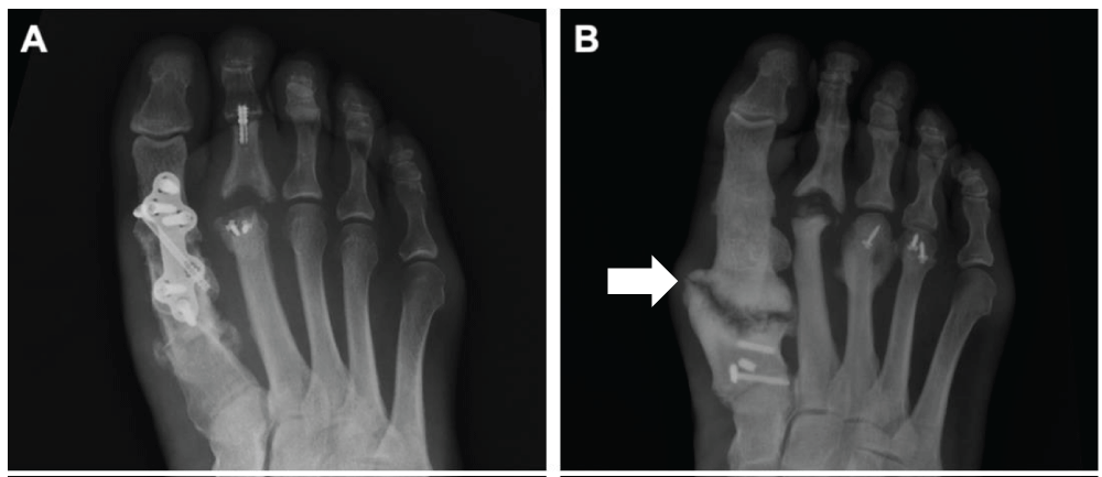
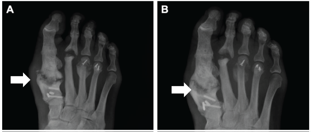
A 58-year-old male patient with no remarkable comorbidity with the initial diagnosis of osteoarthritis was presented with a possible stress fracture in the first metatarsal bone of the right foot that had resulted in chronic pseudoarthrosis after 4 separate surgical interventions including plates and screws, platelet-rich plasma, and autograft. The fracture occurred less than 1 year after the first surgical intervention. The patient complained of severe pain and was unable to put any weight on the affected foot. Following four series of failed fusion procedures including the implantation of autologous bone, which resulted in chronic pain and also broken plate, amputation was considered the last resort. The plate and screws were removed from the affected site (Figure 1). HCCP (Osteo-P® BGS) was mixed with autologous bone marrow (CellFuse) in a sterile cup. HCCP/CellFuse (radiolucent) was pushed through an 8-gauge needle using a metal rod into the fracture site, which was backfilled with additional autologous bone marrow as much as possible under fluoroscopic guidance. No instrumentation or reduction was performed at the implantation site, and no external brace was placed on the patient. The entire procedure was performed in an outpatient setting. X-ray was performed at 5- and 12-weeks post-surgery and showed full bridging at the site of implantation (Figure 2). The patient showed no abnormal palpable motion at the proximal first metatarsal at the 5-week post-surgery visit compared to palpable motion at the site pre-surgery. The patient was able to bear weight by 5 weeks post-surgery and returned to routine activities with greater than 90% improvement in the pain score, to date.
Discussion
According to the US Food and Drug Administration, non-union is considered to be established after a minimum of nine months since the injury with the fracture site showing no observable and progressive signs of fusion for a minimum of three months [9]. Multiple factors may compromise fracture healing and influence the progression to pseudoarthrosis and be categorized into patient-dependent and patient-independent factors. Patient-dependent factors may include age, gender, congenital and genetic comorbidities, smoking, and use of pharmacological agents. Patient-independent factors may include the type and extent of fractures, infections, and adequacy of treatment [9,10]. However, the patient included in this report showed no known risk factors and was provided with adequate treatment. It is possible that the patient may have a genetic factor. A recent study investigated the existence of a genetic predisposition to fracture non-union which, when combined with other risk factors, could synergistically increase the susceptibility of a patient to develop non-union. This study indicated the existence of a potential genetically predetermined impairment within the BMP signaling cascade, initiated after a fracture and when combined with other risk factors could synergistically increase the susceptibility of a patient to develop non-union [11]. Although the patient included in this report was not investigated for a possible genetic cause, pseudoarthrosis following a series of surgical treatments including autograft implantation is highly significant. It is also interesting to note that the patient showed fibrous non-union in the first metatarsal while exhibiting callus formation, which might be a potential reason for different outcomes observed between the metatarsal heads, in addition to potential differences in vascularization and inflammation at the fracture sites.
Working et al. reported that sustained high-energy injuries to the forefoot and midfoot resulted in an amputation rate of 13.9% within 30 days post-injury and rose to 18.9% in one year [12]. This study also showed a statistically significant increase in the rate of amputation following the high-energy fracture of the first metatarsal bone compared to other risk factors such as midfoot dissociation, distal tibia fracture, tobacco use, or BMI; suggesting that the first metatarsal fracture is a significant predictor of amputation along with the fracture of all five metatarsals. Chronic pain caused by pseudoarthrosis in the first metatarsal of the patient included in this case report might have led to amputation. Other than the first metatarsal fracture, the patient showed no other known risk factors for amputation. Although factors such as infection, inadequate blood flow, biomechanics, and patient biology (e.g., metabolic disorder) may be used for the prognosis, clinical observations and imaging modalities are the main methods for monitoring bone healing after a traumatic event and determining nonunion or pseudoarthrosis. However, they are poor prognostic markers as these methodologies only provide conclusive diagnosis many months, if not years, after post-intervention. It creates additional issues for patients who have already failed to fuse and are offered additional surgical intervention, which may or may not lead to a successful fusion and clinical outcome.
Growth factors, specifically, Bone Morphogenic Protein 2 (BMP-2) and Transforming Growth Factor-Beta 1 (TGF-β1), play key roles in bone fracture healing by signaling various osteoinductive pathways. Hara et al. investigated the peak expression of circulating BMP-2 and TGF-β1 as a predictor circulating marker for bone union [13]. The study showed a one-week delay in the peak expression of circulating BMP-2 and TGF-β1 in patients who experienced nonunion when compared to those with successful bone fusion. Urinary cross-linked n-telopeptide (uNTx) was also investigated for its significance as a predictive marker of nonunion, and a low level of the preoperative level of uNTx was implicated in the eventual development of pseudoarthrosis at 6 months and 1-year post-surgical intervention [14]. The development of quantitative markers that can accurately predict successful bone fusion and/or pseudoarthrosis, coupled with advanced bone fusion technology, may provide a significantly improved solution for patients who show a poor prognosis.
This study highlights the capability of Hyper-Crosslinked Carbohydrate Polymer (HCCP) and Bone Marrow Aspirate Concentrate (BMAC) to promote bone repair, regeneration, and bridging at the site of a bone fracture that has been hampered by pseudoarthrosis even after autograft implantation. HCCP has been shown to successfully bridge a critical-sized bone defect statistically equivalent to autologous bone in an animal model [15] and proposed to generate a conducive microenvironment for stem and progenitor cells [16]. Osteoprogenitor cells and growth factors in BMSC may have contributed to new bone formation in the patient included in this report. BMAC has an excellent safety profile and has demonstrated promise as an orthobiologic adjuvant in foot and ankle bone fusion procedures [17]. Further clinical studies are needed to investigate the mechanism of bone repair and regeneration by HCCP and/or BMAC alone or in combination as observed in this study. In the case of an atrophic nonunion, the underlying conditions such as metabolic abnormalities, vascular issues, or infection may need to be addressed in conjunction with the HCCP/BMAC approach. It would be interesting to investigate further whether this approach could address at least some of the causes of atrophic nonunion.
Conclusion
Non-union or pseudoarthrosis after a series of failed surgical interventions over the course of several years has significant clinical consequences in addition to the possibility of irreversible biological changes that may hamper subsequent treatment and bone healing. There is a need for sensitive and quantitative diagnostic modalities to better predict and manage bone fusion and identification of appropriate tools to treat and manage pseudoarthrosis, specifically, before more invasive approaches such as amputation are considered. Additional controlled studies are needed to evaluate the safety and efficacy of HCCP and/or BMAC for addressing pseudoarthrosis in patients with metatarsal fractures.
Conflict of interest
CCL is the inventor of the Hyper-Crosslinked Carbohydrate Polymer (HCCP) used in Osteo-P® BGS and founder and CEO of Molecular Matrix, Inc., which has licensed the HCCP technology from the University of California, Davis, for commercialization.
- Eiff MP, Saultz JW. Fracture care by family physicians. J Am Board Fam Pract. 1993;6(2):179-81. Available from: https://pubmed.ncbi.nlm.nih.gov/8452070/
- Hatch RL, Rosenbaum CI. Fracture care by family physicians: a review of 295 cases. J Fam Pract. 1994;38(3):238-44. Available from: https://pubmed.ncbi.nlm.nih.gov/8126403/
- Hatch RL, Alsobrook JA, Clugston JR. Diagnosis and management of metatarsal fractures. Am Fam Physician. 2007;76(6):817-26. Available from: https://pubmed.ncbi.nlm.nih.gov/17910296/
- Takahara S, Niikura T, Lee SY, Iwakura T, Okumachi E, Kuroda R, Kurosaka M. Human pseudoarthrosis tissue contains cells with osteogenic potential. Injury. 2016;47(6):1184-90. Available from: https://doi.org/10.3390/cimb44110377
- Nolte P, Anderson R, Strauss E, Wang Z, Hu L, Xu Z, et al. Heal rate of metatarsal fractures: a propensity-matching study of patients treated with low-intensity pulsed ultrasound (LIPUS) vs. surgical and other treatments. Injury. 2016;47(12):2584-90. Available from: https://doi.org/10.1016/j.injury.2016.09.023
- Victoria G, Petrisor B, Drew B, Dick D. Bone stimulation for fracture healing: what's all the fuss? Indian J Orthop. 2009;43(2):117-20. Available from: https://doi.org/10.4103/0019-5413.50844
- Wu J, Guo H, Liu X, Li M, Cao Y, Qu X, et al. Percutaneous autologous bone marrow transplantation for the treatment of delayed union of limb bone in children. Ther Clin Risk Manag. 2018;14:219-24. Available from: https://doi.org/10.2147/tcrm.s146426
- Bryant T, Beck DM, Daniel JN, Pedowitz DI, Raikin SM. Union rate and rate of hardware removal following plate fixation of metatarsal shaft and neck fractures. Foot Ankle Int. 2018;39(3):326-31. Available from: https://doi.org/10.1177/1071100717751183
- Pountos I, Georgouli T, Pneumaticos S, Giannoudis PV. Fracture non-union: can biomarkers predict outcome? Injury. 2013;44(12):1725-32. Available from: https://doi.org/10.1016/j.injury.2013.09.009
- Calori GM, Albisetti W, Agus A, Iori S, Tagliabue L. Risk factors contributing to fracture non-unions. Injury. 2007;38 Suppl 2:S11-18. Available from: https://studiocalori.it/images/pdf/pubblicazioni/054.pdf
- Dimitriou R, Carr IM, West RM, Markham AF, Giannoudis PV. Genetic predisposition to fracture non-union: a case control study of a preliminary single nucleotide polymorphisms analysis of the BMP pathway. BMC Musculoskelet Disord. 2011;12:44. Available from: https://link.springer.com/article/10.1186/1471-2474-12-44
- Working ZM, Elliott I, Marchand LS, Jacobson LG, Presson AP, Stuart A, et al. Predictors of amputation in high-energy forefoot and midfoot injuries. Injury. 2017;48(3):536-41. Available from: https://doi.org/10.1016/j.injury.2016.12.005
- Hara Y, Ghazizadeh M, Shimizu H, Matsumoto H, Saito N, Yagi T, et al. Delayed expression of circulating TGF-β1 and BMP-2 levels in human nonunion long bone fracture healing. J Nippon Med Sch. 2017;84(1):12-8. Available from: https://www.jstage.jst.go.jp/article/jnms/84/1/84_12/_article
- Steinhaus ME, Hill PS, Yang J, Feuchtbaum E, Bronheim RS, Prabhakar P, et al. Urinary N-Telopeptide can predict pseudarthrosis after anterior cervical decompression and fusion: a prospective study. Spine. 2019;44(12):770-6. Available from: https://journals.lww.com/spinejournal/abstract/2019/06010/urinary_n_telopeptide_can_predict_pseudarthrosis.5.aspx
- Koleva PM, Keefer JH, Ayala AM, Lorenzo I, Han CE, Pham K, et al. Hyper-crosslinked carbohydrate polymer for repair of critical-sized bone defects. Biores Open Access. 2019;8(3):111-20. Available from: https://doi.org/10.1089/biores.2019.0021
- Lee CC, Hirasawa N, Garcia KG, Ramanathan D, Kim KD. Stem and progenitor cell microenvironment for bone regeneration and repair. Regen Med. 2019;14(9):693-702. Available from: https://doi.org/10.2217/rme-2018-0044
- Harford JS, Dekker TJ, Adams SB. Bone marrow aspirate concentrate for bone healing in foot and ankle surgery. Foot Ankle Clin. 2016;21(4):839-45. Available from: https://doi.org/10.1016/j.fcl.2016.07.005
Article Alerts
Subscribe to our articles alerts and stay tuned.
 This work is licensed under a Creative Commons Attribution 4.0 International License.


 Save to Mendeley
