Open Journal of Orthopedics and Rheumatology
Myositis Ossificans Secondary to Lowlying Supracondylar Fracture of the Humerus in a Child: A Case Report
Janaki Medical College and Teaching Hospital, Janakpur, Nepal
Author and article information
Cite this as
Regmi B, Rauniyar S, Pandit DK. Myositis Ossificans Secondary to Lowlying Supracondylar Fracture of the Humerus in a Child: A Case Report. Open J Orthop Rheumatol. 2025;10(1):032-035. Available from: 10.17352/ojor.000054
Copyright License
© 2025 Regmi B, et al. This is an open-access article distributed under the terms of the Creative Commons Attribution License, which permits unrestricted use, distribution, and reproduction in any medium, provided the original author and source are credited.Abstract
Introduction: Myositis ossificans (MO) is a form of heterotopic ossification that develops within muscles or soft tissues, usually after trauma. It is a benign tumor-like lesion commonly seen in young adults and adolescents, and rarely in children. MO can be classified into two forms: myositis ossificans progressiva (genetic) and myositis ossificans traumatica (acquired). Its exact pathophysiology remains unclear, though trauma is a major contributing factor.
Case Presentation: We present a case of a 6-year-old girl who developed MO in her right elbow following a minor trauma caused by a tractor injury. Initial imaging diagnosed a non-displaced supracondylar humerus fracture and she was managed with immobilization. Despite this, she developed persistent pain and stiffness, followed by physiotherapy which worsened her condition. A soft-tissue mass appeared with limited elbow mobility. Radiographic imaging revealed a calcified mass consistent with heterotopic ossification. Due to limited resources and parental refusal, further imaging like MRI was not performed. Conservative management with NSAIDs and rest led to notable improvement over 4 weeks.
Discussion: MO is characterized by zonal ossification and typically follows trauma or inappropriate rehabilitation. It commonly affects large skeletal muscles like the quadriceps and brachialis. Diagnosis is often clinical and radiological, with CT being the gold standard. In resource-limited settings, radiography remains a practical alternative.
Conclusion: This case underscores the importance of considering MO in cases of post-traumatic stiffness and pain, particularly in children. Conservative treatment can be effective, and physiotherapy should be cautiously approached.
Introduction
Myositis ossificans (MO) is a form of heterotopic ossification that develops within muscles or soft tissues, usually after trauma. Myositis ossificans (MO) is a benign tumor-like lesion, which is predominantly seen in young adults and adolescents. Pediatric MO cases are even rarer [1]. It is well known in sports activities. It is mostly found in skeletal muscle as a solitary lesion [2]. Common site for myositis ossificans are quadriceps muscle of thigh and the brachialis muscle of upper arm.
It is associated with multiple etiologies, such as injury, genetic pre-disposition, post-infection, or undetermined causes, whereas injury is an important factor in its pathogenesis [3]. Myositis ossificans is classified in two different groups: myositis ossificans progressiva (MOP) which describes a genetic autosomal dominant rare disease and myositis ossificans traumatica (MOT) [4]. One being genetic type and other autosomal or non-genetic types. The specific cause for myositis and its pathophysiology remain unclear. Proliferation of connective tissue occurs after a muscle injury that differentiates into mature bone. Such ossification preferably occurs in muscles that are repeatedly strained or injuried [5].
Myositis ossificans can be managed conservatively with NSAIDS for the pain in most of the case until the overgrowing mass affect the daily activities of the patient.
Case presentation
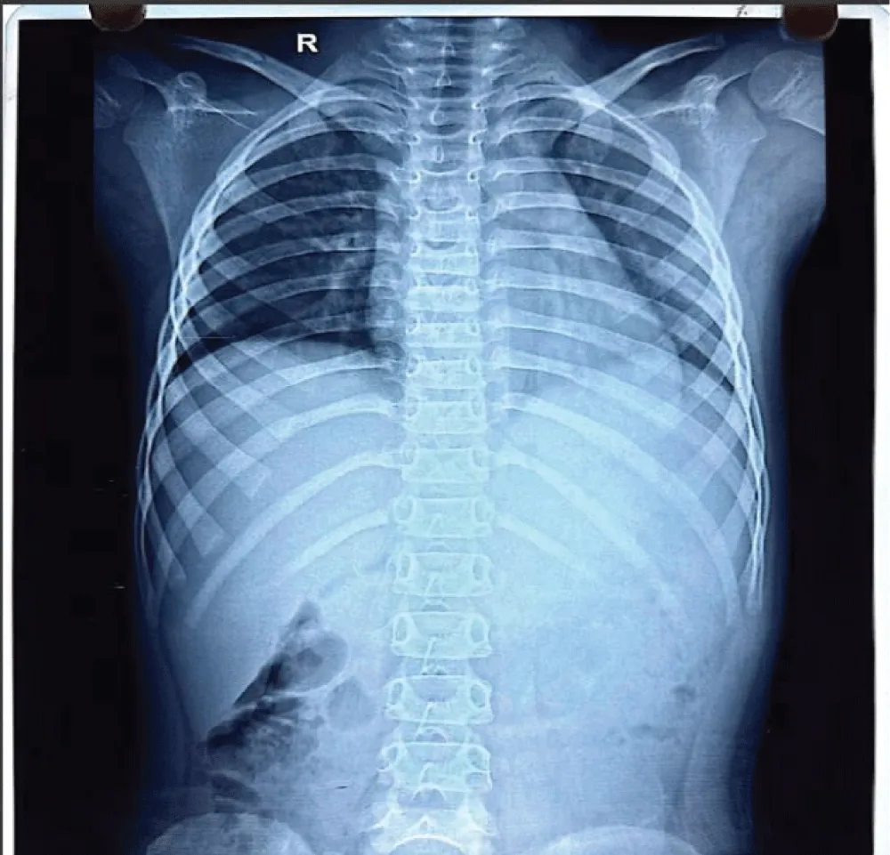
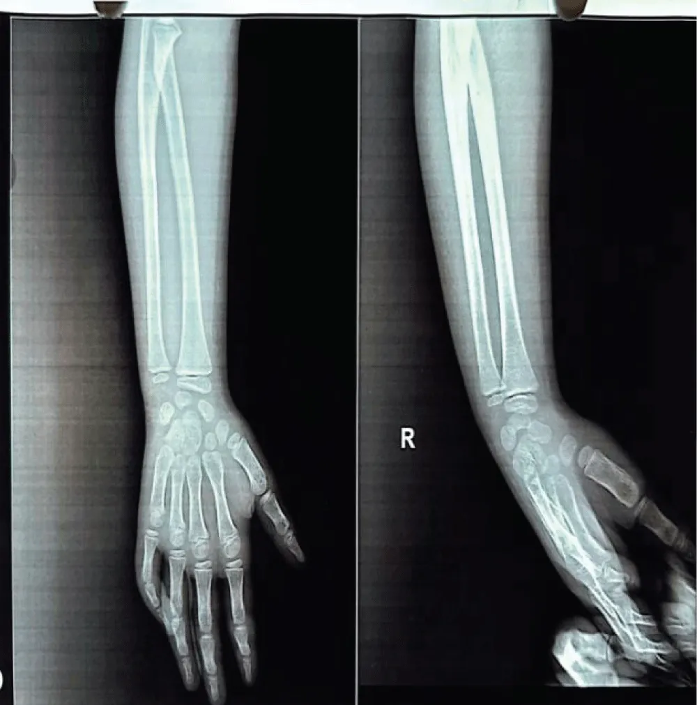
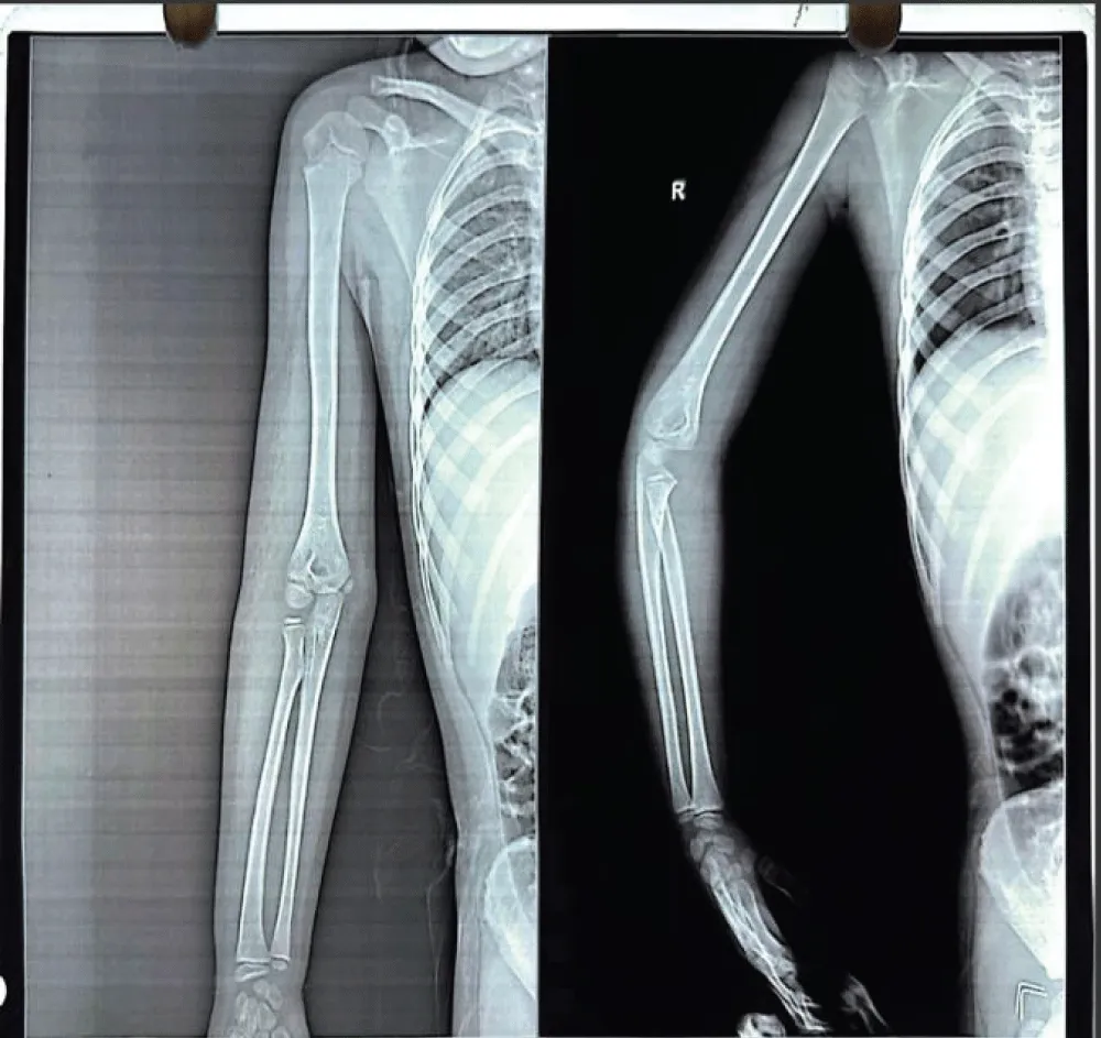
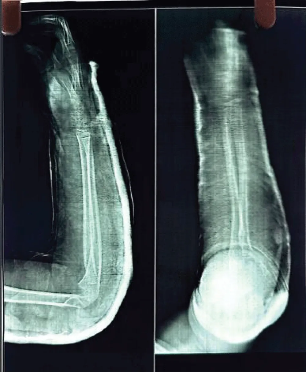
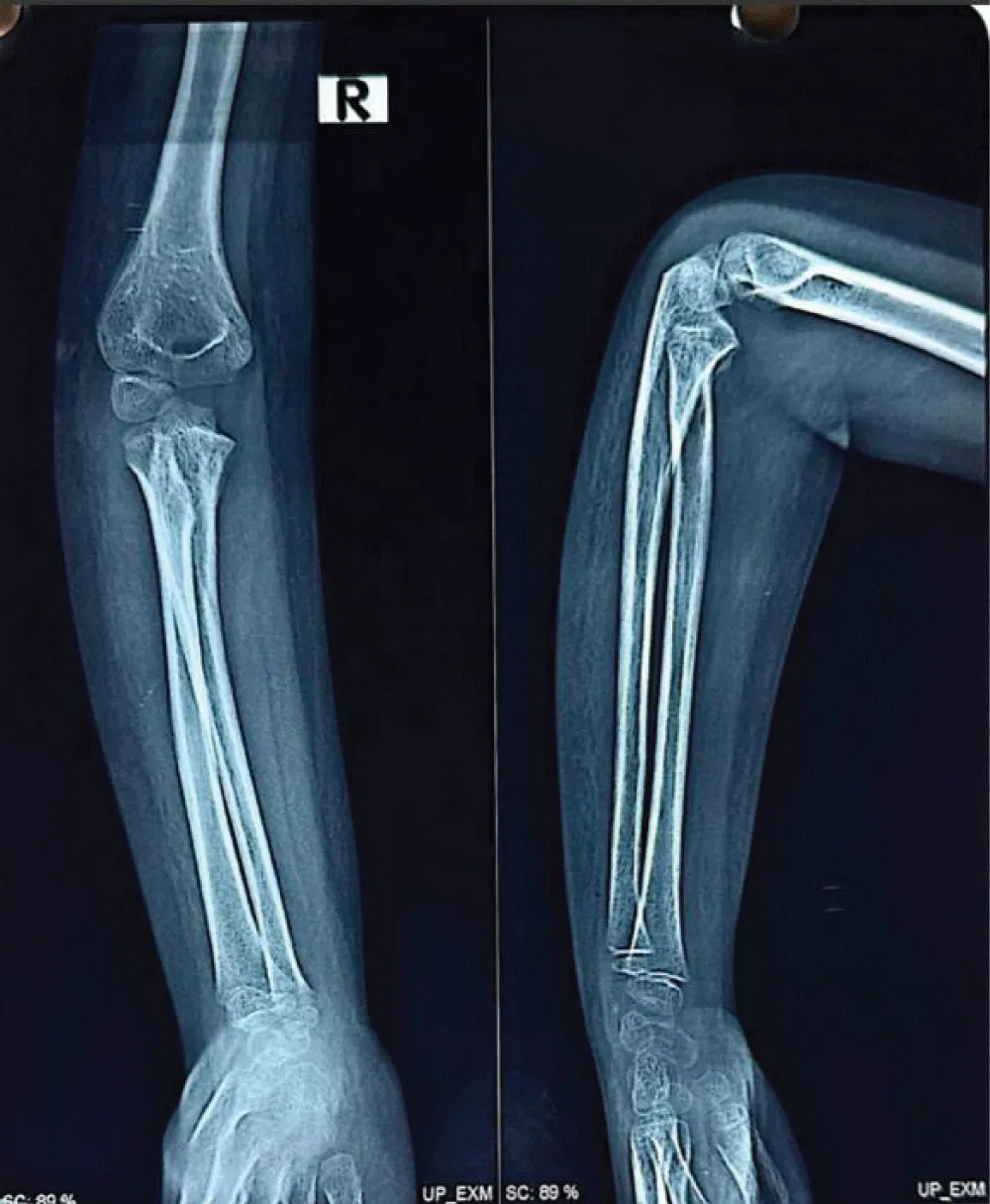
A mother with 6 year old female child presented to our Orthopedic Out Patient Department. She had not been able to move her right elbow in full range with restricted active and passive movements and appeared distressed. According to the informant (mother), she had been apparently well 2 months back when she was hit by rear wheels of a tractor while playing on the roadside. She developed pain and swelling in the elbow and was immediately rushed to the nearby hospital. Routine investigations, CT scan of abdomen and pelvis and X-ray of right elbow and hand were done. (Figures 1-3). All reports were normal except X-ray arm diagnosing undisplaced low lying supracondylar fracture of the right humerus (Figure 3). The child was admitted to the hospital and right upper limb was immobilized in an above-elbow plaster slab, with the elbow in 90o flexion (Figure 4). She was discharged after 4 days with reportedly normal radiographic findings.
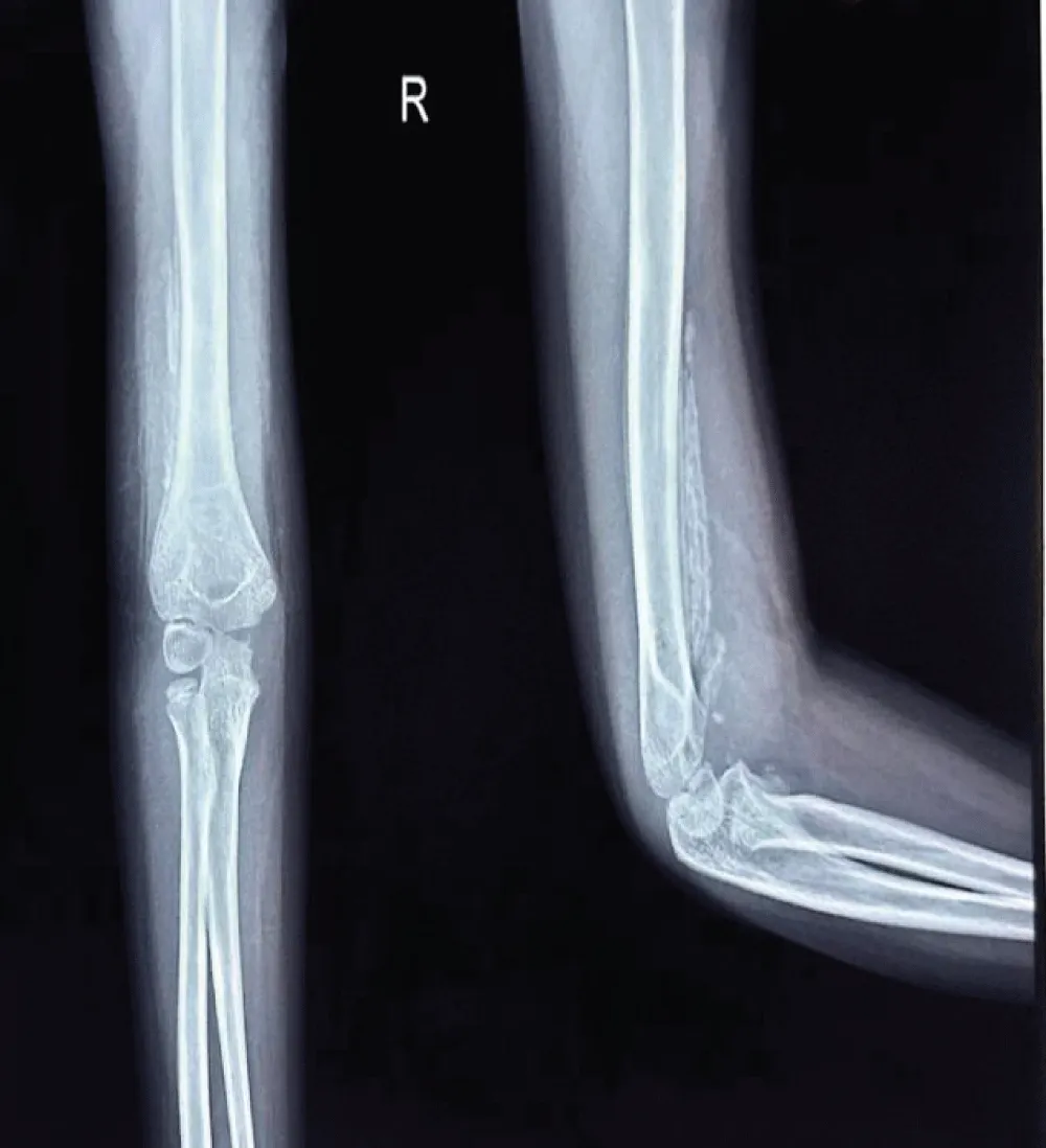
However, the child continued to experience persistent pain and stiffness in the elbow region, progressively limiting the range of motion. This prompted another hospital visit, during which an X-ray of the right elbow was obtained (Figure 5). Subtle calcific changes-early signs of heterotopic ossification-were present but went unnoticed. Physiotherapy was initiated under the assumption of post-immobilization stiffness. However, within a few days of physiotherapy, the patient developed worsening stiffness and a painful, enlarging subcutaneous lump in the elbow region.
Then she was presented to our centre and physical examination revealed a non-discrete soft-tissue mass, approximately 2 × 4 cm in size, that was deep to muscle and tender on palpation (Figure 6). Grip strength in the right hand was normal; right biceps and triceps strength were rated as 3/5. Range of motion at the elbow was limited due to pain, and the patient was unable to lift her left arm over her head.
Laboratory test results at presentation showed a white blood cell count of 7,900/µL and the C-reactive protein was negative ruling out infectious or inflammatory component. Additionally, urine analysis was unremarkable, showing no evidence of proteinuria, hematuria, or infection, further supporting the localized nature of the condition.
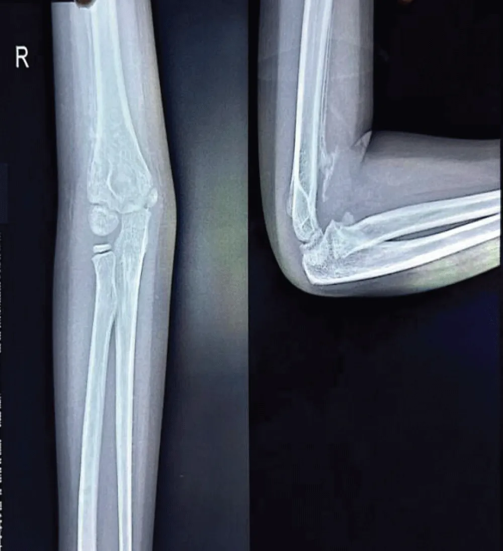
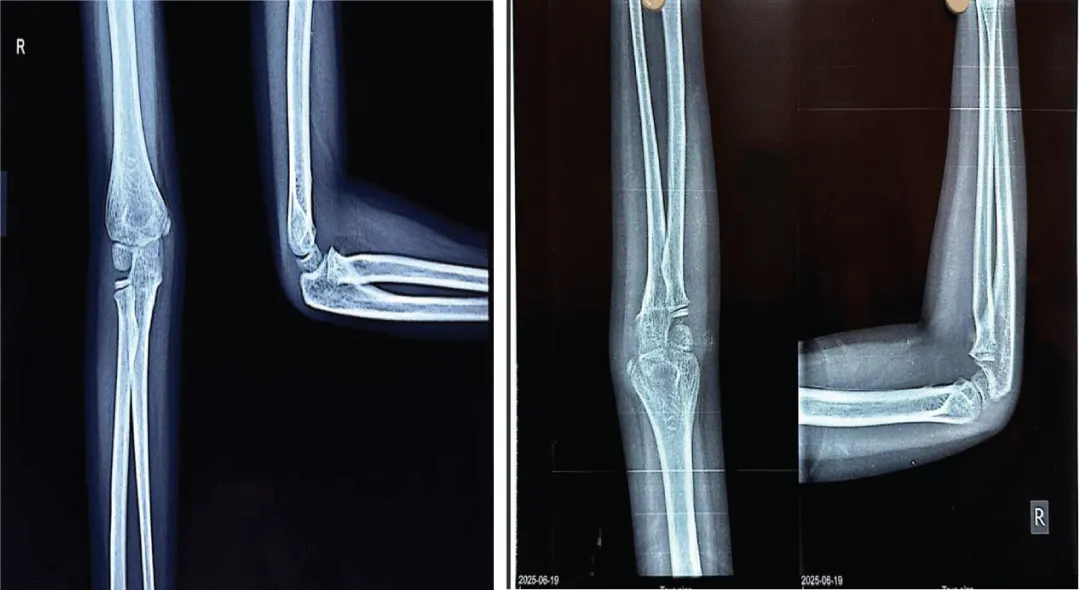
Radiograph of the elbow joint revealed a well-defined calcified mass parallel to the humerus, with a convex shape extending along the supracondylar region with the classical features of heterotopic ossification. The patient guardians declined further advanced imaging studies, including MRI, citing personal reasons. Consequently, the diagnosis of Myositis Ossificans Traumatica was made based exclusively on radiographic findings.
At our hospital, conservative management was initiated, including rest and NSAIDs for symptoms relief. Physiotherapy was discontinued. On follow-up of 4 weeks, there was reduction in swelling and improved in range of motion of the elbow, although residual stiffness persisted.
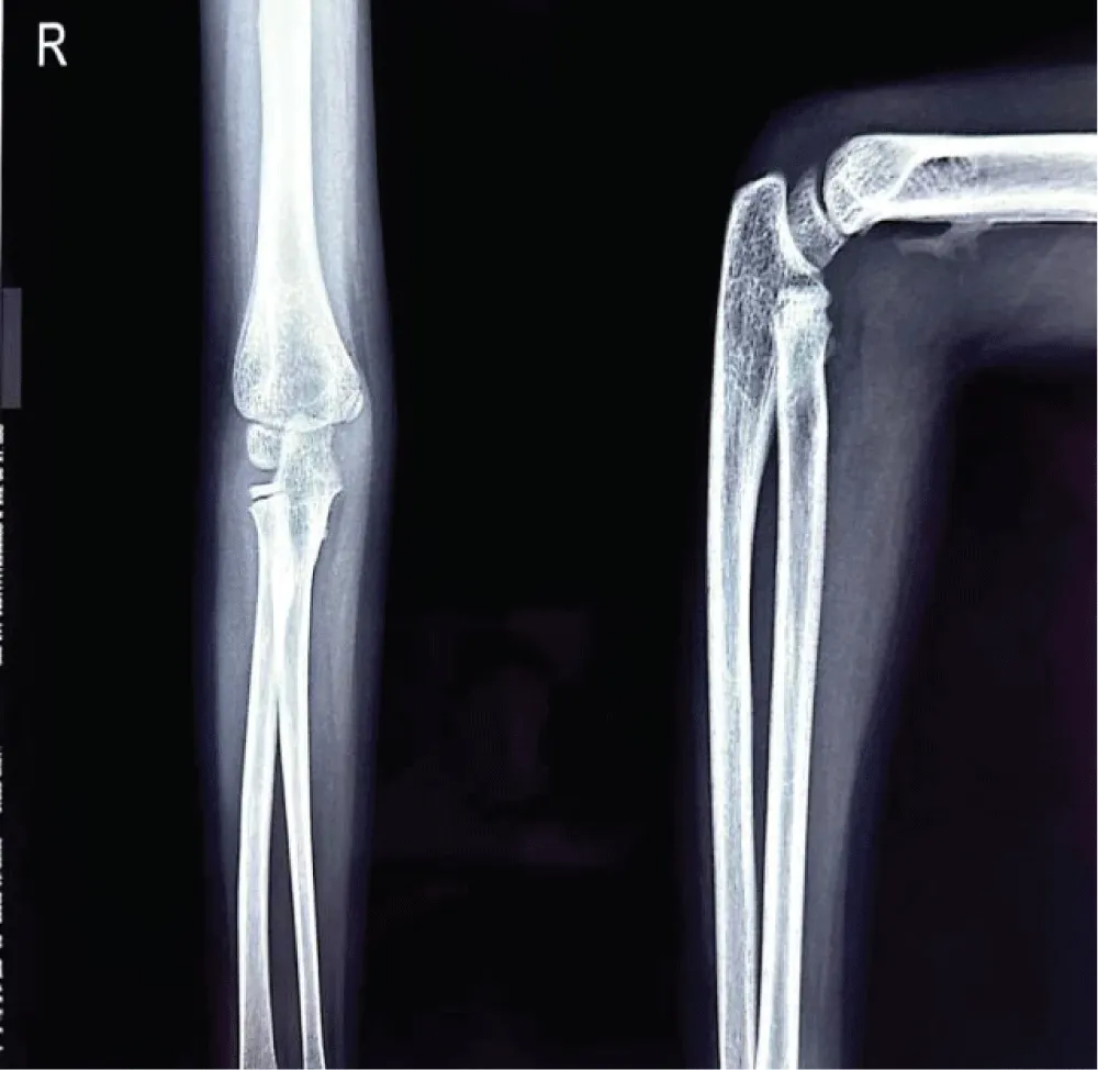
Radiological imaging showed residual heterotopic ossification along the humerus, indicating incomplete resolution. Multiple radiographs of the right upper limb-including the arm, forearm, and elbow joint were taken during outpatient visits at the patient’s convenience and were compared over time to monitor progress (figure 7,8). A well-visualized radiograph obtained 10 months after the initial injury showed near-complete resolution of the heterotopic ossification (Figure 9).
“All radiographs have been anonymized for publication and presented with patient/guardian consent.”
Discussion
Myositis ossificans (MO) is a form of self-localized, non-neoplastic, heterotopic ossification that develops within muscles or soft tissues, usually after trauma. It is most common in adolescents and young adults, especially athletes, but rare in children under 10 and less common in females. Myositis ossificans commonly occurs in large skeletal muscles of the extremities, with the quadriceps (thigh) and brachialis (upper arm) muscles being the most frequently affected areas.Other common sites include the hip, buttocks, and elbow. Histologically, MO is essentially metaplasia of the intramuscular connective tissue resulting in extraosseous bone formation, without inflammation. It has a zonal organisations:
- Peripheral, well-organised mature lamellar bone composed by mature bone.
- Intermediate osteoid region containing immature osteoid tissue and islands of cartilage.
- Central immature non-ossified cellular (fibroblasts) focus characterised by proliferative fibroblastic cells with haemorrhage and necrosis inside the muscle [6].
The case of Myositis Ossificans is diagnosed clinically and radiologically. Gold standard for diagnosing MO is generally by CT imaging.
We present a case of a 6-year-old girl who developed MO after an elbow injury and excessive physiotherapy. In resource-limited settings like Nepal, where patients often decline advanced imaging or biopsy, diagnosis was made solely through radiographic findings. The patient was managed conservatively, with follow-up imaging showing resolution.
Although the patient was discharged after 4 days with reportedly normal radiographs, it is possible that early soft tissue damage was underestimated. Subsequent early physiotherapy, initiated without adequate healing, may have precipitated heterotopic ossification, contributing to the development of myositis ossificans.
Conclusion
This case highlights the importance of early recognition of MO, which can be overlooked after fracture treatment, and demonstrates that conservative management with radiography alone can be effective in such settings. Although spontaneous resolution of myositis ossificans has been reported in up to 38% of lesions [3].
It also highlights that persistent post-fracture pain and stiffness do not always warrant physiotherapy, and MO should be considered as a differential diagnosis.
References
- Cao J, Zheng HJ, Sun JH, Zhu HY, Gao C. Case Report: Unusual Presentation of Myositis Ossificans of the Elbow in a Child Who Underwent Excessive Postoperative Rehabilitation Exercise. Front Pediatr. 2021:9. Available from: https://doi.org/10.3389/fped.2021.757147
- Cherry I, Mutschler M, Samara E, Merckaert S, Zambelli PY, Tschopp B. Myositis ossificans in the pediatric population: a systematic scoping review. Front Pediatr. 2023:11. Available from: https://doi.org/10.3389/fped.2023.1295212
- Chen J, Li Q, Liu T, Jia G, Wang E. Bridging Myositis Ossificans After Supracondylar Humeral Fracture in a Child: A Case Report. Front Pediatr. 2021:9. Available from: https://doi.org/10.3389/fped.2021.746133
- Hanisch M, Hanisch L, Fröhlich LF, Werkmeister R, Bohner L, Kleinheinz J. Myositis ossificans traumatica of the masticatory muscles: etiology, diagnosis and treatment. Head Face Med. 2018:14(1). Available from: https://doi.org/10.1186/s13005-018-0180-6
- Kanthimathi B, Udhaya Shankar S, Arun Kumar K, Narayanan VL. Myositis Ossificans Traumatica Causing Ankylosis of the Elbow. Clin Orthop Surg. 2014;6(4):480. Available from: https://doi.org/10.1186/s13005-018-0180-6
- Landolsi M, Mrad T. Traumatic myositis ossificans circumscripta (MOC). BMJ Case Rep. 2017. Available from: https://doi.org/10.1136/bcr-2017-219422
Article Alerts
Subscribe to our articles alerts and stay tuned.
 This work is licensed under a Creative Commons Attribution 4.0 International License.


 Save to Mendeley
