Open Journal of Orthopedics and Rheumatology
Congenital pseudarthrosis of the clavicle in a 7-year-old girl: A case report treated with smooth elastic intramedullary pinning without bone grafting and literature review
Ibrahima Farikou1,2*, Handy Eone Daniel1, Fokam Pius2, Nana Chunteng Theophil1, Sosso Maurice Aurélien1
2Department of Orthopedic Surgery, Douala General Referral Hospital, Douala, Cameroon
Cite this as
Dung TT, Le KT, Van BH, Son TP, Ngoc MH, et al. (2017) Congenital pseudarthrosis of the clavicle in a 7-year-old girl: A case report treated with smooth elastic intramedullary pinning without bone grafting and literature review. Open J Orthop Rheumatol 2(1): 023-024. DOI: 10.17352/ojor.000011The authors report a case of congenital pseudarthrosis of the right clavicle in a 7-year-old girl. It is a rare congenital malformation. Unanimity is far from being achieved on the best therapeutic method to adopt. This case benefited from a stable elastic medullary pinning without graft with an encouraging result.
Introduction
Congenital pseudarthrosis of the clavicle is a rare clinical entity. The literature reports to date only 200 clinical cases in the world. The first known description of this pathology dates back to 1910. It is mostly described in girls and often affects the right clavicle. Bilateral, familial forms have been reported. The diagnosis is both clinical and radiological. Concensus is still far from clear to the therapeutic attitude to adopt. We report the case of a 7-year-old girl, the first one encountered in our practice for nearly 20 years, and the second case described in Africa to the best of our knowledge. We treated it surgically with a satisfactory anatomical and functional result.
Case Presentation
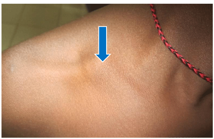
We report the case of a 7-year-old girl borne normally at term but with a low birth weight of 2,200kg. There is no family history of this pathology among the parents and sibblings. She was brought by her parents to our outpatient department complaining of pain and swelling of the left wrist without prior trauma. On the systematic examination of this girl, a swelling of the posterior aspect of the left wrist strongly suggesting a synovial cyst was noticed. On further examination, we noticed a swelling around the middle 1/3 of the right clavicle visible and palpable under the skin. The parents reported that this swelling has been existing since the birth of the child. (Figure 1).
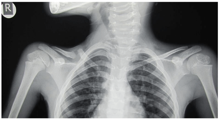
The comparative radiographic assessment of the two wrists showed no bone pathology (Figure 2).
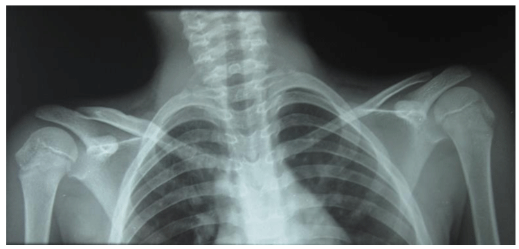
However, the comparative x-ray of the 2 clavicles revealed a congenital pseudarthrosis of the right clavicle (Figure 3).
We prescribed an analgesic treatment to relieve the pain caused by the left wrist cyst. We as well suggested and discussed surgical treatment of this pseudoarthrosis of the clavicle and explained its necessity if she complains of pain, functional or aesthetic discomfort. We reviewed this child after about 1 year, complaining of an aesthetic discomfort that became more and more apparent as the child grew up. We then proposed surgical treatment and discussed it with the parents. The procedure consisted in performing a direct approach to the pseudarthrosis focus under general anesthesia. We then excised the fibrosis exposing the fresh ends of the bone fragments followed by reduction and internal fixation with smooth flexible intramedullary pins (Figure 4), this construct was supplemented by an immobilization of the shoulder with an elbow-to-body sling.
The shoulder sling was removed 6 weeks after surgery.
The clinical evolution was marked by a satisfactory consolidation of the pseudarthrosis focus, this motivated us to perform implant removal on day 305 postoperatively. The radiograph of the clavicle done on day 435 shows a complete consolidation of the pseudarthrosis focus (Figure 5).
Discussion
Epidemiological aspect
The most important published series emphasized the predominance of the female sex: 0.3 and 0.8 respectively according to Cadilhac et al. [1], and Di Gennaro et al. [2]. The age of our patient, 7 years, corroborates with the average age of diagnosis of congenital pseudarthrosis which varies from 7.1 years on average for Studer et al. [3] to 11.5 years for Cadilhac et al. [1].
Aetiological aspect
Congenital pseudarthrosis of the clavicle is due, according to Lloyd-Roberts et al. [4], to a defect of fusion of the 2 points of ossification of the clavicle. But the real mechanism of the origin of this pathology is unknown. The theory advanced by these authors would be the constant and abnormally high pressure exerted by the subclavian artery which is normally high on the right side. This pressure could be increased by the presence of the first ribs on this side.
The rare involvement of the left clavicle would be linked to a dextrocardia and the existence of an abnormally large cervical rib. Lombard [5] also evokes a malposition of the fetus.
The familial cases reported by Cadilhac et al. [1], and Price et al. [6], and in twins by Akman et al. [7] suggest a genetic cause.
Clinical aspect
Pseudarthrosis of the clavicle is most often an incidental discovery [8] as in our case, and rarely symptomatic. It can be revealed by pain, neurological disturbances [9] or by a disgraceful cosmetic appearance that leads parents to consult [10]. It rarely presents with complication such as the thoracic outlet syndrme [9, 11, 12 and 13]. This last eventuality would be the result of a long evolution in the adolescent or the adult age. Gamier et al. [14] reported 3 arterial complications of thoracic paralysis syndrome and recommended Doppler ultrasound of the subclavian or selective arteriography. A simple comparative X-ray of the 2 clavicles often makes it possible to make the diagnosis of congenital pseudarthrosis. In case of doubt, a CT scan with 3D reconstruction images confirms the diagnosis and helps in the choice of therapeutic methods [15]. However, congenital pseudarthrosis of the clavicle may have the appearance of post-traumatic pseudarthrosis. This eventuality, in addition to the asymptomatic aspect of our case, prompted us to observe our case for a few months before the surgical treatment which finally was very necessary to remedy the aesthetic prejudice.
Therapeutic aspect
Most authors agree on a therapeutic abstention in cases of asymptomatic pseudarthrosis with an acceptable cosmetic aspect [16, 17]. The treatment modalities depend on the age and severity of congenital pseudarthrosis. Some authors recommend excision of the fibrosis, autologous cortico-spongy graft with internal osteosynthesis (DCP plate, pins) or external fixation (external fixator) [2, 10, 13, 18- 22]. Grogan et al. [23]. carried out only an excision of the pseudarthrosis while preserving the periosteum without the addition of graft or internal fixation with a satisfactory consolidation in 14 weeks in 8 children. Chandran [24] advocates excision-grafting and internal fixation by a DCP plate for faster consolidation with fewer complications compared to the use of threaded pins in a comparative study of the 2 methods of open reduction and internal fixation.
In our case we carried out a thorough excision of the fibrosis and internal fixation with intramedullary pins according to the technique described by Métaizeau [25] reinforced by immobilization of the shoulder by a elbow-to-body sling, which made it possible to obtain adequate consolidation without complication.
Conclusion
The diagnosis of pseudarthrosis of the clavicle requires a complete history, a brief clinical examination to appreciate the symptomatic, functional and aesthetic repercussions. Therapeutic abstention is still a part of the therapeutic arsenal, but literature and our case teach us that surgery in children can produce quite satisfactory results with very few complications.
- Cadilhac C, Fenoll B, Peretti A, Padovani JP, Pouliquen JC, et al.. (2000) Pseudarthrose congénitale de clavicule. Etude de 25 cas chez l’enfant. Rev Chir Orthop Reparatrice Appar Mot. 86: 575-580. Link: https://goo.gl/adfMCF
- Di Gennaro Gl, Gravino M, Martinelli A, Berardi E, Rao A, et al.. (2017) Congenital pseudarthrosis of the clavicle : a report on 27 cases. J Shoulder Elbow Surg 26: e65-e70. Link: https://goo.gl/ZVW4Eq
- Studer K, Baker MP, Krieg AH (2017) Operative treatment of congenital pseudarthrosis of the clavicle : a single-centre experience. J Pediatr Orthop B 26: 245-249. Link: https://goo.gl/kMCqZ2
- Lloyd-Roberts GC, Apley AG, Owen R (1975) Reflections upon the aetiology of congenital pseudarthrosis of the clavicle. With a note on cranio-cleido-dysostosis. J Bone Joint Surg Br 57: 24-29. Link: https://goo.gl/L421NX
- Lombard JJ (1984) Pseudarthrosis of the clavicle. A case report. S Afr Med J 66: 151-153. Link: https://goo.gl/EiZdQx
- Price BD, Price CT (1996) Familial congenital pseudarthrosis of the clavicle: case report and literature review. Iowa Orthop J 16: 153-156. Link: https://goo.gl/DoXjHa
- Akman YE, Doğan A, Uzümcügil O, Azar N, Dalyaman E, et al.. (2008) Congenital pseudarthrosis of the clavicle in two siblings. Acta Orthop traumatol Turc 42: 377-381. Link: https://goo.gl/FfA4av
- Sung TH, Man EM, Chan AT, Lee WK (2013) Congenital pseudarthrosis of the clavicle: a rare and challenging diagnosis. Hong Kong Med J 19: 265-267. Link: https://goo.gl/iBU8YG
- Watson HI, Hopper GP, Kovacs P (2013) Congenital pseudarthrosis of the clavicle causing thoracic outlet syndrome. BMJ case Rep pil: bcr2013010437. Link: https://goo.gl/Z82ukj
- Galanopoulos I, Ashwood N, Garlapati AK, Fogg Q (2012) Congenital pseudarthrosis of the clavicle: illustrated operative technique and histology findings. BMJ Case Rep pli : bcr2012006908. Link: https://goo.gl/1U4HAK
- Sales de Gauzy J, Baunin C, Puget P, Cahuzac JP (1999) Congenital pseudarthrosis of the clavicle and thoracic outlet syndrome in adolescence. J Pediatr Orthop B 8: 299-301. Link: https://goo.gl/4MBg3c
- Valette H (1995) Pseudarthrose congénitale de la clavicule et syndrome de la traversée thoraco-brachiale. J Mal Vasc 20: 51-52. Link: https://goo.gl/653mAe
- Young MC, Richards RR, Hudson AR (1988) Thoracic outlet syndrome with congenital pseudarthrosis of the clavicle: treatment by brachial plexus decompression, plate fixation and bone grafting. Can J Surg 31: 131-133. Link: https://goo.gl/xkJ9Xk
- Gamier D, Chevalier J, Ducasse E, Modine T, Espagne P, et al.. (2003) Complications artérielles du syndrome du défilé thoraco-brachial et pseudarthrose de clavicule. J Mal Vasc 28: 79-84. Link: https://goo.gl/BBHUVz
- Sloan A, Paton R (2006) Congenital pseudarthrosis of the clavicle: the role of CT-scanning. Acta Orthop Belg 72: 356-358. Link: https://goo.gl/B1CtHz
- O’Leary E, Elsayed S, Mukherjee A, Tayton K (2008) Familial pseudarthrosis of the clavicle: does it need treatment? Acta Orthop Belg 74: 437-440. Link: https://goo.gl/Yy3go6
- Kolodziej L, Bohatyrewicz A, Kotrych D (2008) Congenital pseudarthrosis of the clavicle: is operative treatment necessary? A report of four cases and literature review. Chir Narzdow Ruchu Otop Pol 73: 277-280, 252-256. Link: https://goo.gl/j6Mknj
- 18 . Beslikas TA, Dadoukis DJ, Gigis IP, Nenopoulos SP, Christoforides JE (2007) Congenital pseudarthrosis of the clavicle: a case report. J of Orthop Surg 15: 87-90 Link: https://goo.gl/7j2n9Q
- Etti V, Wild A, Krauspe R, Raab P (2005) Surgical treatment of congenital pseudarthrosis of the clavicle: a report of three cases and review of the literature. Eur J Pediatr Surg 15: 56-60. Link: https://goo.gl/44MtH2
- Persiani P, Molayem I, Villani C, Cadilhac C, Glorion C (2008) Surgical treatment of congenital pseudarthrosis of the clavicle : a report of 17 cases. Acta Orthpp Belg 74: 161-166. Link: https://goo.gl/guKdAH
- Lorente Molto FJ, Bonete Lluch DJ, Garrido IM (2001) Congenital pseudarthrosis of the clavicle: a proposal of early surgical treatment. J Pediatr Orthop 21: 689-693. Link: https://goo.gl/3iQ7ZJ
- Lelei LK (2005) Congenital pseudarthrosis of the clavicle: a case report. East Afr Med J 82: 270-272. Link: https://goo.gl/rFTGrt
- Grogan DP, love SM, Guidera KJ, Ogden JA (1991) Operative treatment of congenital pseudarthrosis of the clavicle. J Pediatr Orthop 11: 176-180. Link: https://goo.gl/zze52a
- Chandran P, George H, James LA (2011) Congenital clavicular pseudarthrosis: a comparison of two treatments methods. J Child Orthop 5: 1-4. Link: https://goo.gl/59Ftc7
- Métaizeau JP (1987) L'embrochache centro-médullaire coulissant. Application au traitement des formes graves d'ostéogenèse imparfaite. Chir Pédiatr 28: 240- 243.
Article Alerts
Subscribe to our articles alerts and stay tuned.
 This work is licensed under a Creative Commons Attribution 4.0 International License.


 Save to Mendeley
