Open J Orthop Rheumatol
Outcome of Treatment of Displaced Intrartcular Fracture Calcaneus by Plate and Screws
Shwan Mohammed Mustafa* and Las Jamal hwaizi
Cite this as
Mustafa SM, hwaizi LJ (2018) Outcome of Treatment of Displaced Intrartcular Fracture Calcaneus by Plate and Screws. Open J Orthop Rheumatol 3(1): 012-019. DOI: 10.17352/ojor.000015Introduction
Calcaneum fractures account for 2% of all fractures, 60% of tarsal bone fractures.10% of fractures are bilateral and 75% are intra articular [1]. 10% of fractures are associated with spine fractures. Mechanism of injury in majority of patients is axial loading i.e. fall from height. Other mechanisms are car accident i.e. brake pedal and high velocity trauma. Current development in imaging technology has allowed better understanding of this complex fracture pathology. Sanders classification [2] of intra articular Calcaneum fractures is widely used now a days because of its proven correlation with management and prognosis. Treating calcaneum fracture is controversy and challenging for orthopedic surgeon. Treatment options either non operative or operative methods [3,4]. Some studies show advantage to internal fixation whereas some show no difference [5,6]. Hence, this study has been carried out with the aim to assess the functional outcome of operatively treated intra articular calcaneal fractures.
Anatomy
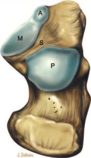
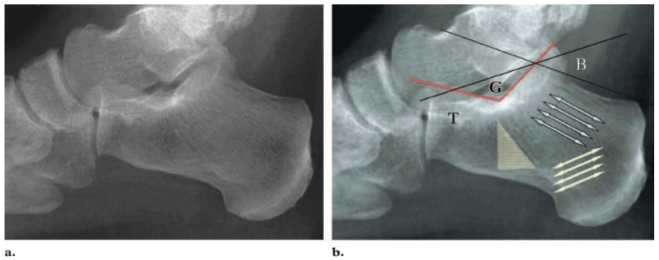
The calcaneus is designed to withstand the daily stresses of weight bearing. It’s important to understanding of the anatomy of the calcaneus in order to determining the patterns of injury and treatment goals and options. The calcaneus has a relatively thin cortex. Traction trabeculae radiate from the inferior cortex, and compression trabeculae converge to support the anterior and posterior articular facets (Figure 1) [7], leaving a “neutral triangle” between them with sparse trabeculations [7] (Figure 2). The cortical bone just inferior to the posterior articular facet is condensed to approximately 1 cm and is called the thalamic portion. Thickening of the cortex is also seen in the regions of the sustentaculum tali, medial wall, and critical angle of Gissane.
The calcaneus has four articulating surfaces, three superior and one anterior. The superior surfaces the posterior, middle, and anterior facets articulate with the talus. The posterior facet is separated from the middle and anterior facets by a groove that runs posteromedially, known as the calcaneal sulcus. The canal formed between the calcaneal sulcus and the talus is called the sinus tarsi. The middle calcaneal facet is supported by the sustentaculum tali and articulates with the middle facet of the talus. The anterior calcaneal facet articulates with the anterior talar facet and is supported by the calcaneal beak. The triangular anterior surface of the calcaneus articuates with the cuboid [7].
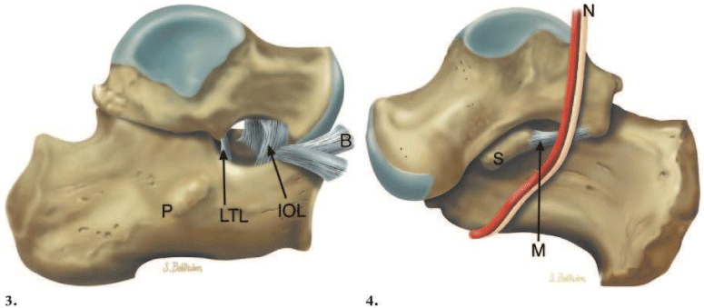
The lateral surface is flat and subcutaneous, with a central peroneal tubercle for the attachment of the calcaneofibular ligament centrally. The lateral talocalcaneal ligament attaches anterosuperiorly to the peroneal tubercle [7] (Figure 3).
Medially, the talus is held to the calcaneus firmly by the interosseous ligament and the thick medial talocalcaneal ligaments [8]. The sustentaculum tali is seen at the anterior aspect of the medial surface. The groove inferior to it transmits the flexor hallucis longus tendon. The neurovascular bundle runs adjacent to the medial border of the calcaneus (Figures 3). The neurovascular bundle may be injured during trauma or during surgery by the reduction of the sustentacular fragment, which is a key element in the surgical management of calcaneal fractures [9].
Mechanism of injury, fracture type, and classification
Fractures of the calcaneus have been described since the times of Hippocrates. They have historically had a poor prognosis, since they are usually caused by direct trauma that injures the articular surfaces, calcaneal fat pad, and peroneal tendons, with resultant change in mechanical forces acting at the ankle joint. Calcaneal fractures are divided into two major categories, intraarticular and extraarticular. Accurate description of calcaneal fractures, including the position and displacement of fracture fragments, is extremely useful to surgeons, with significant implications for the management of these fractures [10].
Intraarticular fractures
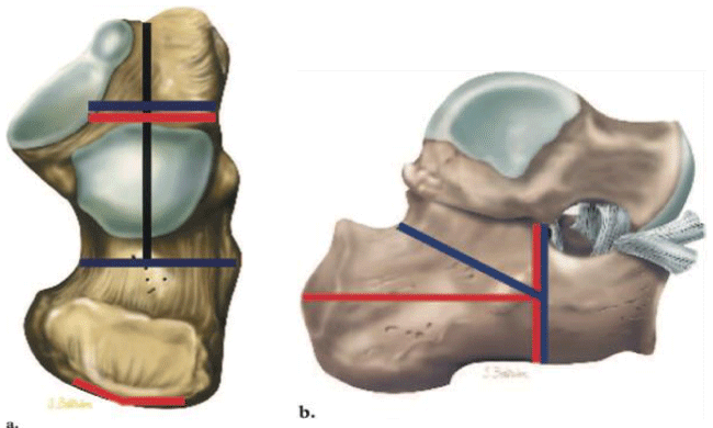
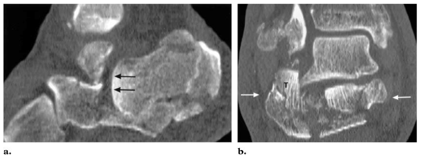
Approximately 75% of calcaneal fractures are intraarticular and result from axial loading, which produces two separate fracture lines: shear and compression [10]. A shear fracture occurs in the sagittal plane and runs through the posterior facet, dividing it into anteromedial and posterolateral fragments (Figure 4). The fracture line may extend anteriorly to involve the cuboid facet. The position of this fracture line depends on the position of the foot at the time of the shear force. If the hindfoot is in varus position, the line extends more anteromedially; if the hindfoot is in valgus position, the line tends to be more posterolateral. If the foot is in extreme valgus position, the fracture line may be lateral to the posterior facet and extraarticular. A sagittal shear fracture splits the calcaneus into two fragments: the anteromedial or “sustentacular” fragment and the posterolateral or “tuberosity” fragment [11]. The medial fragment is not substantially displaced relative to the talus because of the medial talocalcaneal and interosseous ligaments. The lateral fragment is dislocated laterally and remains impacted following release of the axial load, leading to a “step off” in the posterior facet (Figures 5a,b). Occasionally, the talus continues to impact on the lateral edge of the medial fragment, creating a “double split” in it (Figure 5c). The resultant fragment is called the “middle fragment” and typically is displaced by about 1–2 mm.
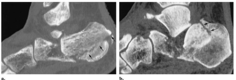
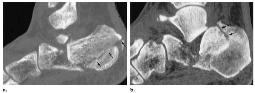
The compression fracture line is produced by wedging of the anterolateral process of the talus into the angle of Gissane. The resultant compression fracture runs through the coronal plane and can extend medially to split the middle facet and the anteromedial fragment (Figure 4a). As mentioned, significant displacement of this fracture is prevented by the medial talocalcaneal and interosseous ligaments [11]. When viewed laterally, the compression fracture appears as an inverted “Y,” with the posterior limb extending horizontally toward the tuberosity as a “tongue type” fracture, or more vertically as a “joint depression type” fracture [11] (Figure 4b). The primary difference between these two fracture types is the connection of the tuberosity or posterolateral fragment to the lateral portion of the posterior facet, which is present in the tongue type and absent in the joint depression type [4] (Figure 6). The anterior limb of this fracture results in the formation of an anterolateral fragment, which is connected to the cuboid facet. This fracture causes buckling of the lateral wall, resulting in loss of height and marked widening of the calcaneus. Intraarticular calcaneal fractures produce typical features, including (a) loss of height due to impaction and rotation of the tuberosity fragment, (b) increase in width due to lateral displacement of the tuberosity fragment, and (c) disruption of the posterior facet of the subtalar joint (Figure 7).
Classification
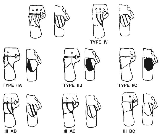
The Sanders classification is used more commonly and is based on the pathophysiology proposed by Soeur and Remy; it relies on sagittally reconstructed CT images reformatted parallel and perpendicular to the posterior facet of the subtalar joint [3,12,13]. Type I fractures are nondisplaced. Type II fractures (two articular pieces) involve the posterior facet and are subdivided into types A, B, and C, depending on the medial or lateral location of the fracture line (more medial fractures are harder to visualize and reduce intraoperatively). Type III fractures (three articular pieces) include an additional depressed middle fragment and are subdivided into types AB, AC, and BC, depending on the position and location of the fracture lines. Type IV fractures (four or more articular fragments) are highly comminuted (Figure 8) [12]. The Sanders classification has been shown to have good interobserver variability, making it useful in clinical practice [3]. The Sanders classification system is useful not only in treatment planning but in helping to determine prognosis [11]. In their series of 120 intraarticular calcaneal fractures, type I fractures were treated without surgery [14]. Patients with type II and type III fractures who underwent surgery experienced excellent or good clinical results in 73% and 70% of cases, respectively [12]. Alternatively, only 9% of patients with type IV fractures had excellent or good clinical results after surgical treatment. Sanders et al have shown that although anatomic reduction is necessary for a good clinical outcome, success is not guaranteed, possibly related to cartilage necrosis at the time of injury [12] (Table 1).
Patients & Methods
This was a prospective study, which was carried out over a period of 18 months (January 01, 2016 to august 01, 2017) in the west emergency and erbil teaching hospital, all surgeries done by same surgical team. Inclusion criteria patients with intra articular fracture calcaneus, were accepted to be enrolled for the study. All the patients above 18 years of age, with (Sanders Types II, III, IV), all patients are active and mobile before injury and they accepted surgery. Exclusion criteria patients with medically unstable, polytrauma and nonambulatary are excluded from the study.
A total of 24 patients with fracture calcaneus were admitted in the Orthopedics Department at west emergency and erbil teaching hospital, who fulfilled the inclusion criteria were enrolled in this study.
The demographic profile, complete histories, systemic, local and neurovascular examinations, patients were subjected to the following radiological investigations - X-ray foot (anteroposterior, lateral and axial), spine (anteroposterior and lateral), pelvis (anteroposterior), computed tomography scan (CT scan) of foot; other relevant investigations were performed wherever required. Broad-spectrum antibiotics, anti-inflammatory drugs and painkiller, were administered as needed. The temporary compressive bandage, ice packing was provided and the affected part was elevated. The patient was planned for surgery once the edema subsides, and wrinkle sign develops.
Procedure of surgery
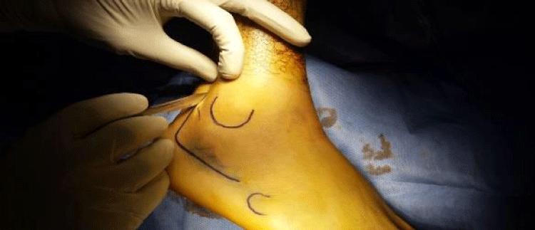
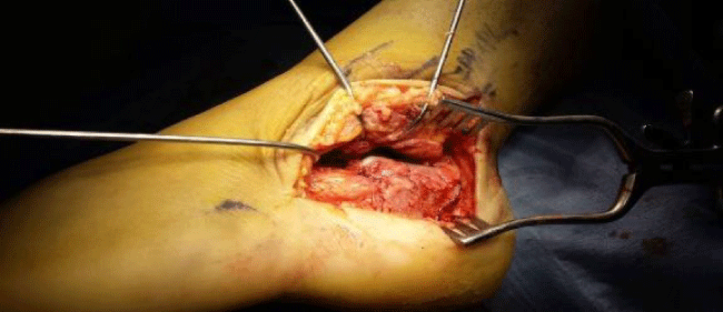
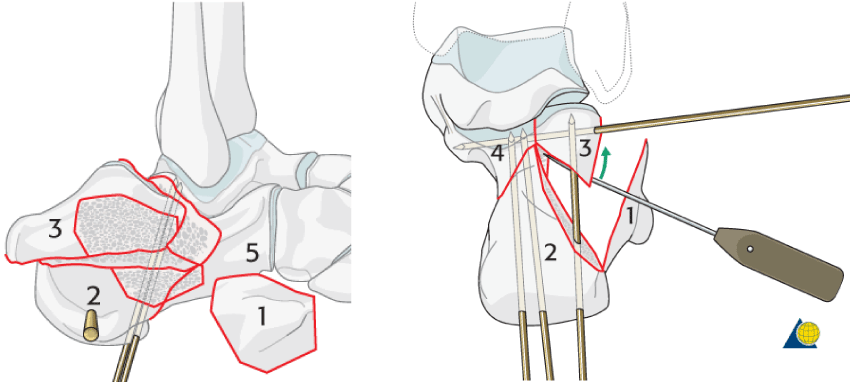
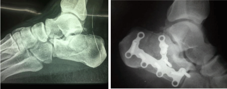
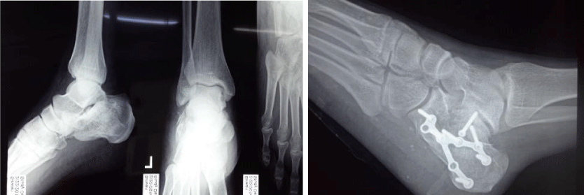
Surgery were performed under spinal or general anesthesia, with the following techniques, ORIF with reconstruction of the articular surface of the posterior facet, in the present study, 26 calcaneus were operated with this technique. All of the patients were operated in lateral decubitus position under pneumatic tourniquet, IV antibiotic giving one hour before induction. Using lateral approach, after taking all aseptic precaution tourniquet was inflated. After that an extensile right-angled lateral incision was done (Figure 9). Taking the flap down to the periosteum, full thickness flap was elevated and temporarily held with 2 to 3 k-wires (tip of distal fibula, talus and cuboid bones) (Figure 10). Lateral wall of the calcaneus was exposed up to subtalar and calcaneocuboid joint. A Schanze-screw with grip is inserted in the calcaneal tuberosity aiming lateral and plantar. The first reposition is done, using this grip, with the forefoot in maximal plantar flexion (taking advantage of ligament axis). In type C medial fracture type were sustentaculum tali was displaced first reduced and held by temporary K-wires (Figure 11), The different pieces of cortex can be put together using fine instruments like hooks and the small deperiosteur. When an anatomical restoration of the lateral and/or medial (sustentaculum) facets of the calcaneus is achieved, temporary fixation with a thick K-wire (tuber calcanei) and thinner K- wires (different fragments) is done. When radiological control shows an optimal reposition, fixation using singular or multiple locked-plates follows (Figure 12). Followed by closure of the wound after achieving hemostasis, the mean duration of surgery 85 minutes.
Follow-up
After surgery, limb elevation was maintained, injectable antibiotics (injection ceftriaxone 1 g intravenous (IV) BD) for 4 days, sterile dressing was done using butadiene solution, drain was removed after 24 h of surgery. Ankle was mobilized on the second post-operative day. Sutures were removed after two week. Gradual weight bearing was started from 10 to 12 weeks.
All the patients were called for 6, 12 and 18 month follow-up on outdoor basis and examined and evaluated according to Maryland foot score [15,16].
Physiotherapy is also very important for the rehabilitation of the patients all the patients went through a course of active physiotherapy, inversion - eversion exercise from 2nd postoperative day.
Statistical analysis
Where appropriate, the results were tested for significance using linear regression tests. p value < 0.05 was considered to be statistically significant.
Results
Twenty-four patients (22 unilateral and 2 bilateral), the mean age being 35.41 years (range 18-60 years), consisting of 3(12.5%) female and 21(87.5%) male were included in the study. Most common mode of injury was fall from height in 23(95.8%) patients, while road traffic accident was in 1(4.16%) patients. Three patients had associated spinal injuries, two in L1 vertebra and one in L3, none of them had neurological deficit.
The mean duration between injury and surgery was 11.66 days (range 6- 16 days). The mean duration of surgery was 85 minutes (range 60 to 110 minutes).
The average follow-up was 16 months (range 14 to 18 months). After 10 week more than 96% of the patients had no or only mild occasional pain with no limitation of daily activity and no gait abnormality and were able to walk at least more than 300 m with only some difficulty on uneven surface. All patients had normal ankle joint with all having dorsiflexion and plantar flexion more than 20°. The average subtalar range of motion was 18°, with only 2 cases having near normal restriction (75%-100%), and 1 having severe restriction (<25%).
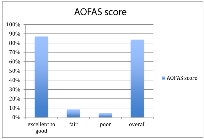
Average AOFAS score at 12-week follow-up was 85.3(range 74 to 96), with 87% having excellent to good results and 2 (8.3%) and 1 (4.16%) had fair and poor results respectively (Figures 13,14) (Table 2).
Two patients had flap necrosis at incision site and two had superficial infection, both of which healed by extended antibiotics. Subtalar arthritis was seen in 3 patients which were treated by subtalar arthrodesis, whereas sural nerve hypoaesthesia in 1 patient, broadening heal 2 patient, varus 1 patient (Table 3). None of the patients had heel pad problem, compartment syndrome, peroneal tendinitis or implant failure. All fractures united and none needed secondary bone grafting. Patients returned to work on an average of 13.45 week.
The patients were allowed gradual weight bearing after 10-12 weeks, after clinicoradiological signs of union. All patients started full weight results were evaluated according to the criteria of the AOFAS score and “Maryland foot score.” (Internal consistency [Cronbach’s alpha, reliability] for the ministeriums für staatssicherheit [MFS] is 0.82) [16] (Table 4).
Discussion
The calcaneum is the most commonly fractured tarsal bone. It accounts for approximately 2% of all fractures and 60 -65 % of all tarsal fracture [1-3]. Calcaneus fractures results in a varus deformity with heel widening, loss of calcaneal height, and subtalar joint incongruency [17,18]. Open reduction of the fracture fragments with anatomic reduction and rigid internal fixation plays important role in management of calcaneum fracture.
In the series reported by Farrell [19], 90% males were involved. In both the study, male more than female. Our study is going with the literature. More number of male patients can be explained as more number of males is working in industries and construction work.
According to Mostafa et al. surgery was performed after an average of duration of 4.83 days from admission [17]. Fracture fixation should be performed within fourteen days from the injury as fracture reduction is difficult after this period due callus formation.
There is no consensus of the surgical approach for fixation in calcaneum fractures. Stephenson et al. has used a combined medial and lateral approach [20]. McReynolds et al. has recommended the lateral approach for tongue type fracture and joint depression fractures [21].
The inappropriate surgical scar healing may lead to wound breakdown in calcaneum fractures [6-16,22-24]. The key step which may prevent scar healing problems is proper thickness of skin flap and minimal handling of the flap. In post-operative period patient should be encouraged for minimal walking and limb elevation.
Conclusions of Bajammal who analyzed 20 publications dealing with operative vs. conservative treatment showed significant benefits of surgical therapy for females, young males, patients with lighter workload, and patients with initially high B-angle or with simple, minimally dislocated fractures, whereas older males and those who have an occupation involving heavy workload had benefited from osteosynthesis with and without bone grafting and did not find any significant difference [25].
Rak et al. concluded that there were less complications and better results related to treatment with locking compression plates [26]. Their results showed that locking plates could be used for fixation in all types of intraarticular calcaneal fractures.
In this study, we have followed Sanders classification, which is CT, based. The CT also helps in designing a surgical plan.
Meantime duration between injury and surgery in our series is 11.6 days. We waited for the wrinkle sign to appear before taking the patient to surgery. This is recommended in the literature and in order to prevent wound complication. The approach used was lateral in all patients where ORIF was performed.
The conservative therapy [27]. Buckley (analysis of 559 calcaneal fractures) [28] and Tufescu reported similar findings [29] and both definitely recommended operative treatment.
In our opinion, there is no indication for bone grafting when LCP are used except for the Sanders IV type injuries with huge defects after the reduction. Longino had compared postoperative radiological and clinical results of LCP (Tables 5,6) [12,30,31].
Using SPSS software and multiple linear regression analysis was used to identify factors which affecting the general outcome. Detrimental host factors which can affect outcomes in this study had been excluded and there was no bias in the surgeon factors because only one qualified Ankle and Foot surgeon had performed this surgery. There were 2 main factors found to be affecting the patient’s outcome
There was a significant positive linear relationship found between compliance of no weight bearing and the MFS (p=0.004). The patients who had well compliance of not weight bearing for more than 10 weeks had higher MFS outcome compared to those started early weight bearing before 10 weeks.
There was a significant negative linear relationship found between weeks before they were able to RTW and the MFS (p=0.001).
Conclusion
ORIF with restoring the articular surface and congruity with locking plate and screw is the ideal treatment for joint depression type and Sanders Type II/III and IV. Even Sanders Type IV (which was thought to be associated with poor results) had a good outcome in short-term follow-up. CT scan preoperatively very important for surgical planning and classification, Use of proper surgical timing/technique/asepsis can lead to good or excellent results in more than 90% of patients and avoiding the majority of the complications. Thus ORIF in these patients should be encouraged.
Recommendation
1. Surgery should be delay until swelling subside and wrinkle sign appear to avoid flap necrosis.
2. Preopertive CT scan very important for classification and planning surgery.
3. The principle of treating intra-articular fractures especially in a weight bearing joints are anatomical reduction, rigid fixation of intra-articular fractures and early mobilisation for good treatment outcome [30].
- Fitzgibbons TC, McMullen ST, Mormino MA (2001) Fractures and dislocations of the calcaneum. In: Bucholz RW and Heckman JD Eds Rockwood and Green's Factures in adults, 5th ed. Philadelphia: Lippincott Williams & Wilkins 3: 2133-2179.
- Sanders R, Fortin P, DiPasquale T, Walling A (1993) Operative treatment in 120 displaced calcaneal fractures: results using a prognostic computed tomography scan classification. Clin Orthop 290: 295.
- Sanders R (1996) Intra-articular fractures of the calcaneum: present state of the art. J Orthop Trauma 6: 252-265. Link: https://goo.gl/JYcjps
- Eastwood DM, Phipp L (1997) Intra-articular fractures of the Calcaneum: why such controversy?. Injury 28: 247–259. Link: https://goo.gl/hiG3zr
- Jarvholm U, Korner L, Thoren O, Wiklund LM (1984) Fractures of the calcaneum: a comparison of open and closed treatment. Acta Orthop Scand 55: 652-656. Link: https://goo.gl/ij9LMs
- Buckley RE, Meek RN (1992) Comparison of open versus closed reduction of Intraarticular calcaneal fractures: a matched cohort in workmen. J Orthop Trauma 6: 216-222. Link: https://goo.gl/mdhUPz
- Gray H (2000) Anatomy of the human body. Philadel- phia, Pa: Lea & Febiger, 1918.
- Carr JB (1993) Mechanism and pathoanatomy of the intraarticular calcaneal fracture. Clin Orthop Relat Res 290: 36-40. Link: https://goo.gl/EFmeGK
- Burdeaux BD (1983) Reduction of calcaneal fractures by the McReynolds medial approach technique and its experimental basis. Clin Orthop Relat Res 177: 87–103. Link: https://goo.gl/CKPfi4
- Linsenmaier U, Brunner U, Schoning A, Rieger J, Krotz M, et al. (2003) Classification of calcaneal fractures by spiral computed tomography: implications for surgical treatment. Eur Radiol 13: 2315-2322. Link: https://goo.gl/61UM6s
- Eastwood DM, Phipp L (1997) Intra-articular fractures of the calcaneus: why such controversy?. Injury 28: 247-259.
- Sanders R, Fortin P, DiPasquale T, Walling A (1993) Operative treatment in 120 displaced intraarticular calcaneal fractures: results using a prognostic com- puted tomography scan classification. Clin Orthop Relat Res 290: 87–95. Link: https://goo.gl/9wgwi9
- Sanders R, Gregory P (1995) Operative treatment of in- tra-articular fractures of the calcaneus. Orthop Clin North Am 26: 203-214. Link: https://goo.gl/vC78Ht
- Essex-Lopresti P (1952) The mechanism, reduction technique, and results in fractures of the os calcis. Br J Surg 39: 395-419. Link: https://goo.gl/3pH68x
- Sanders R (1992) Intra-articular fractures of the calcaneus: Present state of the art. J Orthop Trauma 6: 252-265. Link: https://goo.gl/rpZTbq
- Zwipp H, Tscherne H, Thermann H, Weber T (1993) Osteosynthesis of displaced intra-articular fractures of the calcaneus. Results in 123 cases. Clin Orthop Relat Res 290: 76-86. Link: https://goo.gl/gmL6A3
- Mostafa MF, El-Adl G, Hassanin EY, Abdellatif M (2010) Surgical treatment of displaced intra- articular calcaneal fracture using a single small lateral approach. Strategies Trauma Limb Reconstr 5: 87-95. Link: https://goo.gl/TUsnQR
- Letournel E (1993) Open treatment of acute calcaneal fractures. Clin Orthop Relat Res 290: 60-67. Link: https://goo.gl/HLd5m2
- O’Farrell DA, O’Byrne JM, McCabe JP, Stephens MM (1993) Fractures of the os calcis: Improved results with internal fixation. Injury 24: 263-265. Link: https://goo.gl/vMx4r6
- Stephenson JR (1987) Treatment of displaced intra- articular fractures of the calcaneus using medial and lateral approaches, internal fixation, and early motion. J Bone Joint Surg Am 69: 115-130. Link: https://goo.gl/y9R6Bd
- McReynold IS (1982) Trauma to the Os Calcis and heel cord. In: Jahss MH eds Disorders of the Foot, 4th ed. Philadelphia: WB Saunders 1497-1542.
- Harty M (1973) Anatomic considerations in injuries of the calcaneus. Orthop Clin North Am 4: 179-183. Link: https://goo.gl/E8jwkk
- Carr JB, Hamilton JJ, Bear LS (1989) Experimental intraarticular calcaneal fractures: anatomic basis for a new classification. Foot Ankle 10: 81-87. Link: https://goo.gl/FLwzTi
- Furey A, Stone C, Squire D, Harnett J (2003) Os calcis fractures: analysis of interobserver variability in using Sanders classification. J Foot Ankle Surg 42: 21–23. Link: https://goo.gl/mKkLr5
- Longino D, Buckley RE (2001) Bone graft in the operative treatment of displaced intraarticular calcaneal fractures: Is It Helpful?. J Orthop Trauma 15: 280-286. Link: https://goo.gl/bRbE2M
- Rak V, Ira D, Masek M (2009) Operative treatment of intra-articular calcaneal fractures with calcaneal plates and its complications. Indian J Orthop 43: 271-280. Link: https://goo.gl/GMn2uF
- Bajammal S, Tornetta P III, Sanders D, Bhandari M (2005) Displaced intra-articular calcaneal fractures. J Orthop Trauma 19: 360-364. Link: https://goo.gl/kVmpvz
- Buckley R (2002) Letters to the Editor. J Orthop Trauma 16: 210-211. Link: https://goo.gl/NWoLnk
- Tufescu TV, Buckley R (2001) Age, gender, work capability, and worker's compensation in patients with displaced intraarticular calcaneal fractures. J Orthop Trauma 15: 275–279. Link: https://goo.gl/7pSWLh
- Muller ME, Allgower M, Schneider R, Willenegger H (1979) Manual of Internal Fixation. Techniques recommended by AO Group, 2nd edition. New York: Springer. Link: https://goo.gl/SiDVk8
- Gülabi D, Sari F, Sen C, Avci CC, Saglam F, et al. (2013) Mid-term results of calcaneal plating for displaced intra articular calcaneus fractures. Ulus Travma Acil Cerrahi Derg 19: 145-151. Link: https://goo.gl/9omXDi
- Schepers T, Heetveld MJ, Mulder PG, Patka P (2008) Clinical outcome scoring of intra-articular calcaneal fractures. J Foot Ankle Surg 47: 213-218. Link: https://goo.gl/szprcn
- Tornetta P III (1998) The Essex-Lopresti reduction for calcaneal fractures revisited. J Orthop Trauma 12: 469-473. Link: https://goo.gl/cX8Xny
Article Alerts
Subscribe to our articles alerts and stay tuned.
 This work is licensed under a Creative Commons Attribution 4.0 International License.

 Save to Mendeley
