Open Journal of Orthopedics and Rheumatology
Anatomical and Technical Considerations of "Dual Subsartorial Block" (DSB), A Novel Motor-sparing Regional Analgesia Technique for Total Knee Arthroplasty
Kartik Sonawane1*, Hrudini Dixit2, Tuhin Mistry1 and J. Balavenkatasubramanian3
2Fellow in Regional Anesthesia, Department of Anesthesiology, Ganga Medical Centre & Hospitals, Pvt. Ltd., Coimbatore, Tamil Nadu, India
3Senior Consultant, Department of Anesthesiology, Ganga Medical Centre & Hospitals, Pvt. Ltd., Coimbatore, Tamil Nadu, India
Cite this as
Sonawane K, Dixit H, Mistry T, Balavenkatasubramanian J (2021) Anatomical and Technical Considerations of "Dual Subsartorial Block" (DSB), A Novel Motor-sparing Regional Analgesia Technique for Total Knee Arthroplasty. Open J Orthop Rheumatol 6(1): 046-056. DOI: 10.17352/ojor.000038Background: The modernization of arthroplasty has paved the way for the resurgence of ultrasound-guided regional analgesia (RA) techniques. The evolution of newer RA techniques aids in reducing postoperative pain considerably as well as facilitates early ambulation and discharge. “Dual Subsartorial Block (DSB)” is recently described as a novel procedure-specific, motor-sparing, and opioid-sparing RA technique for total knee arthroplasty (TKA) surgery. This review article highlights the innervations covered by DSB based on the anatomical considerations and its suitability for providing analgesic coverage in TKA with medial approaches.
Methodology: We describe anatomical considerations based on the available literature about the anatomy related to the femoral triangle, adductor canal, and subsartorial region. The technical consideration of the DSB is based on our observations of the ongoing study on patients undergoing TKA with medial approaches. However, other details of the study are not part of this article.
Results: After studying the anatomical and technical aspects of the DSB, it is possible to cover almost all procedure-specific innervations of TKA surgeries with the DSB. Our observations and statistical analysis found DSB as a procedure-specific, motor-sparing, and opioid-sparing RA technique.
Discussion: We describe the complex anatomy of the femoral triangle and adductor canal block along with sonoanatomical variations of various subsartorial regions. We also elaborate on the technical details, analgesic coverage, and possible complications of DSB.
Abbreviations
DSB: Dual Subsartorial Block; TKA: Total Knee Arthroplasty; RA: Regional Analgesia; ERAS: Enhanced Recovery After Surgery; FT: Femoral Triangle; ALM: Adductor Longus Muscle; STM: Sartorius Muscle; SN: Saphenous Nerve; FN: Femoral Nerve; FA: Femoral Artery; VMM: Vastus Medialis Muscle; NVM: Nerve To Vastus Medialis; LFCN: Lateral Femoral Cutaneous Nerve; AC: Adductor Canal; AMM: Adductor Magnus Msucle; VAM: Vasoadductor Membrane; LA: Local Anesthetic; PNS: Peripheral Nerve Stimulator
Introduction
Total Knee Arthroplasty (TKA) is one of the most commonly performed elective orthopedic surgery in the modern world. This surgery offers the patient a life-changing experience due to increased joint mobility and painless ambulation. However, one deterrent which precluded a patient from undergoing TKA is the significant immediate postoperative pain. Thanks to the regional analgesia (RA) resurgence aided by ultrasound, which enables a considerable reduction in immediate postoperative pain. Most arthroplasty surgeons are keen to mobilize the patient soon after the surgery, necessitating the evolution of new RA techniques that would aid early ambulation with optimal analgesia.
Dual Subsartorial Block (DSB) is a procedure-specific technique for TKA that would aid in early ambulation due to its motor-sparing and opioid-sparing effect [1]. The DSB covers almost all the innervations of the pain-generating components of the anterior and posterior knee joint. Therefore, knowing the anatomy of the knee joint, types of the subsartorial blocks and their neural components, pain generators before and after TKA surgeries, and innervation of those pain generators is essential to understand the technical aspect of the DSB.
The knee joint is complexly innervated by the main branches of the lumbar and sacral plexus. These include the femoral nerve (anterior knee), obturator nerve (posteromedial knee), and sciatic nerve (posterior knee) [2-4]. The blockade of all three nerves provides complete analgesia in their respective territories. However, it is associated with unwanted motor blockade of quadriceps (femoral nerve), adductors (obturator nerve), and hamstring muscles (sciatic nerve). This causes the risk of falls, delayed mobility, and prolonged hospital stay [5]. For these reasons, such blocks are not recommended in enhanced recovery after surgery (ERAS) protocol for knee surgery.
Many alternative motor-sparing regional analgesia techniques have been described to overcome these unwanted side effects. However, the ideal RA technique should cover all the essential innervations of the knee joint involved in each surgical step without causing motor blockade to support early mobility and discharge. Considering all these requirements, we had designed DSB in 2018 and practicing it in all patients undergoing TKA with medial approaches since then. We named it “Dual Subsartorial Block,” as it is given at two different locations under the sartorius muscle.
We hypothesize that the DSB is motor-sparing, procedure-specific, and opioid-sparing RA technique for TKA. This technical report highlights the innervations covered by DSB based on the anatomical considerations and its suitability in providing analgesic coverage in all TKA procedures with medial approaches. Motor-sparing and procedure-specific RA techniques are the future of RA practices, which induces the analytical thought process in applying everchanging knowledge of anatomy, techniques, and methodologies for providing the best patient care. Therefore, this review article mainly focuses on the anatomical and technique aspect of DSB that will make the readers understand the intricacies of this novel motor-sparing technique.
Relevant anatomy
The “DSB” combines two subsartorial blocks, the distal femoral triangle block and the adductor canal block. This combination is given to block most of the innervations of the pain generating structures involved in TKA surgery. Various literatures have made the demarcation between the femoral triangle and adductor canal clear [6-9]. It is essential to understand the extent and content of both these prominent landmarks of the thigh.
Femoral triangle:
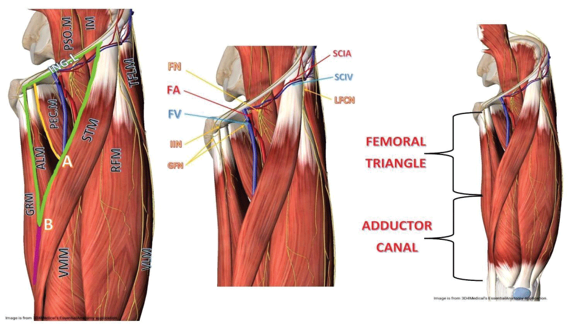
The femoral triangle (FT), also called Scarpa’s triangle, is a subfascial space below the inguinal ligament that lies in the upper third of the thigh [10]. The FT base is formed by the inguinal ligament, the medial border by the medial border of the adductor longus muscle (ALM), and the lateral border by the medial border of the sartorius muscle (STM) (Figure 1). The FT roof is formed by the skin, superficial fascia, and deep fascia (fascia lata). The superficial fascia contains the superficial inguinal lymph nodes, femoral branch of the genitofemoral nerve, branches of the ilioinguinal nerve, superficial branches of the femoral artery with accompanying veins, and upper part of the great saphenous vein. The deep fascia has a saphenous opening covered by the cribriform fascia [11]. The FT floor is formed by the pectineus and ALM medially and iliopsoas muscle laterally [11].
There are two intersecting points of the medial and lateral walls of the FT.
1. The apex of the iliopectineal fossa [12,13] (a proximal subset of FT), where the lateral border of ALM and medial border of STM intersect [Point A in Figure 1].
2. The apex of FT [7,8] (lower extent of FT), where the medial border of ALM and medial border of STM intersect [Point B in Figure 1]. The apex of the triangle is continuous with the adductor canal [11].
Content: [Figure 1]
1. Saphenous Nerve (SN) lies anterolateral to the femoral artery (FA) between the STM and vastus medialis muscle (VMM).
2. Nerve to vastus medialis (NVM) lies lateral to the SN in the groove between STM and VMM.
3. Lateral femoral cutaneous nerve (LFCN) crosses the lateral angle of the FT.
4. Femoral nerve (FN) enters the FT by passing beneath the inguinal ligament, just lateral to the FA. In the thigh, it lies in a groove between the iliacus and psoas major muscles, outside the femoral sheath, and lateral to the FA. After a short course in the thigh, the FN is divided into anterior and posterior divisions, separated by the lateral femoral circumflex artery [11].
5. Nerve to pectineus arises from the FN just above the inguinal ligament. It passes behind the femoral sheath to reach the anterior surface of the pectineus muscle [11].
6. The femoral sheath (enclosing upper 4 cm of the femoral vessels) contains a femoral branch of the genitofemoral nerve, FA and its branches, femoral vein and its tributaries, and deep inguinal lymph nodes.
Adductor canal
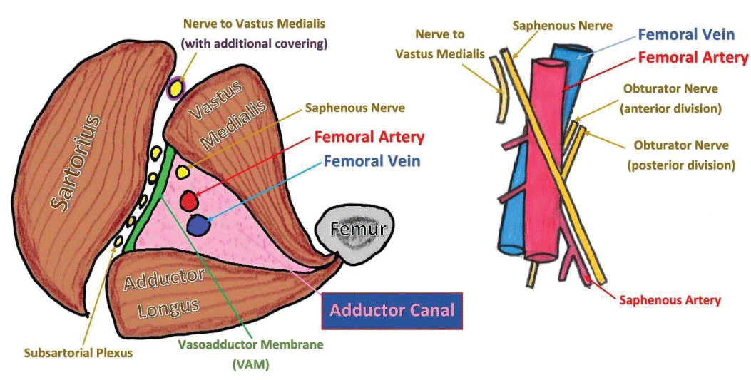
The Adductor Canal (AC), also known as Hunter’s canal or the subsartorial canal, is a musculoaponeurotic tunnel in the middle third of the thigh. It is about a 15 cm long tunnel that serves as a passageway for structures moving between the anterior thigh and posterior leg [14]. It extends from the apex of the FT above to the tendinous opening in the adductor magnus muscle (AMM), called adductor hiatus, below. The AC is triangular in a cross-section bounded anterolaterally by VMM, posteromedially by ALM proximally and AMM distally, and medially by the vasoadductor membrane (VAM), a strong fibrous membrane joining anterior and posterior walls (Figure 2). The AC roof is formed by the skin, subcutaneous tissue, and STM (lying above the VAM). The subsartorial plexus lies in this region sandwiched between STM and VAM. (Figure 2) It is formed by an infrapatellar branch from the SN, anterior division of obturator nerve, medial femoral cutaneous nerve, and NVM. It supplies the skin over the anteromedial aspect of the knee joint, retinaculum, collateral ligaments, and capsule of the knee joint [15].
The AC consists of three foramina: superior, anterior, and inferior. The femoral vessels and the SN enter AC through the superior foramen. The SN and descending genicular vessels exit AC through the anterior foramen, piercing the VAM. Finally, the femoral vessels exit AC via the inferior foramen, also called the adductor hiatus (a gap between the oblique and medial head of AMM). After exiting through this hiatus, femoral vessels become popliteal vessels [16].
Content
1. Femoral artery.
2. Femoral vein (posterior to the FA in the upper part and lateral in the lower part).
3. The SN lies lateral to the FA in the proximal AC and superior to the FA before leaving the AC in the mid-adductor canal area.
Subsartorial region and blocks: [Figure 3]
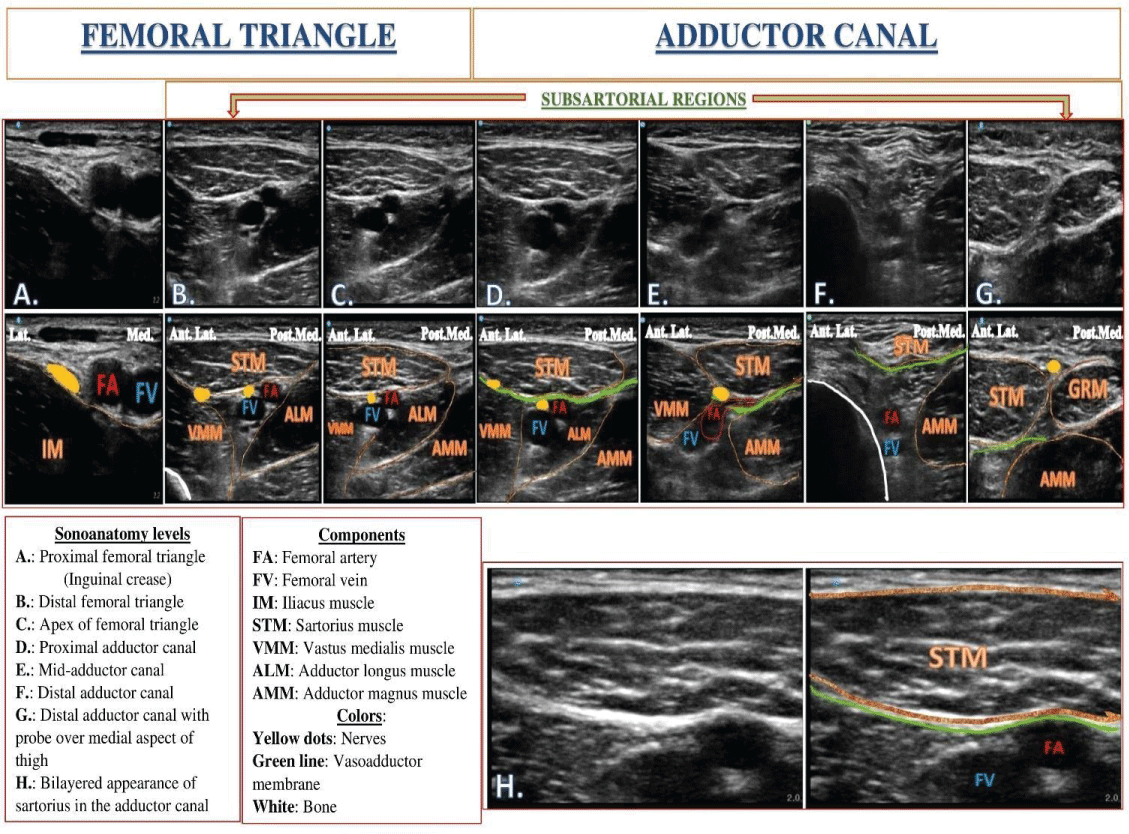
The apex of the FT that separates the FT from the AC usually lies distal to the mid-thigh point [6] It can be identified under ultrasound where medial borders of ALM and STM overlie (Figure3C), forming a figure of “3,” also called a “kissing sign.” The area just proximal to the apex of FT is the distal femoral triangle, and the area just distal to the apex is the proximal adductor canal. The mid-thigh level block is called the femoral triangle block, as this level usually lies at the distal part of FT.
The sartorius muscle is a common landmark for both femoral triangle and adductor canal blocks while using ultrasound. Therefore, the area under the sartorius in these regions can also be called a subsartorial area. Thus, the femoral triangle block and adductor canal block can be considered variants of the subsartorial blocks. The regional block effect in these subsartorial areas differs from each other due to the varied neurovascular anatomy. The variants of subsartorial block are,
A. Femoral triangle block
It is given at the distal-most part of FT, just (1-2 cm) proximal to the apex of FT. Sonoanatomy of this region shows ALM posteromedially, VMM anterolaterally, and STM medially (Figure 3B). In this region, there is no VAM like in AC. The SN lies just lateral to the FA, whereas the NVM lies lateral to SN in the intermuscular fascial planes between STM and VMM. The required local anesthetic (LA) volume for this block is 10-20cc. A higher volume (more than 40cc) causes proximal spread that may include the femoral nerve and its branches. The drug injected into the distal FT spreads distally into the AC along with the vessels, and most of the drug spreads distally under the STM but above the VAM in the AC region. The distal spread of the drug below STM and above VAM involves the subsartorial plexus, which is sandwiched between STM and VAM [17-20].
B. Proximal adductor canal block
It is given just (1-2 cm) distal to the apex of FT. Sonoanatomy of this region shows ALM posteromedially, VMM anterolaterally, VAM medially, and STM above VAM (Figure 3D). Due to the presence of VAM, the lower border of STM appears bilayered under ultrasound [6,8] (Figure 3H). Only SN lies into the adductor canal lateral to the FA, whereas the NVM, along with its additional fascial covering, lies above the VAM in the intermuscular fascial planes between STM and VMM [8,21,22]. The required LA volume for this block is 10-20cc. A drug injected into the proximal adductor canal below VAM travels along with the femoral vessels. It enters into the adductor hiatus involving the posterior division of the obturator nerve, and then it goes backside of the knee to include the popliteal plexus [23,24].
C. Mid-adductor canal block
It is given distal to the proximal adductor canal, where the ALM is replaced by AMM posteromedially. In this region, the SN pierces VAM along with the descending genicular artery (branch of FA) and lies below STM and above the VAM (Figure 3E). Due to the presence of VAM, the lower border of sartorius appears bilayered. The NVM lies above VAM in its fixed location between STM and VMM. The required LA volume for this block is 10-20cc. A drug injected into the mid-adductor canal region below the VAM travels along with the femoral vessels and finally enters the popliteal region to involve the popliteal plexus.
D. Distal adductor canal block
It is given in the lower one-third of the adductor canal. At this level, femoral vessels dip into the opening of the adductor hiatus to become popliteal vessels. Sonoanatomy of this region shows AMM posteromedially, VMM anterolaterally, and VAM (with STM above) medially (Figure 3F). No nerves lie in this part of the adductor canal. The SN lies above VAM initially between STM and AMM. Later, it crosses the adductor canal from the anterior to the medial side, becoming superficial by piercing the deep fascia of the thigh, and finally lies between STM and gracilis muscle (Figure 3G). The required LA volume for this block is 10-20cc. A drug injected into the distal adductor canal below the VAM directly enters into the popliteal region along with the femoral vessels to involve the popliteal plexus.
Dual Subsartorial Block (DSB)
The DSB is a hybrid form of subsartorial block, in which the combination of two subsartorial blocks (distal femoral triangle and adductor canal block) is used to cover all procedure-specific innervations of pain generators involved in TKA surgery. In this block, the local anesthetic drug is deposited under the sartorius muscle at two different places, so it is termed as a “Dual Subsartorial Block.” The DSB is given immediately after the surgery with two different injections at two different locations.
Indications and contraindications
The DSB is a newly introduced motor-sparing RA technique for TKA surgeries. This block can be used for any knee surgery with a medial approach (medial parapatellar, mid-vastus, or subvastus). This block may not work in TKA surgeries with other approaches. Similarly, it may not provide complete analgesia in revision TKA surgeries or knee debridement surgeries as the incision is not always medial parapatellar. The DSB can be an essential component of multimodal analgesia protocol to provide postoperative analgesia without causing any motor blockade. This technique should not be used as a supplementary RA technique in the presence of other RA techniques like femoral nerve block or epidural analgesia. In such a scenario, the motor-sparing advantage of this block will be lost.
Choice of local anesthetics
The DSB requires a larger volume (20-30 mL) of LA solution to adequately cover all the innervation of the postoperative pain-generating components of the TKA surgery. The choice of type, volume, and LA concentration should be based on the patient’s size and general condition. We recommend using lower concentrations and relatively safer LA like 0.2% ropivacaine or 0.25%-0.125% levobupivacaine along with dexamethasone (8 mg) as an additive known to improve the quality and duration of analgesia [25].
Equipments
• Ultrasound machine with high-frequency (6–13 MHz) linear array transducer, sterile sleeve, and gel
• Standard nerve block tray
• One 20-mL syringe containing LA mixture
• A 100- or 120-mm, 21-gauge, short-bevel, insulated stimulating needle
• Peripheral nerve stimulator
• Sterile gloves
• Injection pressure monitor if available
Landmark and patient positioning
The DSB is performed with the patient in the supine position. The hip is abducted, and the thigh is externally rotated to facilitate ultrasound probe and needle placement. If nerve stimulation is to be used simultaneously, thigh exposure is required to observe the motor response. In addition, exposure of the entire thigh will help appreciate the distance from the groin to the knee to determine the middle portion of the thigh.
Probe position and sonoanatomy
A high-frequency linear array ultrasound probe placed horizontally over the anteromedial aspect of the thigh will reveal the prominent musculatures (sartorius, vastus medialis, adductor longus) of the thigh. Beneath the sartorius muscle are the femoral vessels which can be identified with color doppler for orientation. The VMM is identified anterolaterally to the femoral vessels, and the ALM is identified posteromedially to the vessels. The hyperechoic rim of the femur can be seen beneath the vastus intermedius muscle shadow.
By moving the ultrasound probe in the craniocaudal direction, the apex of the FT can be identified where the medial borders of the ALM and STM overlie, forming a figure of “3”. The area just (1-2cm) proximal to the apex of FT is the distal femoral triangle region, where the SN appears hyperechoic just lateral to the FA. The NVM usually lies lateral to the SN in the intermuscular plane between VMM and STM.
The area just (1-2cm) distal to the apex of FT is the proximal adductor canal, where the SN lies in the same location (anterolateral) in relation to the artery. However, the NVM lies above VAM in the intermuscular plane between VMM and STM. Due to the presence of VAM in the adductor canal region, the lower border of the STM appears bilayered.
On moving the probe more distally, it can be seen that the SN moves from lateral to anterior of the artery and becomes superficial by piercing the thigh’s deep and superficial fascia. During its course, the SN lies initially between the STM and VMM, then between STM-ALM, STM-AMM, and finally between STM and gracilis muscle[8]. Throughout the adductor canal region, the NVM can be identified as a hyperechoic structure above the VAM between STM and VMM.
Goal
During the first injection, the goal is to place the needle tip immediately adjacent to the NVM and SN in the intermuscular plane between the VMM and STM. During the second injection, the goal is to place the needle tip below the VAM, distal to the apex of the FT, immediately adjacent to the FA.
Technique
The DSB can be given by following 3 simple steps,
1. Identifying the apex of the femoral triangle
2. First injection (distal femoral triangle block)
3. Second injection (adductor canal block)
1. Identifying the apex of the femoral triangle:
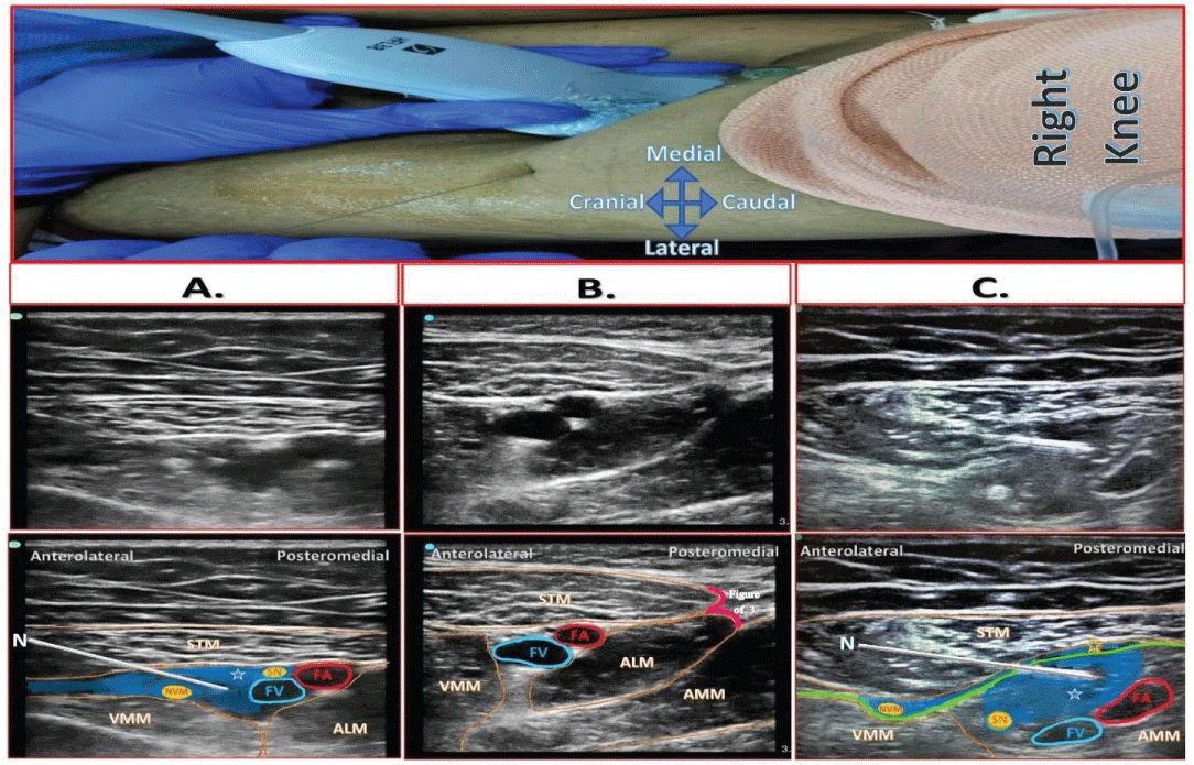
The high-frequency linear ultrasound probe is placed over the mid-thigh. The anterolateral VMM, posteromedial ALM, and medial STM are identified. The apex of the femoral triangle is identified where the medial border of STM overlies the medial border of ALM, forming a sign of “3” or “kissing Sign,” as shown in Figure 4B.
2. Distal femoral triangle block: [Figure 4A]
After identifying the apex of the FT, an ultrasound probe is moved slightly (1-2 cm) proximally. The STM is identified above, VMM anterolaterally, and ALM posteromedially. Below the STM, femoral vessels are identified using color doppler. The SN nerve lies lateral to the FA and appears as a hyperechoic structure under ultrasound. The NVM lies lateral to the SN most of the time in the intermuscular plane between STM and VMM. If the nerves are not immediately apparent, sliding and tilting the probe proximally or distally can be useful to improve the contrast and bring the nerves “out of the background” from the musculatures [26]. A 100 mm Stimuplex needle is inserted in-plane from the lateral aspect of the thigh or out-of-plane and advanced toward the intermuscular plane between VMM and STM to target NVM and SN.
An in-plane approach is more practical and feasible due to the relatively superficial location of the target structures and better resolution of the high-frequency linear probe. Identifying both target nerves (NVM and SN) is very important to block them successfully in this injection. Identifying the NVM is further confirmed by stimulating the nerve using the peripheral nerve stimulator (PNS), which can cause twitching of the VMM. Direct stimulation of the VMM can be differentiated from nerve stimulation by reducing the current between 0.3-0.5 mA. The muscles require higher currents than the nerves to produce an appreciable motor response. The NVM can also be blocked without using PNS by depositing the LA solution and separating the plane between STM and VMM, as it is consistently seen in that location.
After confirming the proper needle tip position, 1-2ml of LA is injected to confirm adequate drug spread and help delineate the NVM within its muscular tunnel. In an adult patient, 10-20 ml (5-10 ml each for NVM and SN) of LA is usually adequate for the successful blockade. It may always be beneficial to inject two-three smaller aliquots at different locations to ensure LA spread around the target nerves (NVM and SN). After confirming the NVM stimulation, 5-10ml of prepared LA mixture is injected around it, and another 5-10 m of LA mixture is injected around SN.
3. Second injection (adductor canal block): [Figure 4C]
The high-frequency linear ultrasound probe is placed just (1-2 cm) distal to the FT apex, i.e., below the sign of “3”. Here lower border of STM appears bilayered (Figure 3H) due to the presence of VAM below the STM fascia. A 100 mm Stimuplex needle is inserted inplane from lateral to the medial direction in the plane between STM and VMM. A 10-20 cc of LA mixture is deposited next to the FA under the VAM, typically seeing a disappearing spread of drug and compression of the vessel (Figure 4C). Such that injecting the drug compresses the femoral artery, and when the injection is stopped, the drug seems to disappear, thus restoring the femoral vessels to their original position. This block can be given at any distance distal to the apex of FT. However, we recommend giving this block in the proximal adductor canal area to avoid proximity to the sterile surgical site.
Distribution of analgesia
The first injection (distal femoral triangle block) will target SN and NVM directly (Figure 4A). Next, the distal spread of the drug under STM but above the VAM targets the subsartorial plexus, which is sandwiched between VAM and STM in the adductor canal region (Figure 4C).
In the second injection (adductor canal block), no nerves are targeted. Simply depositing the drug perivascularly (around FA) under the VAM is sufficient to obtain the desired outcome. A drug injected into the adductor canal below VAM will travel along the femoral vessels and enter the adductor hiatus to reach the posterior aspect of the knee joint. It blocks the popliteal plexus formed by the articular branches from the posterior division of the obturator nerve, tibial, common peroneal, and sciatic nerve. Larger LA volumes (30-40 ml) in the proximal AC (Hi-PAC block)[27] or distal AC have the potential to involve the sciatic nerve indirectly (required for below-knee surgeries)[28,29].
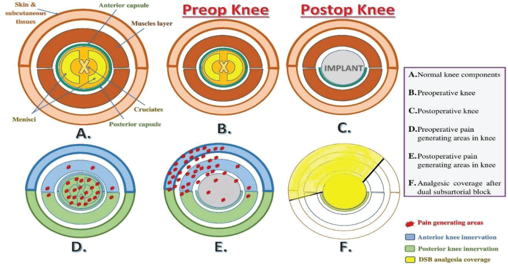
Thus, DSB involves blockade of SN, NVM, subsartorial plexus, the medial half of the peripatellar plexus, and the popliteal plexus. This results in sensory blockade over the anteromedial aspect of the knee up to the tibial tuberosity, medial retinacular complex, intraarticular region (popliteal plexus) with the exception of the skin over the anterolateral (supplied by lateral half of peripatellar plexus) and posterior aspect (supplied by the posterior femoral cutaneous nerve of the thigh) of the knee (Figure 5). Unless the surgical incision involves these spared areas, the lack of analgesia in its distribution is of little clinical consequence. The DSB does not cover tourniquet pain, which is mainly inflammatory pain due to muscle ischemia rather than pain due to pressure over the skin. If given precisely with the recommended LA concentrations and volumes, the DSB may not cause any motor blockade and provide optimal analgesia by blocking the procedure-specific target nerves.
Since DBS is a new technique, it is pertinent to compare it with the existing RA techniques for TKA surgeries (Table 1). It is also essential to assess the possible complications and measures to improve its safety profile (Table 2). More analysis and discussions are required in the future on these two crucial aspects of this block.
Summary
Many available RA options are known to provide the best perioperative analgesia for TKA surgeries. Among these options, it is more suitable to use a motor-sparing modality in this modern era as a part of ERAS protocol to promote early mobilization and reduce hospital stay. Furthermore, after studying the anatomical technical aspect of the DSB, it is almost possible to cover all procedure-specific innervations of TKA surgeries with the DSB (Table 3).
Motor-sparing and procedure-specific RA techniques are the future of RA practices, which induces the analytical thought process in applying ever-changing knowledge of anatomy, techniques, and methodologies for providing the best patient care. Therefore, to understand DSB better, knowing its anatomical and technical aspects is very important.
This novel technique can potentially provide excellent analgesia and aid in early ambulation by covering all the procedure-specific innervations. However, the analgesic efficacy, motorsparing, and opioid-sparing effect of DSB need more extensive sample-sized comparative studies.
We thank 3D4Medical from Elsevier for granting permission to use and modify images from the Essential Anatomy Application.
We would like to express our tremendous gratitude to all researchers enlightening us with their cadaveric studies, anatomical studies, comparative studies, dye studies, imaging studies, and descriptive reviews about femoral triangles and subsartorial regions.
Contributorship statement
KS: Designed and implemented a strategy for this new innovative technique. With the guidance and approval of JB, termed this technique as “Dual Subsartorial Block.” Drawn the illustrations of the manuscript. Involved in reference collection and sorting work with the help of HD and TM. Designed manuscript content and co-wrote the paper. Took the lead in manuscript writing.
HD & TM: Involved in anatomical description and information collection required for the manuscript writing. Co-wrote and proofread the manuscript. Took the second lead in manuscript writing.
JB: Approved idea by KS and provided scientific guidance for manuscript writing. Provided guidance for the content of the manuscript and Co-wrote the paper. Approved final version of the manuscript.
All authors provided critical feedback and helped shape the manuscript.
- Sonawane K, Dixit H, Balavenkatasubramanian J, Goel VK (2021) “Dual Subsartorial Block (DSB)”: An innovative procedure-specific, motor-sparing and opioid-sparing regional analgesia technique for Total knee replacement surgery - A pilot study. Journal of Clinical Anesthesia 69: 110149. Link: https://bit.ly/3xYp7aO
- Gardner E (1948) The innervation of the knee joint. Anatomical Record 101: 109–130. Link: https://bit.ly/3dpUgw2
- Kennedy JC, Alexander IJ, Hayes KC (1982) Nerve supply of the human knee and its functional importance. American Journal of Sports Medicine 10: 329–335. Link: https://bit.ly/360aOXy
- DaCosta JC, Spitzka EA (1908) Anatomy, descriptive and surgical: Henry Gray; 17th. Ed. Lea & Febiger, Philadelphia 970.
- Elkassabany NM, Antosh S, Ahmed M, Nelson C, Israelite C, et al. (2016) The Risk of Falls After Total Knee Arthroplasty with the Use of a Femoral Nerve Block Versus an Adductor Canal Block: A Double-Blinded Randomized Controlled Study. Anesthesia and Analgesia 122: 1696–1703. Link: https://bit.ly/3hkitoA
- Bendtsen TF, Moriggl B, Chan V, Pedersen EM, Børglum J (2014) Defining adductor canal block. Regional Anesthesia and Pain Medicine 39: 253–254. Link: https://bit.ly/3drmAy5
- Bendtsen TF, Moriggl B, Chan V, Børglum J (2015) Basic Topography of the Saphenous Nerve in the Femoral Triangle and the Adductor Canal. Regional Anesthesia and Pain Medicine 40: 391–392. Link: https://bit.ly/3h5kFRS
- Bendtsen TF, Moriggl B, Chan V, Børglum J (2016) The Optimal Analgesic Block for Total Knee Arthroplasty. Regional Anesthesia and Pain Medicine 41: 711–719. Link: https://bit.ly/3h30RPd
- Cunningham DJ, Robinson A. Cunningham’s Text-book of Anatomy. Edinburgh: Henry Frowde; 1915
- Moore Keith L (2013) Clinically oriented anatomy (7th ed.). Philadelphia: Wolters Kluwer/Lippincott Williams & Wilkins Health.
- Garg K (2010) “Front of the thigh (Chapter 3)”. BD Chaurasia’s Human Anatomy (Regional and Applied Dissection and Clinical) Volume 2 - Lower limb, abdomen, and pelvis (Fifth ed.). India: CBS Publishers and Distributors Pvt Ltd. 51: 55.
- Cowlishaw P, Kotze P (2015) Reply to Dr Bendtsen. Regional anesthesia and pain medicine 40: 392–393. Link: https://bit.ly/3x3n31a
- Bjørn S, Nielsen TD, Moriggl B, Hoermann R, Bendtsen TF (2019) Anesthesia of the anterior femoral cutaneous nerves for total knee arthroplasty incision: randomized volunteer trial. Regional Anesthesia and Pain Medicine. Link: https://bit.ly/3A57Rml
- Songo Lolomari. The Adductor Canal. Link: https://bit.ly/3h5kZ32
- Vas L, Pai R, Khandagale N, Pattnaik M (2014) Pulsed radiofrequency of the composite nerve supply to the knee joint as a new technique for relieving osteoarthritic pain: a preliminary report. Pain Physician 17: 493–506. Link: https://bit.ly/360KegU
- IMAIOS. Adductor Canal - Canalis Adductorius. Link: https://bit.ly/2U4p8eJ
- Johnston DF, Black ND, Cowden R, Turbitt L, Taylor S (2019) Spread of dye injectate in the distal femoral triangle versus the distal adductor canal: a cadaveric study. Regional Anesthesia and Pain Medicine 44: 39-45. Link: https://bit.ly/3y0JrbJ
- Kopp SL, Børglum J, Buvanendran A, Horlocker TT, Ilfeld BM, Memtsoudis SG, et al. (2017) Anesthesia and Analgesia Practice Pathway Options for Total Knee Arthroplasty: An Evidence-Based Review by the American and European Societies of Regional Anesthesia and Pain Medicine. Regional Anesthesia and Pain Medicine 42: 683–697. Link: https://bit.ly/2Th39kK
- Pascarella G, Costa F, Del Buono R, Agrò FE (2019) Adductor canal and femoral triangle: Two different rooms with the same door. Saudi journal of anaesthesia 13: 276–277. Link: https://bit.ly/3AbVkNZ
- Swenson JD, Davis JJ, Loose EC (2015) The Subsartorial Plexus Block: A Variation on the Adductor Canal Block. Regional Anesthesia and Pain Medicine 40: 732–733. Link: https://bit.ly/2T8uqG5
- Andersen HL, Zaric D (2014) Adductor canal block or midthigh saphenous nerve block: same same but different name!. Regional Anesthesia and Pain Medicine 39: 256–257. Link: https://bit.ly/3doXK1W
- Ozer H, Tekdemir I, Elhan A, Turanli S, Engebretsen L (2004) A clinical case and anatomical study of the innervation supply of the vastus medialis muscle. Knee Surgery, Sports Traumatology, Arthroscopy 12: 119–122. Link: https://bit.ly/3w3Tnjg
- Andersen HL, Andersen SL, Tranum-Jensen J (2015) The spread of injectate during saphenous nerve block at the adductor canal: a cadaver study. Acta Anaesthesiologica Scandinavica 59: 238–245. Link: https://bit.ly/3Aa3Gpe
- Runge C, Moriggl B, Børglum J, Bendtsen TF (2017) The Spread of Ultrasound-Guided Injectate From the Adductor Canal to the Genicular Branch of the Posterior Obturator Nerve and the Popliteal Plexus: A Cadaveric Study. Regional Anesthesia and Pain Medicine 42: 725–730. Link: https://bit.ly/3h2ZZtM
- Choi S, Rodseth R, McCartney CJ (2014) Effects of dexamethasone as a local anaesthetic adjuvant for brachial plexus block: a systematic review and meta-analysis of randomized trials. British Journal of Anaesthesia 112: 427–439. Link: https://bit.ly/3AbVHYT
- Ultrasound-Guided Sciatic Nerve Block. Link: https://bit.ly/3xYqUg2
- Sonawane K, Dixit H, Mistry T, Gurumoorthi P (2021) A high-volume proximal adductor canal (Hi-PAC) block - an indirect anterior approach of the popliteal sciatic nerve block. Journal of Clinical Anesthesia 73: 110348. Link: https://bit.ly/3hn5umm
- Roy R, Agarwal G, Pradhan C, Kuanardr D, Mallick D (2018) Ultrasound guided 4 in 1 block – a newer, single injection technique for complete postoperative analgesia for knee and below knee surgeries. Anaesthesia, Pain and Intensive Care 22: 87-93. Link: https://bit.ly/3w4LMkk
- Roy R, Agarwal G, Pradhan C, Kuanar D (2020) Total postoperative analgesia for total knee arthroplasty: Ultrasound guided single injection modified 4 in 1 block. Journal of Anaesthesiology, Clinical Pharmacology 36: 261–264. Link: https://bit.ly/3xazkRK
Article Alerts
Subscribe to our articles alerts and stay tuned.
 This work is licensed under a Creative Commons Attribution 4.0 International License.

 Save to Mendeley
