Journal of Cardiovascular Medicine and Cardiology
The prevalence, clinical profile and surgical outcomes of children presenting with vascular ring and pulmonary sling
Chinawa JM1*, Agarwal V2, Garekar S3, Gaikwad S4 and Trivedi B5
2Chief Pediatric Cardiac Surgeon, Fortis Hospital limited, Mulund goregaon Link road, Bhandup (West), Mumbai, India
3Consultant Pediatric Cardiologist, Fortis Hospital limited, Mulund goregaon Link road, Bhandup (West), Mumbai, India
4Pediatric Cardiac Surgeon, Fortis Hospital limited, Mulund goregaon Link road, Bhandup (West), Mumbai, India
5Pediatric Cardiologist, Fortis Hospital limited, Mulund goregaon Link road, Bhandup (West), Mumbai, India
Cite this as
Chinawa JM, Agarwal V, Garekar S, Gaikwad S, Trivedi B (2018) The prevalence, clinical profile and surgical outcomes of children presenting with vascular ring and pulmonary sling. J Cardiovasc Med Cardiol 5(4): 067-072. DOI: 10.17352/2455-2976.000075Background: Vascular anomalies are rare abnormalities which present with inspiratory stridor and recurrent respiratory tract infection. They are the commonest causes of mortality and morbidity in children due to misdiagnosis. They comprise about less than 4% of congenital heart diseases. The commonest of these anomalies were vascular ring and pulmonary sling.
Objectives: The objective of this study was to determine the prevalence, pattern of presentation and surgical outcomes of children presenting with vascular ring and pulmonary sling.
Methods: A cross-sectional retrospective study in which a review of the records of all children attending Fortis hospital over a 3-year period (2012- and 2015) was undertaken.
Data were analyzed using SPSS 20. Frequencies, rates and proportions were represented in tables. Children with confirmed diagnosis of vascular rings and pulmonary slings are included while other causes of stridor were excluded from this study.
Result: A total of 1200 children had open heart surgery in the hospital over a three-year period. Of these, 2 had vascular ring and 3 had pulmonary sling giving a prevalence of 0.4%. Out of the 5 cases, 3 (60 %) were male and 2 (40%) female. Male to female ratio was 1.5:1. The mean age of presentation was 2.46± 3.52 months. There were three neonates and two infants. All presented with stridor. The mean number of days spent postoperatively was 8±3 days and tracheal stenosis was the only complication noted.
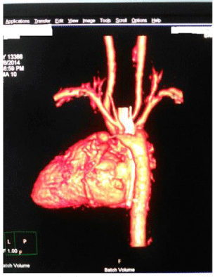
Computerized axial tomography (CT) scan of one of the subjects with vascular ring showed a dilated Kommerells diverticulum (KD).
Conclusion: Vascular ring and pulmonary sling are rare congenital abnormalities seen in our center, early identification and repair will help avert numerous complications that follow it.
Abbreviation
CT: Computerized axial Tomography; KD: Kommerells Diverticulum; Ligamentum arteriosum; LPA: Left Pulmonary Artery; PDA: Patent Ductus Arteriosus; VSD: Ventriculo Septal Defect
Background
A vascular ring is a congenital anomaly with abnormal formation of the arch, associated vessels, which encircles the trachea and esophagus, forming a complete or incomplete ring around them [1]. They are unique and occur early in the development of the aortic arch and great vessels [1]. The primary symptom associated with vascular rings stems from the arising structures that are encircled by the ring especially the trachea, and esophagus. Early diagnosis and treatment of this anomaly is therefore lifesaving [1].
Anomalous origin of the left pulmonary artery from the right pulmonary artery is an uncommon congenital anomaly first described on autopsy by Glaevecke and Doehle in 1897 [2]. Pulmonary artery sling is formed by an unusual origin of the left pulmonary artery from the posterior aspect of the right pulmonary artery [3].
Their etiology is related to embryopathies of the aortic arches. These vascular rings may be complete or incomplete. The severity of symptoms depends on the degree of compression of the trachea and esophagus [4]. Compression caused by the pulmonary sling can also give rise to obstructive symptoms such as emphysema and atelectasis [5]. Thus, early recognition and diagnosis of pulmonary artery sling is crucial [6].
Vascular rings encompass only 1-3% of all congenital heart disease [6]. Some vascular rings are associated with other congenital heart lesions while others are isolated defects. Aberrations of tracheobronchial trees are rarely seen with vascular rings but are common in pulmonary artery slings [7].
The diagnosis of vascular rings and pulmonary sling are protean. Barium swallow is the diagnostic procedure of choice. An anterior indentation of the esophagus on the lateral projection is diagnostic of pulmonary artery sling [8].
Furthermore, magnetic resonance imagery or angiography, computerized axial tomography scanning, or a combination can be helpful in delineating the details of the anatomy, as well as 3-dimensional reconstruction of the anatomy of the pulmonary sling as it relates to the airway anatomy [8]. Echocardiography remains the less invasive diagnosis of choice [9-11]. However, examination of the right pulmonary artery reveals the left pulmonary artery arising from its posterior surface.
This retrospective study is crucial so as to enable the pediatrician, cardiologists and cardiac surgeons have a high index of suspicion of its existence (especially in children presenting with stridor at an early age) and to be equipped with the skills to tackle the management and understand the numerous complications that follow the disease. Early identification and appropriate intervention can significantly improve the quality of life.
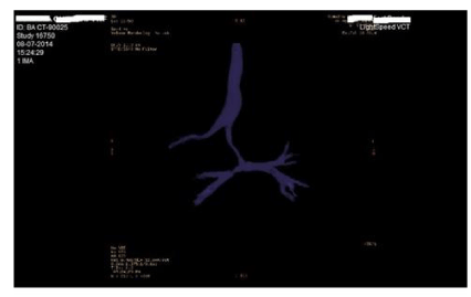
Prevalent studies of vascular rings and pulmonary slings anomalies are useful to establish baseline rates and to monitor trends over time. They may also help in health services planning and evaluating antenatal screening in populations with high risk. We are not aware of any study of this nature in Fortis hospital, Mumbai. It is hoped that this study will add to the body of knowledge available on these disorders and may stimulate further research in the area on the subject (Figures 1,2).
Material and Methods
The aims of this study were to determine the prevalence of vascular rings and pulmonary slings among children who attended and then admitted in Fortis hospital, Mumbai. It also aims at determining the pattern of presentation, surgical outcomes and follow up states of the subjects.
The study was conducted at the Pediatric cardiac surgery department, Fortis Hospital over a three-year period, from 2012 to 2015. The department of Pediatric cardiac surgery in Fortis is a budding department which is just three years old. Diagnosis was made with the use of Computerized axial tomography scan and Echocardiography.
Study design
A cross-sectional retrospective study in which a review of the records of all children attending Fortis hospital over a 3-year period (2012- and 2015) was undertaken. The diagnosis of pulmonary sling and vascular rings were based on clinical evaluation, echocardiogram and computerized axial tomography. The prevalence rate was estimated as a ratio of the total number of children diagnosed with pulmonary sling and vascular rings to the number of children with open heart surgery in the hospital at duration of study.
Children with confirmed diagnosis of vascular rings and pulmonary slings are included while other causes of stridor were excluded from this study.
Consent
Both written and informed consent were obtained from the parents and caregivers before admission, investigations and surgical intervention on these subjects.
Data analysis
Data were analyzed using SPSS 20. Frequencies, rates and proportions were represented in tables.
Results
A total of 1200 children were operated in the hospital over a three-year period. Of these, three were diagnosed with pulmonary sling and 2 with vascular ring giving a prevalence of 0.4%. There were three neonates and two infants. All presented with stridor. The mean number of days spent postoperatively was 8±3days and tracheal stenosis was the only complication noted.
Age group of subject ranged from birth to 9 months. Table 1 and 2 showed clinical summaries of Vascular ring and pulmonary sling respectively.
The mean age of presentation was 2.46± 3.52 months. Ventriculo septal defect (VSD), Patent ductus arteriosus (PDA) and Cotriatum sinitrum (CS) all occurred in equal distribution of 20%. Over half (50%) of the subjects with vascular sling had these associated anomalies. Stridor is the commonest symptom 4 (80.0%) The mean age at diagnosis and mean duration of extubation were 5±4 days and 3.07±3,75 days respectively. Tracheal stenosis was the only complication noted (Table 3)
Computerized axial tomography (CT) scan of one the subjects with vascular ring showed a dilated Kommerells diverticulum (KD) and ligamentum arteriosum arising from the KD and inserting onto the left pulmonary artery (LPA). This formed a vascular ring which was compressing the oesophagus and the trachea. The other case with vascular ring showed a right aortic arch and an aberrant left subclavian artery with a ligamentum arteriosum inserting on left pulmonary artery on Computerized axial tomography scan. This forms a vascular ring leading to narrowing of the left bronchus at its origin. Echocardiography of cases with pulmonary sling showed pulmonary sling while Computerized axial tomography scan showed pulmonary sling in addition to tracheal and left bronchial stenosis.
Surgical details of pulmonary sling repair
All the pulmonary slings cases were done on Cardiopulmonary bypass (CPB) through median sternotomy. The subjects were cooled to 32°C, and a normal sinus rhythm was maintained. The left pulmonary artery origin was identified and was seen to arise posteriorly from the right pulmonary artery (RPA). The patient was put on Cardiopulmonary bypass and the left pulmonary artery was dissected along its entire course up to the hilum. Heart was arrested with Saint Thomas cardioplegia given in aortic root. On the arrested heart, left pulmonary artery was transected from right pulmonary artery. Opening in right pulmonary artery was closed directly with 6 0 proline 13mm sutures. The left pulmonary artery which was coursing behind trachea was brought in front of trachea i.e. between trachea and aortic arch. Opening was made in MPA. End to side anastomosis of left pulmonary artery to main pulmonary artery was done using 6 0 10 mm proline sutures.
In case 1, table 2, in view of cotriatum sinistrum, right atrium was opened, the membrane was visualized and excised.
In case 3, table 2, in view of documented tracheal compression on Computerized axial tomography and peri-operative bronchoscopy, aortopexy was done where in after coming off Cardiopulmonary bypass and decannulation, the aortic arch was hitched up on the underside of sternum. Also in case 3, table 2, who had Patent ductus arteriosus (PDA), this was divided and ligated while the Ventriculo septal defect (VSD) was also closed.
Surgical details of Vascular Ring Repair: In case 1, table 1. The left subclavian artery was well mobilized up to the origin of its branches. Descending aorta was mobilized and looped. Kommerells diverticulum and ligamentum arteriosum dissected and looped. Ligamentum arteriosum ligated with no.2 silk. Cross clamp was placed across the KD at its origin from the descending aorta. Kommerells diverticulum was incised and divided between clamp and Ligamentum arteriosum ligature. Cut end of Kommerells diverticulum was also sutured with 6.0 prolene 10mm suture and reinforced with 6.0 prolene 13mm running suture.
For case 2, table 2, For Vascular Ring with aberrant subclavian artery and Right aortic arch, a Left postero-lateral thoracotomy through 4th intercostal space was made. The left subclavian artery was well mobilized up to the origin of its branches. Descending aorta was mobilized and looped. Ligamentum arteriosum was dissected and looped. Ligamentum arteriosum ligated with no.2 silk and divided. ’C’ Clamp was applied at the base of left subclavian artery and arch, ostia were opened along the anterior surface. Ostial shelf visualized, dissected with direct anastomosis done using 7‘o’ prolene suture.
Discussion
Vascular rings and pulmonary slings are uncommon anomalies that make up less than 1% of all congenital cardiac defects. They occur with about equal frequency in both sexes [10]. This rarity is also confirmed in our setting, having seen five cases in three years with a prevalence also less than 1% (0.4%). Backer [12] et al in his series, described pulmonary artery sling as a rare congenital vascular anomaly and noted only twelve cases in thirty-seven years among infants.
Most of our series presented at neonatal age or early infancy with respiratory symptoms especially stridor. These features were also recorded by Turner [13] et al, though he noted that a minority still presents much later.
Symptoms of airway obstruction are usually seen in patients with this anomaly especially within first few years of life. They include slow breast or bottle feeding, fatigue with feeding, continuous regurgitation, and aspiration pneumonias [14]. Most of our series also presented with poor suck at birth. The respiratory symptoms elicited by these patients could be due to either external tracheal compression or severe tracheal stenosis following complete rings [14]. It is pertinent to note that some vascular rings are associated with congenital heart defects [11]. This is corroborated in this study where 40% of children with pulmonary sling presented with various congenital heart diseases. These include Patent ductus arteriosus (PDA), Ventriculo septal defect (VSD) and cotriatum sinistrum. Andrew [15] and Louknov et al [16] also noted atrial septal defect, ventricular septal defect in their series. The reason for this association is not known but embryological attributes may be contributory.
Symptomatic newborns and infants with these complex lesions have a high mortality rate without surgical intervention. The ideal operation of pulmonary sling and vascular rings remains debatable, with issues rising from focusing on the need for left pulmonary artery re-implantation and the technique of tracheal reconstruction.
We noted just one case of congenital tracheal stenosis in our study. Congenital tracheal stenosis is a rare condition seen in 30-50% of cases, interestingly, a left pulmonary artery sling may be found with a short segmental stenosis.
Though we noted associated maternal diabetes or hypothyroidism with pulmonary sling, we could not explain if there is any link between these disease and pulmonary sling. This association is also lacking in literatures. [12-14]
There appears a male predominance in both vascular rings and pulmonary sling as seen in our study. Yoon [17] et al also confirmed this gender dominance. The reason for this male predominance could not easily be ascertained, though female double endowment of chromosome X could be suggestive.
It is important to take cognizance of the fact that though echocardiography could be helpful in investigating vascular rings and pulmonary slings. It may not have been able to delineate appropriately if there is any bronchial obstruction or bronchial rings.
This is buttressed by the fact that our case on aberrant subclavian artery was missed with.
Echocardiography. The use of Computerized axial tomography scan and magnetic resonance imagery remains a gold standard as supported in our findings. This was also corroborated in other studies [16-19].
Definitive managements of vascular rings and pulmonary slings are surgical. Our cases underwent surgery, spent less than two weeks in the hospital after surgery, except one case who spent three weeks because of respiratory obstruction after surgery. However, she was discharged and is presently doing well.
Surgery should be performed promptly after the diagnosis is made, especially in patients with stridor, apnea, or other symptoms of respiratory distress [18]. Delay in operative intervention can result in complications of a serious nature [18].
Left thoracotomy is the surgical approach of choice for the division of a vascular ring in the majority of cases. Anomalous left pulmonary artery has been corrected using the left thoracotomy; however, the use of median sternotomy and cardiopulmonary bypass are known to yield better outcomes [18]. The extremely rare configurations associated with left aortic arch and right descending thoracic aorta are the lesions that should be approached via a right thoracotomy for division of the ring [18].
The major contributor to postoperative mortality is the high frequency of bronchial and tracheal abnormalities in this group of patients [20]. Only one of our patient with pulmonary sling had bronchial obstruction and audible stridor which has gradually reduced after treatment.
We noted Kommerell diverticulum (KD) in one of our series. This is a bulb-like out- pouching at the origin of the subclavian artery and ligamentum arteriosum [21]. The major complication of KD is the presence of aneurysm of aorta observed in 3 to 8% of the patients with aberrant subclavian artery [8]. The risk for rupture is variable with incidence of 19 to 53% reported in some series [21].
The importance of bronchoscopy as both a diagnostic modality and a monitor of successful surgical manipulation is vital. As noted in this study, failure to diagnose and/or to treat the pre or intra-operative development of airway narrowing may result in respiratory failure in the very postop and failure to wean the patient from the mechanical ventilation [22,23]. We used bronchoscopy for the aortopexy in order to reduce the need for reoperation and to make intraoperative surgical decision-making easier. Intraoperative bronchoscopy is known to improve the prognosis for favorable clinical outcomes in children with vascular rings and airway obstruction.
The rationale for aortopexy depends on the severity of the condition being treated [22]. In children with severe respiratory failure due to compression of the trachea, aortopexy is the only dependable treatment option.
Conclusion
Vascular ring and pulmonary sling are rare congenital abnormalities seen in our center, early identification and repair will help avert numerous complications that follow it.
Declaration
Ethics approval and consent to participate: This complies with national guidelines [23]. All procedures performed in studies involving human participants were in accordance with the ethical standards of the institutional and/or national research committee and with the 1964 Helsinki declaration and its later amendments or comparable ethical standard. Fortis hospital gave permission to use the data.
Informed written consent was also granted by the parents/caregivers of subjects, before they were operated upon.
We acknowledge those that work in records department for retrieving all necessary documents.
Author contributions
JMC, VA and GS conceived and designed this study while SG and BT helped in diagnosis and, critical revision of the article. JMC also did the Data analysis/interpretation.
- Jai PS, Mohan M, Suresh K, Kirti C, Pradeep S, et al. (2017) Journal of evidenced based medicine and Healthcare 4: 1568-1571.
- Humphrey C, Duncan K, Fletcher S (2006) Decade of experience with vascular rings at a single institution. Pediatrics 117: 903-908. Link: https://goo.gl/aAgQJo
- Greiner A, Perkmann R, Rieger M, Neuhauser B, Fraedrich G (2003) Vascular ring causing tracheal compression in an adult patient. Ann Thorac Surg. 75: 1959-1960. Link: https://goo.gl/hDpoQP
- Sade RM, Rosenthal A, Fellows K (1975) Pulmonary artery sling. J Thorac Cardiovasc Surg 69: 333-346.
- Newman B, Cho YA (2010) Left Pulmonary Artery Sling-Anatomy and Imaging. Semin Ultrasound CT MR 31: 158-170. Link: https://goo.gl/aZYMbz
- Stem bronchus.JAMA (1998) 1954. 155:1409.pulmonary slings: the role of MRI. Magnetic Resonance Imaging 16:137-45.
- Backer CL, Russell HM, Kaushal S, Rastatter JC (2012) Pulmonary artery sling: Current results with cardiopulmonary bypass. J Thorac Cardiovasc Surg 143:144-151. Link: https://goo.gl/nGcYec
- Morrow R, Huhta J (1990) Aortic arch and pulmonary artery anomalies. The Science and Practice of Pediatric Cardiology 1: 1444-1447.
- Bai S, Li XF, Liu CX (2012) Surgical treatment for vascular anomalies and tracheoesophageal compression.Chin Med J (Engl) 125: 1504-1507. Link: https://goo.gl/UGGBzT
- Axt-Fliedner R, Kawecki A, Enzensberger C, Wienhard J (2011) Fetal and Neonatal Diagnosis of Interrupted Aortic Arch: Associations and Outcomes. Fetal Diagn Ther 30: 299–305. Link: https://goo.gl/Ng1Eia
- Newman B, Cho YA (2010) Left Pulmonary Artery Sling-Anatomy and Imaging. Semin Ultrasound CT MR 31: 158-170. Link: https://goo.gl/d5dnnA
- Backer CL, Russell HM, Kaushal S, Rastatter JC, Rigsby CK, et al. (2012) Pulmonary artery sling: Current results with cardiopulmonary bypass. J Thorac Cardiovasc Surg 143: 144-151. Link: https://goo.gl/XFFch3
- Turner A, Gavel G, Coutts J (2005) Vascular rings--presentation, investigation and outcome. Eur J Pediatr 164: 266-70. Link: https://goo.gl/XJxygq
- Humphrey C, Duncan K, Fletcher S (2006) Decade of experience with vascular rings at a single institution.Pediatrics 117: 903-908. Link: https://goo.gl/D75dx3
- Fiore AC, John WB, Thomas R (2003) Weber, Mark WTurrentine. Surgical Treatment of Pulmonary Artery Sling and Tracheal Stenosis .Presented at the Fiftieth Annual Meeting of the Southern Thoracic Surgical Association, Bonita Springs, FL, 13–15. Link: https://goo.gl/3cBWB6
- Loukanov T, Sebening C, Springer W (2005) Simultaneous management of congenital tracheal stenosis and cardiac anomalies in infants. J Thorac Cardiovasc Surg 130: 1537-1544. Link: https://goo.gl/qfXPb1
- Suh YJ, Kim GB, Kwon BS, Bae EJ, Noh CI, et al. (2012) Clinical Course of Vascular Rings and Risk Factors Associated With Mortality. Korean Circ J 42: 252–258. Link: https://goo.gl/uCBXQj
- Michał P, Łukasz C, Jarosław DK, Ludomir S, Mirosław T (2014) “The Aberrant Right Subclavian Artery (Arteria Lusoria): The Morphological and Clinical Aspects of One of the Most Important Variations—A Systematic Study of 141 Reports,” The Scientific World Journal. Link: https://goo.gl/3vMh5k
- Ronny C, Pablo L, Christine G, Lizmer D, Brooks M (2012) Syncope as Initial Presentation of Kommerell Diverticulum. Int J Angiol 21: 111–116. Link: https://goo.gl/QFLqfp
- Yong MS, D'Udekem Y, Robertson CF, Butt W, Brizard CP, et al. (2014) Konstantinov IE. Tracheal repair in children: reduction of mortality with advent of slide tracheoplasty. ANZ J Surg 84: 748-754. Link: https://goo.gl/uXnhwm
- Lee JH, Yang JH, Jun TG (2015) Video-assisted thoracoscopic division of vascular rings. Korean J Thorac Cardiovasc Surg 48: 78-81. Link: https://goo.gl/pGw3bC
- Weber TR, Keller MS, Fiore A (2002) Aortic suspension (aortopexy) for severe tracheomalacia in infants and children. Am J Surg 184: 573–577. Link: https://goo.gl/1GDxew
- Fiore AC, Brown JW, Weber TR, Turrentine MW (2005) Surgical treatment of pulmonary artery sling and tracheal stenosis. Ann Thorac Surg 79: 38-46. Link: https://goo.gl/eV7xwg

Article Alerts
Subscribe to our articles alerts and stay tuned.
 This work is licensed under a Creative Commons Attribution 4.0 International License.


 Save to Mendeley
