Journal of Cardiovascular Medicine and Cardiology
Coronary angiographic profile in patients with failed thrombolysis
Saravanan M, Vinodkumar Balakrishnan* and Muralidharan TR
Cite this as
Saravanan M, Balakrishnan V, Muralidharan TR (2019) Coronary angiographic profile in patients with failed thrombolysis. J Cardiovasc Med Cardiol 6(4): 069-073. DOI: 10.17352/2455-2976.000095Background: Acute Myocardial Infarction (MI) is a life threatening condition responsible for over 85% of the deaths all over the world. The management of MI has to be done on an emergency footing, without losing any time. The standard protocol involves thrombolysis in these patients. However, not all the thrombolysis procedures turn out to be successful. This study was done to explore the prevalence and risk factors associated with the success of thrombolysis.
Methods: This cross sectional study was carried out among 59 patients who were admitted to our tertiary care hospital with a diagnosis of acute ST segment elevation MI and started on thrombolysis. Patients who were contraindicated for thrombolysis were excluded from the study. A 12 lead electrocardiography was done prior and 60 minutes after initiation of thrombolysis. Echocardiogram was done to evaluate the left ventricle function. Coronary angiogram was carried out to assess the vascular abnormalities.
Results: Out the total 59 patients in the study group, 31 patients belonged to successful thrombolysis group and 28 patients belonged to the failed thrombolysis group. It was observed that patients with diabetes mellitus had a significant rise in the number of failed thrombolysis compared to those without diabetes mellitus (p<0.005). It was observed that anterior wall MI was significantly at risk for a failure in thrombolysis (82.1%) compared to inferior wall MI (17.8%).
Conclusions: Failed thrombolysis was more common in patients who had diabetes as risk factor. Moreover anterior wall MI proved to be increasingly at risk for failure of thrombolysis. This study proves that screening of risk factors prior to the initiation of thrombolysis will help in devising alternative treatment procedures so that the failure rates can be minimized and resources may be redirected to effective treatment modalities.
Introduction
Acute Myocardial Infarction (MI) is one of the leading causes of death and disability throughout the world. There are several therapeutic options which have been implemented the past decades and there is a continuing need to improve the techniques in order to improve the outcome of patients, especially in elderly and among patients in low socio economic groups. In the recent past the treatment of acute myocardial infarction has been thrombolysis with fibrin non specific agents. The parameter used to evaluate the outcome of the thrombolysis was patency rate and TIMI flow. Studies are proven that the patency rate of the infarct artery was better with fibrin specific agent compared to fibrin non specific agent. Therefore it has been observed that use of non specific agent is associated with increased failure of thrombolysis [1]. In the current scenario in countries like India thrombolysis is the most commonly used mechanism of infarct management.
Currently the primary management guideline of myocardial infarction is primary Per Cutaneous Intervention (PCI). PCI is performed only by experienced team and the patency rate and grade of thrombolysis is said to be better with PCI. A Patent artery has better prognosis when performed on patients with occluded vessels [2-5]. There are several factors which influences the outcome of primary PCI which include delay in the presentations of the patients, non availability of cath lab and cost involved in the entire procedure. Considering the disadvantages of using PCI in resource limited setting as in emerging economies like India, thrombolysis continues to prevail as the preferred choice of treatment. Even with the recently available thrombolytic drugs the patency of the coronary artery was found to be 60%. It has been proven that more than one-third of the patients who undergo thrombolysis develop spontaneous recanalization within 12 to 24 hours thereby decreasing the mortality by 50% [6]. More than the thrombolytic agent used, it is important to note that the time between the onset of MI and access to health care facility plays a crucial role in determining the outcome of the patients. Every 30 minute delay increases the relative risk of one year mortality by 8 times and fibrinolysis has shown to improve both short term and long term survival [7].
Although thrombolysis is commonly performed, the procedure is often prone for failures. It is essential to explore the reasons for the failure in the given situation. It is a well established fact that thrombolysis has better prognosis and an adequate analysis of the failed thrombolysis will help in exploring the factors which significantly play the role in the successes of thrombolysis. An in-depth analysis of such factors will also help in minimizing the risk factors which leads to a failed thrombolysis.
Objectives
1. To evaluate the risk factors of successful and failed thrombolysis.
2. To estimate the prevalence of no reflow phenomenon in patients with successful thrombolysis.
Methodology
Study setting
This study was conducted as a cross sectional study in the Department of Cardiology of our tertiary care hospital for a period of one year between 2013 and 2014.
Study population
Patients who got admitted to our tertiary care center with symptoms of Acute myocardial infarction and in whom the diagnosis of ST segment elevation MI was made during the study period were selected for the study. Among the selected patients, those who were thrombolysed were included for this study. A total of 59 patients participated in the study and were selected by convenient sampling.
Inclusion criteria
Electro cardio graphic evidence of acute myocardial infarction.
Exclusion criteria
1. Patients with evolved myocardial infarction [New pathologic Q waves on serial ECGs, ischemic changes on ECG, rise and fall of biomarker with no anginal symptoms].
2. Patients with history of old myocardial infarction.
3. Patients with left bundle branch block associated with MI.
4. Patients who died within 60 minutes of streptokinase therapy.
5. Patients with Chronic Kidney Diseases.
6. Patients with any other systemic complications and are contraindicated for coronary angiogram.
Ethical approval and informed consent
Approval was obtained from the Institutional Ethics Committee prior to the commencement of the study. Each participant was explained in detail about the study and informed consent was obtained prior to the data collection.
Data collection
A structure interview schedule was used to elicit the history regarding onset of pain and the time of presentation to the hospital. Other history including the risk factors for coronary heart disease and the treatment history, systemic diseases history were also elicited. Hemo dynamic parameters were assessed and all patients were continuously monitored during the period of thrombolysis. The participants were connected to 12 lead Electrocardiography (ECG) initially on presentation. Repeat ECG’s were taken after 60 minutes of initiation of thrombolysis. Based on the ECG findings, patients were classified as anterior wall MI and inferior wall MI depending on the location of ST segment elevation. Presence of rhythm disturbances including idioventricular rhythm and atrioventricular blocks were also assessed.
After evaluating the systemic functions with blood investigations, thrombolysis was initiated with 15 lakh units of streptokinase infusion over 1 hour with close monitoring. After 60 minutes of thrombolysis a repeat ECG was taken. Echocardiographic assessment of LV function was done by modified Simpsons method pre and post thrombolysis. Patients were classified as normal mild, moderate and severe LV dysfunctions based on the ejection fraction (Table 1). Apart from LV dysfunction, regional wall motion abnormalities were also assessed and scored (Table 2). Other mechanical complications of acute MI were also assessed using echocardiography.
All the participants were subjected to coronary angiogram. The angiogram was performed within 72 to 96 hours of admission. It was done by a trained consultant cardiologist within cath lab with TOSHIBA infinix machine. Following pre procedure guidelines, coronary angiogram was done by radial or femoral route. Standard 6F Judkins 3.5 or 4 Judkins left and right catheter was used to engage left coronary artery and right coronary artery respectively. Optimal angiographic views were taken to access the lesion characteristics and coronary arteries were visualized after iodine contrast injection.
Operational definition
ST segment was used evaluate the success or failure of thrombolysis. Patients were classified as a successful thrombolysis if there was more than 50% ST segment resolution in the lead with maximum ST elevation from the time of presentation to 60 minutes after thrombolysis. This was coupled with resolution of chest pain.
Thrombolysis was categorised as failed when ST elevation failed to show greater than 50% resolution at 60 minutes after the thrombolysis. Symptomatic persistence of chest pain was also considered as failed thrombolysis.
Patients on coronary angiogram were considered to have significant coronary artery disease if the diameter of stenosis was more than 50% compared to the normal vessel luminal diameter.
Data analysis
Data was entered an analysed using SPSS version 10 software. Percentages were used to compute the prevalence of successful and failed thrombolysis. Risk factor analysis was done using parametric and non parametric test. P value <0.05 was considered as statistically significant.
Results
All the 59 patients in the study group were analysed statistically in the study. The study group was divided in to two groups as those patients with successful and failed thrombolysis. Out the total 59 patients in the study group, 31 patients belonged to successful thrombolysis group and 28 patients belonged to the failed thrombolysis group. The demographic characteristics of the study participants are given in table 3. It was observed that patients with diabetes mellitus had a significant rise in the number of failed thrombolysis compared to those without diabetes mellitus (p<0.005).
The particulars regarding the onset and type of myocardial infarction are given in table 4. It was observed that anterior wall MI was significantly at risk for a failure in thrombolysis (82.1%) compared to inferior wall MI (17.8%). The observed difference was statistically significant (p<0.05). A majority of the patients with successful thrombolysis reached the health care facility within 3 hours (54.9%).
The echocardiographic findings of the study participants are given in table 5. It was observe that in 50% of the participants with failed thrombolysis, there was a single vessel disease, and in 32.2% of these participants, there was a multivessel disease. Moreover, higher ejection fraction was observed with successful thrombolysis (35.5%) while it was observed only in one patient with failed thrombolysis (3.6%). The observed association was significant statistically (p<0.002).
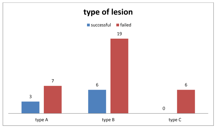
The coronary angiogram findings are depicted in Figure 1. It was observed that type B lesion was found to be maximum among the patients with failed thrombolysis (67.8%) while type A lesion was found to be among 25% of failed thrombolysis and type C lesion was found to be among 21.4% of the failed thrombolysis patients.
Among the study population no reflow phenomenon was seen with failed big participant who had failed thrombolysis. Out of 28 participants, 2 patients had no reflow phenomenon. These patients had normal coronary artery but persistent ST segment elevation. Overall no reflow phenomenon was seen in 5% of the study participants.
Discussion
In the study 52.5% of the patients had successful thrombolysis. The percentage of among the patients with failed thrombolysis was more among patients who had anterior wall myocardial infarction and this was statistically significant. Patients with inferior wall MI were more likely to have a successful thrombolysis.
The incidence of failed thrombolysis varies with studies but roughly ranges from 25% to 45%. In a study done by a Katyal, et al., the prevalence of failed thrombolysis was 34% which is similar to our study. Sudhindra, et al., also showed similar findings in his study [1,8]. In a study done by Richardson S.G, et al., 44% had failed thrombolysis which is similar to our study. [9] Our study shows that diabetes was significant risk factor for failed thrombolysis where in 73.7% of the diabetics had failed thrombolysis compared to 26% who had successful thrombolysis. The observed difference was found to be statistically significant. The our study findings correlated well with study done by Sudhindra, et al., [8], Samith M. Rafulah, et al., also observed similar findings showing a significant association between diabetes and ST segment [10].
Our study also demonstrated that time to thrombolysis also contributes to thrombolysis failure. This was significantly emphasized by studies done by Lee Y.Y, et al., He also observed that for each 60 second delay in the thrombolysis from time of onset of symptoms the failure rate of thrombolysis increased by 10% [11]. Another trial named GISSI-2 showed that patients who present within 6 hours had better prognosis in coronary thrombolysis. This was emphasized by Sudhindra, et al., who showed that door to needle time was more than 5 hours (5.85±2.47 hours) in failed thrombolysis while it was only 4.55±2.4 hours in the successful thrombolysis group. In our study 85% of the patients who presented to the coronary unit within 3 hours had successful thrombolysis.
The other risk factor for a failure thrombolysis was smoking. In angiographic data of GUSTO-1 study showed higher TIMI flow among smokers. In TEAM study patients who were smokers had better possibility TIMI 3 flow and this was related to the increase in thromboses formation in the smokers. A TIMI flow of grade 3 was essential for successful thrombolysis and adequate mortality benefit was achieved when the TIMI had a flow of grade-3. Jeffery, et al., in his study showed that patients with more ejection fraction was found in patients with TIMI 3 flow and was associated with lesser death rate [12]. In our study successful thrombolysis was observed with better TIMI flow compare to patients with failed thrombolysis.
Conclusion
Our study demonstrated a higher prevalence of successful thrombolysis compare to failed thrombolysis. Failed thrombolysis was influenced by diabetes mellitus and Type B coronary lesion. The prevalence of no reflow phenomenon in the failed thrombolysis patient in the study group was only 5%. This study elucidates the need was screening prior to initiation of the thrombolysis. This will help in minimizing the failure rates and also redirecting the resources to effective treatment modalities.
Limitations
Our study was conducted on a small sample of population. A larger analyses of more number of participants over longer period of time would not only evaluate the reasons for failed thrombolysis but also would given an insight into the prognoses of successful thrombolysis. ST segments in our study analysis were dynamic therefore faults can occur in the diagnosis of thrombolysis.
- Katyal VK, Siwach SB, Jagadish, Sharma P, Batra M (2002) Failed thrombolysis and its impact after acute MI. Assoc physicians india 51: 1149-1150.
- Puma JA, Sketch MH Jr, Thompson TD, Simes RJ, Morris DC, et al. (1999) Support for the open-artery hypothesis in survivors of acute myocardial infarction: analysis of 11,228 patients treated with thrombolytic therapy. Am J Cardiol 83: 482-487. Link: http://bit.ly/2NKHMm6
- Fath-Ordoubadi F, Huehns TY, Al-Mohammad A, Beatt KJ (1997) Significance of the Thrombolysis in Myocardial Infarction scoring system in assessing infarct related artery reperfusion and mortality rates after acute myocardial infarction. Am Heart J 134: 62-68. Link: http://bit.ly/2qbLsoa
- French JK, Hyde TA, Patel H, Amos DJ, McLaughlin SC, et al. (1999) Survival 12 years after randomization to streptokinase: the influence of thrombolysis in myocardial infarction flow at three to four weeks. J Am CollCardiol 34: 62-69. Link: http://bit.ly/2qWhPaf
- Cannon CP, Gibson CM, McCabe CH, Adgey AA, Schweiger MJ, et al. (1998) TNK-tissue plasminogen activator compared with front-loaded alteplase in acute myocardial infarction: results of the TIMI 10B trial. Circulation 98: 2805-2814. Link: http://bit.ly/3729uT7
- Kiernan TJ, Gersh BJ (2007) Thrombolysis in acute myocardial infarction: Current status. Med Clin North Am 91: 617-637. Link: http://bit.ly/2NM9PBO
- Thygesen K, Alpert JS, Jaffe AS, Simoons ML, Chaitman BR, et al. (2012) Third Universal Definition of Myocardial Infarction. J Am Coll Cardiol 60: 1581-1598. Link: http://bit.ly/3729Rgt
- Sudhindra Rao M, Patil BS (2012) Predictors of failed thrombolysis in acute myocardial infarction. Int J Bio Res 3: 239-244. Link: http://bit.ly/351hh1A
- Richardson SG, MortanP, Murtagh JG, Scott ME, Barry BO (1988) Relation of coronary artery patency and LV function to ECG changes after streptokinase treatment during acute MI. Am J Cardiol 61: 961-965. Link: http://bit.ly/2rIwZ3u
- Samir M. Rafla, Said M. Kandil, Fatma A. et al. (2007) Non invasive assessment of prognosis after acute myocardial infarction in diabetic and non diabetic patients. JMRI 28: 226-234.
- Lee YY, Tee MH, Zurkurnai Y, Than W, Sapawi M, et al. (2008) Thrombolytic failure with streptokinase in acute myocardial infarction using electrocardiogram criteria. Singapore Med J 49: 304-310. Link: http://bit.ly/2CIjbbn
- Anderson JL, Karagounis LA, Becker LC, Sorensen SG, Menlove RL (1993) TIMI perfusion grade 3 but not grade 2 results in improved outcome after thrombolysis for myocardial infarction. Ventriculographic, enzymatic, and electrocardiographic evidence from the TEAM-3 Study. Circulation 87: 1829-1839. Link: http://bit.ly/33O0Xkz

Article Alerts
Subscribe to our articles alerts and stay tuned.
 This work is licensed under a Creative Commons Attribution 4.0 International License.

 Save to Mendeley
