Journal of Cardiovascular Medicine and Cardiology
Epicardial adipose tissue thickness in patients with urolithiasis
Burak Altun1*, Eyup Burak Sancak2, Berkan Resorlu2, Hakan Tasolar3, Alpaslan Akbas2, Gurhan Adam5, Mustafa Resorlu4 and Mehzat Altun5
2MD, Assistant Professor, Department of Urology, Canakkale Onsekiz Mart University, Canakkale, Turkey
3MD, Department of Cardiology, Adiyaman University Training and Research Hospital, Adiyaman, Turkey
4MD Assistant Professor, Department of Radiology, Canakkale Onsekiz Mart University, Canakkale, Turkey
5Vocational School of Health Services, Canakkale Onsekiz Mart University, Canakkale, Turkey
Cite this as
Altun B, Sancak EB, Resorlu B, Tasolar H, Akbas A, et al. (2020) Epicardial adipose tissue thickness in patients with urolithiasis. J Cardiovasc Med Cardiol 7(1): 024-027. DOI: 10.17352/2455-2976.000106Aim: We aimed to assess the relationship between urinary stone disease which is accepted as a component of metabolic syndrome and epicardial adipose tissue (EAT) thickness.
Methods: The study included 45 patients and 39 healthy controls. EAT thickness was measured by echocardiography in all subjects.
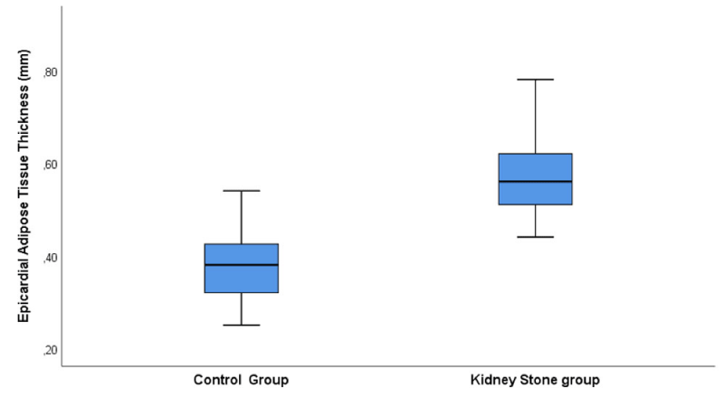
Results: EAT thickness was higher (5.77±0.88 vs. 3.83±0.72mm, p<0.001) in patients than in control subjects. EAT thickness was correlated with age, triglyceride levels, low density lipoprotein cholesterol levels and family history. Regression analysis showed that family history, triglyceride levels and age were independent predictors of EAT thickness in kidney stone patients.
Conclusion: We suggest that urolithiasis should be considered as a component of metabolic syndrome and EAT thickness may be useful to detect early atherosclerosis in urolithiasis.
Introduction
Kidney Stone (KS) disease is a worldwide health problem, and its prevalance is increasing especially in industrialized countries, probably as a result of enviromental factors, such as lifestyle and dietary habits [1]. The etiology of KS is multifactorial, with epidemiologic studies showing that age, genetic factors, nutritional properties, geoghraphical factors and some medical conditions such as diabetes mellitus, hypertension and obesity are associated with urinary stone formation [1-3]. These medical conditions are now collectively named to as Metabolic Syndrome (MS) and large series reported that presence of metabolic syndrome is also associated with the increased risk of urinary stone disease [4-10].
Epicardial Adipose Tissue (EAT) thickness is true visceral fat located around the heart, especially subepicardial coronary vessels. Increases in the thickness of EAT which was measured by echocardiography have been shown to be directly associated with an increased risk of hypertension, coronary artery disease and diabetes mellitus [11-16]. So this test can be used as a predictor of MS and its components. However there is currently no data in the literature regarding the relationship between thickness of EAT and KS disease. In this study, we aimed to assess the relationship between KS disease accepted as a component of MS and EAT thickness.
Materials and methods
Study population
Fifty-tree patients with KS disease and 39 healthy subjects were enrolled in our study. Patients were excluded if they had inadequate view on echocardiography, a history of any kind of cardiovascular disease, active infection and history of uric acid, cystin or struvite stones. For this reasons 8 KS patients were excluded because of cardiovascular disease in 4 and insufficient echocardiographic view in 4. Each participant signed an informed consent form in accordance with the Declaration of Helsinki, and this study was approved by the local ethical committee of Canakkale Onsekiz Mart University.
Measurements
Systolic and diastolic blood pressures were measured after 5 minutes of rest. Laboratory tests included fasting plasma glucose, creatinine, total cholesterol, High-Density Lipoprotein (HDL) cholesterol, Low Density Lipoprotein cholesterol (LDL), triglycerides. Biochemical measurements were made using standard biochemical techniques with a device from Beckman Coulter Ireland Inc., Mervue, Galway, Ireland. Body Mass Index (BMI) was calculated by dividing the weight in kilograms by the squared height in meters. MS diagnosed according to the National Cholesterol Education Program Adult Treatment Panel III [17]. MS was defined as the presence of 3 or more following components: systolic and diastolic blood pressure ≥130/85mmHg, fasting plasma glucose ≥110mg/dL, serum triglyceride ≥150mg/dL, HDL≥40mg/dL, and elevated waist circumference ≥102cm in men and ≥88cm in women.
The diagnosis of KS was established on the basis of the results of urinary ultrasound (Toshiba Aplio XG, Japan) using a 3.5MHz transducer. Renal calcification was classified as a urinary stone if the calcification was located in the collecting system. Stone burden was also determined. All evaluations were performed by experienced radiologists, who did not have any information about metabolic status of the patients.
EAT measurements
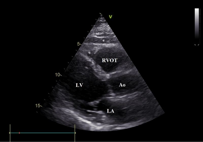
Echocardiograms were performed with a Vivid 7 (Vingmed electronic,GE, Horten, Norway) instrument according to standard techniques. We measured EAT thickness on the free wall of right ventricle from the parasternal long-axis views. EAT was identified as an echo-free space in the pericardial layers on the two-dimensional echocardiography, and its thickness was measured perpendicularly on the free wall of the right ventricle at end-diastole for 3 cardiac cycles [16,18] (Figure 1). An average value from these three measurements was obtained. The offline measurement of EAT thickness was performed by the same cardiologist who was unaware of the clinical data.
Statistical analysis
SPSS 19.0 statistical program (SPSS Inc, Chicago,IL) was used to statistical analysis. All values are given as mean±standard deviation. Kolmogorov–Smirnov test was used normallity of the variances. Descriptive statistics were used for definition of clinical and social demographic variables. Correlation of numerical variables were examined by Pearson correlation. The determinants of the dependent EAT thickness variable were assessed with multiple regression analyses using the following independent variables: Age, LDL cholesterol, triglyceride, and family history in patients group. Regression analysis was performed with a stepwise method.
Results
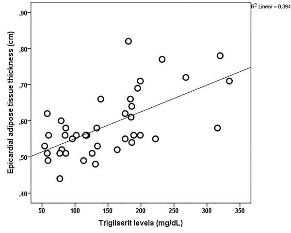
Forty-five patients with KS disease and 39 healthy subjects were included in this study. Of the 84 patients included in the final analysis, 44% were men and 56% were women. Mean age was 50.52±10.4 years. Mean BMI was 25.5±3.4kg/m2. Demographic and laboratory characteristics of the study population are presented in Table 1. EAT thickness were higher (p<0.001) in KS disease patients than in healthy subjects (Figure 2). Multivariable analysis showed that increased EAT thickness was associated with family history of urolithiasis. EAT thickness was also significantly correlated with triglyceride levels (r=0.627, p<0.001, Figure 3), LDL levels, age, and family history. Table 2 shows the correlation between EAT thickness and study parameters in patients.
Multiple linear regression analyses with stepwise method were performed to evaluate independent variables of EAT thickness. Triglyceride levels, age, and family history of urolithiasis were independent predictors of EAT (Table 3).
Discussion
In our study EAT thickness were higher in KS disease than in control subjects and correlated with triglyceride levels, age and family history.
EAT is a component of visceral adiposity and related to MS and cardiovascular risk factors [11-13,16]. Recently, there has been increased interest in EAT thickness as a marker of atherosclerosis. Inline with previous studies, it has been shown to be positively correlated with the severity of coronary artery disease [19-21].
The MS seems to be a risk factor for stone formation, because all components of this syndrome have been demonstrated to be independent risk factor for urinary stone formation [1-4]. National Health and Nutrition Examination Survey III reported a significant positive correlation between the MS traits and stone prevalence [22]. The association between MS and KS disease also confirmed with imaging studies. In a large study in which 2132 Caucasian patients were reviewed, presence of MS was independently found related to ultrasonographic evidence of urolithiasis [23]. Although several studies have reported an association between urolithiasis and individual components of MS, none of them showed an clear evidence about this togetherness [4-10]. Rendina, et al., [5], stated in their study that insulin resistance significantly influences the urinary salts supersaturation, and KS formation results from a phase change in which urinary dissolved salts condense into solids, and all phase changes are driven by salts supersaturation [24]. It has been shown in previous studies [6-10], that, especially its components, not directly MS, may cause kidney stones, however, the underlying mechanisms have not clearly been demonstrated. In our study, we found the EAT thickness higher in KS disease than in control subjects. Furthermore positive family history of urolithiasis is a risk factor for increased EAT thickness, in KS disease patients. Therefore present study is the first to report a clear evidence about relationship between MS and KS disease.
There are several limitations in the present study. First, it is performed with a cross-sectional design. Second, urinary metabolic evaluations were not performed and urinary US was used as the main imaging modality instead of noncontrast computed tomography. However US has many advantages, including wide availability, low cost, and lack of radiation exposure. Finally, our study population is limited in numbers. Therefore, these results should be confirmed by large multicenter studies.
Conclusion
The relationship between KS disease and EAT thickness was researched in this study and EAT thickness was found to be higher in KS disease patients than in controls. We suggest that urolithiasis should be considered as a component of metabolic syndrome and EAT thickness may be useful to detect early atherosclerosis in urolithiasis.
- Ekane S, Wildschutz T, Simon J, Schulman CC (1997) Urinary lithiasis: epidemiology and physiopathology. Acta Urol Belg 65: 1-8. Link: http://bit.ly/37VHRes
- Meydan N, Barutca S, Caliskan S , Camsari T (2003) Urinary Stone Disease in Diabetes Mellitus. Scand J Urol Nephrol 37: 64-70. Link: http://bit.ly/2RXCKoy
- Lieske JC, de la Vega LS, Gettman MT, Slezak JM, Bergstralh EJ, et al. (2006) Diabetes mellitus and the risk of urinary tract stones: a population-based case-control study. Am J Kidney Dis 48: 897-904. Link: http://bit.ly/2RZqtAa
- West B, Luke A, Durazo-Arvizu RA, Cao G, Shoham D, et al. (2008) metabolic syndrome and self-reported history of kidney stones: the National Health and Nutrition Examination Survey (NHANES III) 1998-1994. Am J Kidney Dis 51: 741-747. Link: http://bit.ly/37YZwlF
- Rendina D, Mossetti G, de Filippo G, Benvenuto D, Vivona CL, et al. (2009) Association between metabolic syndrome and nephrolithiasis in an inpatient population in Southern Italy: role of gender, hypertension and abdominal obesity. Nephrol Dial Transplant 24: 900-906. Link: http://bit.ly/3bcICBO
- Jeong IG, Kang T, Bang JK, Park J, Kim W, et al. (2011) Association between metabolic syndrome and the prevalence of kidney stones in a screened population. Am J Kidney Dis 58: 383-388. Link: http://bit.ly/37YaoQG
- Lange JN, Mufarrij PW, Wood KD, Holmes RP, Assimos DG (2012) The association of cardiovascular disease and metabolic syndrome with nephrolithiasis. Curr Opin Urol 22: 154-159. Link: http://bit.ly/2SfE4SB
- Gambaro G, Ferraro PM, Capasso G (2012) Calcium nephrolithiasis, metabolic syndrome and the cardiovascular risk. Nephrol Dial Transplant 27: 3008-3010. Link: http://bit.ly/2GVjgKQ
- Sakhaee K, Capolongo G, Maalouf NM, Pasch A, Moe OW, et al. (2012) Metabolic syndrome and the risk of calcium Stones. Nephrol Dial Transplant 27: 3201-3209. Link: http://bit.ly/3bjW4UO
- Kohjimoto Y, Sasaki Y, Iguchi M, Matsumura N, Inagaki T, et al. (2013) Association of metabolic syndrome traits and severity of kidney stones: results from a nationwide survey on urolithiasis in Japan. Am J Kidney Dis 61: 923-929. Link: http://bit.ly/2GV9HMc
- Peiris AN, Sothmann MS, Hoffmann RG, Hennes MI, Wilson CR, et al. (1989) Adiposity, fat distribution, and cardiovascular risk. Ann Intern Med 110: 867-872. Link: http://bit.ly/394cDC7
- Folsom AR, Kushi LH, Anderson KE, Mink PJ, Olson JE, et al. (2000) Associations of general and abdominal obesity with multiple health outcomes in older women: the Iowa Women’s Health Study. Arch Intern Med 160: 2117-2128. Link: http://bit.ly/2RWrsRz
- Rexrode KM, Buring JE, Manson JE (2001) Abdominal and total adiposity and risk of coronary heart disease in men. Int J Obes Relat Metab Disord 25: 1047-1056. Link: http://bit.ly/37XI9Sb
- Natale F, Tedesco MA, Mocerino R, de Simone V, Di Marco GM, et al. (2009) Visceral adiposity and arterial stiffness: echocardiographic epicardial fat thickness reflects, better than waist circumference, carotid arterial stiffness in a large population of hypertensives. Eur J Echocardiogr 10: 549-555. Link: http://bit.ly/3bjWASK
- Jeong JW, Jeong MH, Yun KH, Oh SK, Park EM, et al. (2007) Echocardiographic epicardial fat thickness and coronary artery disease. Circ J 71: 536-539. Link: http://bit.ly/394dDWT
- Iacobellis G, Ribaudo MC, Assael F, Vecci E, Tiberti C, et al. (2003) Echocardiographic epicardial adipose tissue is related to anthropometric and clinical parameters of metabolic syndrome: a new indicator of cardiovascular risk. J Clin Endocrinol Metab 88: 5163-5168. Link: http://bit.ly/2ttU4Z1
- Grundy SM, Cleeman JI, Daniels SR, Donato KA, Eckel RH, et al. (2005) Diagnosis and management of the metabolic syndrome: an American Heart Association/National Heart, Lung, and Blood Institute. Circulation 112: 2735-2752. Link: http://bit.ly/37SFYzf
- Iacobellis G, Assael F, Ribaudo MC, Zappaterreno A, Alessi G, et al. (2003) Epicardial fat from echocardiography: A new method for visceral adipose tissue prediction. Obes Res 11: 304-310. Link: http://bit.ly/36Xm8l8
- Reaven GM (1988) Role of insulin resistance in human disease. Diabetes 37: 1595-1607.
- DeFronzo RA, Ferrannini E (1991) Insulin resistance: a multifaceted syndrome responsible for NIDDM, obesity, hypertension, dyslipidemia, and atherosclerotic cardiovascular disease. Diabetes Care 14: 173-194. Link: http://bit.ly/2RXgyuY
- Haffner S, Valdez R, Hazuda H, Mitchell BD, Morales PA, et al. (1992) Prospective analysis of the insulin-resistance syndrome (syndrome X). Diabetes 41: 715-722. Link: http://bit.ly/31nw6uS
- West B, Luke A, Durazo-Arvizu RA, Cao G, Shoham D, et al. (2008) metabolic syndrome and self-reported history of kidney stones: the National Health and Nutrition Examination Survey (NHANES III) 1988-1994. Am J Kidney Dis 51: 741-747. Link: http://bit.ly/37YZwlF
- Rendina D, Mossetti G, De Filippo G, Benvenuto D, Vivona CL, et al. (2009) Association between metabolic Syndrome and nephrolithiasis in an inpatient population in southern Italy: role of gender, hypertension and abdominal obesity. Nephrol Dial Transplant 24: 900-906. Link: http://bit.ly/3bcICBO
- Coe FL, Evan A, Worcester E (2005) Kidney stone disease. J Clin Invest 115: 2598-2608. Link: http://bit.ly/2vPdZT0

Article Alerts
Subscribe to our articles alerts and stay tuned.
 This work is licensed under a Creative Commons Attribution 4.0 International License.


 Save to Mendeley
