Journal of Cardiovascular Medicine and Cardiology
Descending thoracic aorta to bifemoral bypass grafting in Aortobiiliac occlusive disease
Suraj Wasudeo Nagre*
Cite this as
Nagre SW (2020) Descending thoracic aorta to bifemoral bypass grafting in Aortobiiliac occlusive disease. J Cardiovasc Med Cardiol 7(3): 277-280. DOI: 10.17352/2455-2976.000152Introduction: The aim of this prospective study is to describe the clinical symptoms, investigation findings and surgical treatment of aortoiliac occlusive disease where abdominal approach not feasible or not possible for aortobifemoral bypass grafting.
Method: From May 2013 to May 2019, ten patients were treated with descending thoracic aorto-bifemoral bypass for aortoiliac occlusive disease at Grant Medical College and JJ group of Hospitals, Mumbai (Maharashtra). Total 20 limbs were re-vascularized. Demographic data, co-morbid factors, per operative findings were noted. Indication for surgery is juxtarenal occlusion of the abdominal aorta, in most of the cases. In postoperative period, all patients were evaluated for appearance of distal pulsations, primary patency, warmness of foot, symptom relief, wound infection, healing of the ulcer, and complications. CT angiography was done in follow-up. Maximal followup of two years and minimum followup of one month was done after surgery.
Results: No mortality. Mean duration of surgery was 2.5-5 h and mean blood loss was 250 - 400 mL. Major morbidity includes one graft occlusion. None of the patients developed proximal propagation of aortic thrombus. Ulcers showed healing. All patients had a good quality of life postoperatively. Postoperative angiography showed good re-vascularization.
Conclusion: Majority of our patients are old age having juxtarenal aortic occlusion so we decided to use DTA as inflow for primary revascularization to avoid entering abdomen and renal injury. Some authors have recommended supraceliac aorta bypass surgery within the retroperitoneal space for juxtarenal aortic occlusive disease but is technically difficult and associated with morbidity. The major limitation to thoracic aorta bifemoral grafting technique is the morbidity rate associated with thoracotomy in a relatively high-risk vascular surgery population. Also, tunnelling in conventional DTA-to-femoral artery bypass is usually a “blind” procedure.For the cardiothoracic surgeon with experience, the use of descending thoracic aorta could be a very good alternative for infrarenal aortic occlusion or extra-anatomic aortoiliac bypass without any morbidity and mortality with good improvement in quality of life.
Introduction
The standard method of aortoiliac re-vascularization for occlusive disease is through a transabdominal approach. When this option is not feasible, descending thoracic aorto-bifemoral bypass grafting is a good alternative. The purpose of this prospective study was to evaluate the effectiveness and results of the descending thoracic aorta as an inflow source for aortoiliac re-vascularization in cases where transabdominal approach was considered to be hazardous. Descending thoracic aorta to femoral artery bypass has been used as a remedial operation after aortic or axillofemoral graft failure or graft infection and other intra-abdominal pathologies not amenable to standard aortofemoral revascularization. It can avoid abdomen approach and has been known as a durable procedure with excellent long-term patency. The aim of study is to describe the clinical symptoms, investigation findings and surgical treatment of aortoiliac occlusive disease where abdominal approach not feasible or not possible for aortobifemoral bypass grafting.
Material and method
From May 2013 to May 2019 , ten patients were treated with descending thoracic aorto-bifemoral bypass for aortoiliac occlusive disease at Grant Medical College and J J group of Hospitals, Mumbai (Maharashtra). Total 20 limbs were re-vascularized. Demographic data, co-morbid factors, per operative findings were noted. Indication for surgery is juxtarenal occlusion of the abdominal aorta, in most of the cases.
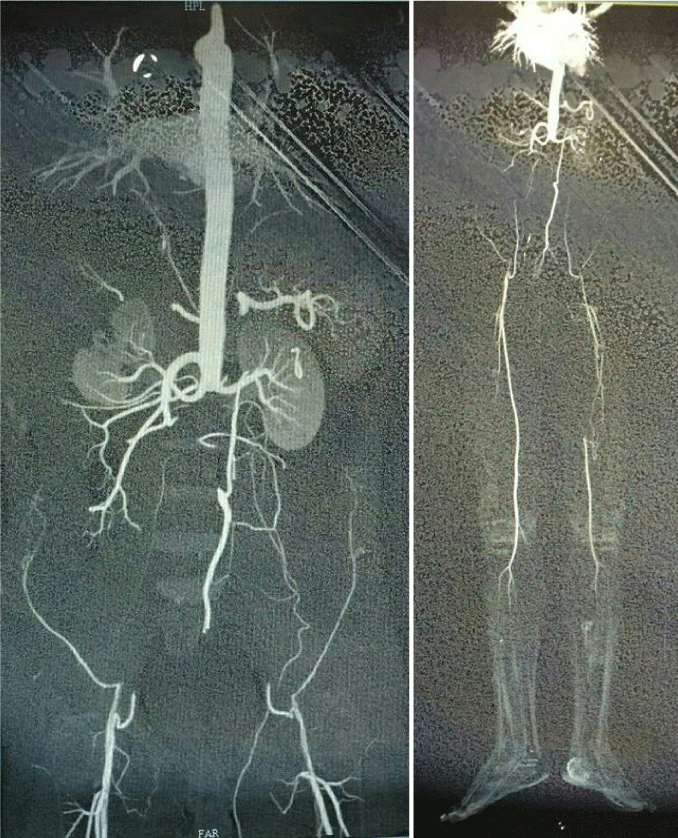
All patients had disabling intermittent claudication or rest pain. Six patients were in Rutherford’s Class IV with rest pain, two patients were in Class V with ischemic ulcers over toes and two patients were in Class III with severe claudication. Computed Tomography (CT) angiography showed juxtarenal aortic occlusion (Figure 1A). in addition two patients has left superficial femoral artery occlusion (Figure 1B) and one patient has right superficial femoral artery occlusion. Abdominal aorto-bifemoral was considered to be hazardous as there was no suitable site for aortic cross clamping as well as grafting. After discussing various surgical alternatives, we decided to perform descending thoracic aorta to both femoral artery bypass surgery.We used Gortex vascular graft for bypass surgery.Patients have various comorbidities that are shown in Table 1.
Surgical technique
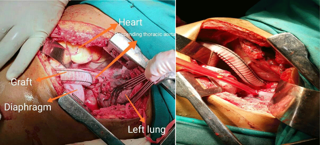
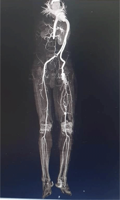
Patients were given general anesthesia with hemodynamic monitoring. With double-lumen endo-tracheal intubation to allow deflation of the left lung, the patient was positioned with the right semi-lateral position with the left hemi thorax elevated 30-45° and the pelvis as flat as possible to allow access to both groins. The chest, abdomen, and both groins were prepared and draped. Left posterolateral thoracotomy was performed through the eighth intercostal space, then, the distal descending thoracic aorta just above diaphragm and both femoral arteries were exposed simultaneously. Leaving the inguinal ligament intact, the oblique and transverse muscles of left side were split and the left retroperitoneal space was entered. Using blunt finger dissection, retroperitoneal tunneling was performed between the left hemithorax and the left suprainguinal preperitoneal space and cross over tunnel between both groin incisions was created similarly. After partial clamping on thoracic descending aorta with systemic heparinization,Proximal anastomosis of a 14 mm × 7 mm Gortex bifurcated graft was performed end-to-side at the lower descending thoracic aorta (Figure 2). The graft limbs were drawn through a tunnel between rectus abdominus muscle and peritoneum to a short midline incision at level of umbilicus, from which each limb of the graft was drawn through a subcutaneous tunnel to each side of the groin and anastomosis to each common femoral artery. A chest tube (28Fr) was placed in the left pleural space and all incisions were closed. In postoperative period, all patients were evaluated for appearance of distal pulsations, primary patency, warmness of foot, symptom relief, wound infection, healing of the ulcer, and complications. CT angiography was done in follow-up.In Figure 3 CT angiography is showing normal flow pattern in graft.
Results
Ten patients with a range of 50-65 years of age, with a mean age of 58.6 years were treated in Grant Government Medical College,Mumbai,Maharashtra during study period. All patients were male.In literature female has less than 1% incidence of aortoiliac occlusive disease [1]. Pulmonary function tests showed mild chronic obstructive pulmonary disease in four patients. Other co-morbidities were coronary artery disease in one patient, hypertension in four patients, renal impairment in three patients, and diabetes in three patients (Table 1).
Indication for surgery is juxtarenal occlusion of the abdominal aorta, in most of the cases. No mortality. Mean duration of surgery was 2.5-5 h and mean blood loss was 250 - 400 mL (Table 2). Major morbidity includes one graft occlusion which was treated successfully with embolectomy . None of the patients developed proximal propagation of aortic thrombus. Ulcers showed healing. All patients had a good quality of life postoperatively. Postoperative angiography showed good re-vascularization.
Discussion
Aorto-femoral bypass grafting is the standard surgical treatment for aortoiliac occlusive disease, partly because of a 5 years patency rate that exceeds 80% in many reports [1]. When abdominal aortic surgery is contraindicated because of severe disease at the inflow site, axillo-femoral bypass or thoraco-bifemoral bypass are alternative procedures [2]. Some authors have recommended supraceliac aorta bypass surgery within the retroperitoneal space for juxtarenal aortic occlusive disease but is technically difficult and associated with morbidity. Also while clamping the supraceliac aorta surgeon may damage renal arteries and hamper the blood supply to kidneys.So better to avoid this technique [2].
However, the results of axillo-femoral bypass are generally less than ideal in terms of patency and quality of life [3].
Thoraco-femoral bypass has major advantages over axillo-femoral bypass because it provides better inflow, requires a shorter graft length, offers better protection of the graft from infection and mechanical trauma, and carries a superior patency rate [4]. Thoraco-femoral bypass was first described by Blaisdell, et al. as an alternative to standard aorto-femoral bypass [3]. Criado, et al. [4] described 16 cases that in addition to those reported in the English language literature, gave a combined surgical mortality rate of 6.4% and the primary graft patency was 98% at 1-year, 88% at 2 years, and 70.4% at 5 years [5].
The risk of subsequent renal artery thrombosis is a concern with the use of thoraco-femoral bypass in patients with juxtarenal aortic occlusion [6]. Using radioisotope renography pro-spectively, Cevese and Gallucci confirmed normal renal perfusion up to 5 years after thoraco-femoral bypass in 6 patients with complete juxtarenal aortic occlusion [7,8]. In our patients, renal function tests were normal postoperatively in all cases.
In our series, one patient having coronary artery disease were advised coronary artery bypass grafting followed by thoraco-femoral bypass. But on patient’s choice, thoraco-femoral bypass was done first.
While most of the patients having mild pulmonary function test impairment, diabetes or hypertension behave normally in the postoperative period and discharged uneventfully.In other series [9-11], the graft was tunneled through posterior aspect of diaphragm to the left short transverse incision from this to both femoral arteries, but in our study we tunneled the graft anteriorly through anterior aspect of the diaphragm to the short midline incision then to both femoral arteries. All patients have good distal pulses.Operative mortality rate was 0-12% in different series [12,13] but in our series of 10 patients, no operative mortality was there and late mortality was 25-36% in other series [14,15] due to myocardial infarction, renal failure, stroke, respiratory insufficiency or septicaemia. In our series no mortality.
Graft failure or occlusion occurred in 4-30% cases on 3-5 years in another series. In our series one graft limb occluded, which was managed by embolectomy, had good distal flow and patient made a good recovery.Our series demonstrated superior inflow, excellent quality of life, and more reliable patency with thoraco-bifemoral bypass. This approach is recommended in selected patients when conventional approaches to the abdominal aorta are considered hazardous.
Conclusion
Majority of our patients are old age having juxtarenal aortic occlusion so we decided to use descending thoracic aorta as inflow for primary revascularization to avoid entering abdomen and renal injury. The major limitation to thoracic aorta bifemoral grafting technique is the morbidity rate associated with thoracotomy in a relatively high-risk vascular surgery population. Also, tunnelling in conventional DTA-to-femoral artery bypass is usually a “blind” procedure.For the cardiothoracic surgeon with experience, the use of descending thoracic aorta could be a very good alternative for infrarenal aortic occlusion or extra-anatomic aortoiliac bypass without any morbidity and mortality with good improvement in quality of life.
Ethical approval
All procedures performed in studies involving human participants were in accordance with the ethical standards of the institutional and/or national research committee and with the 1964 Helsinki declaration and its later amendments or comparable ethical standards.
- Kasap S, Gonenc A, Sener DE, Hisar I (2007) Serum Cardiac Markers in Patients with Acute Myocardial Infarction: Oxidative Stress, C-reactive protein and N-terminal probrain natriuretic peptide. J Clin Biochem Nutr 41: 50-57. Link: https://bit.ly/3bbgwHx
- Thygesen K, Alpert JS, Jaffe AS, Chaitman BR, Bax JJ, et al. (2018) Fourth Universal Definition of Myocardial Infarction. J Am Coll Cardiol 72: 2231-2264. Link: https://bit.ly/2EOm1QA
- Gupta S, Alagona P (2008) Troponins: not always a myocardial infarction. Am J Med 121: e25. Link: https://bit.ly/3gNuLU5
- Chen Y, Tao Y, Zhang L, Xu W, Zhou X (2019) Diagnostic and prognostic value of biomarkersin acute myocardial infarction. Postgrad Med J 95: 210-216. Link: https://bit.ly/3bbVUis
- Wu AH, Feng YJ, Contois JH, Pervaiz S (1996) Comparison of myoglobin, creatine kinase-MB, and cardiac troponin I for diagnosis of acute myocardial infarction. Ann Clin Lab Sci 26: 291-300. Link: https://bit.ly/34Monu4
- Nigam PK (2007) Biochemical markers of myocardial injury. Indian J Clin Biochem 22:10–17. Link: https://bit.ly/3gJa8Zo
- Yousuf O, Mohanty BD, Martin SS, Joshi PH, Blaha MJ, et al. (2013) High-sensitivity C-reactive protein and cardiovascular disease: a resolute belief or an elusive link? J Am Coll Cardiol 62: 397-408. Link: https://bit.ly/31IMB6M
- Hamzic Mehmedbasic A (2016) Inflammatory cytokines as risk factors for mortality after acute cardiac events. Med Arch 70: 252–255. Link: https://bit.ly/2ETtlKs
- Wang J, Tang B, Liu X, Wang H, Xu D, et al. (2015) Increased monomeric CRP levels in acute myocardial infarction: a possible new and specific biomarker for diagnosis and severity assessment of disease. Atherosclerosis 239: 343-349. Link: https://bit.ly/31JdBCM
- Rabitzsch G, Mair J, Lechleitner P, Noll F, Hofmann U, et al. (1995) Immunoenzymometric assay of human glycogen phosphorylase isoenzyme BB in diagnosis of ischemic myocardial injury. Clin Chem 41: 966-978. Link: https://bit.ly/2YRMYKm
- Krause EG, Rabitzsch G, Noll F, Mair J, Puschendorf B (1996) Glycogen phosphorylase isoenzyme BB in diagnosis of myocardial ischaemic injury and infarction. Mol Cell Biochem 160: 289-295. Link: https://bit.ly/2EJohbH
- Mair J (1997) Progress in myocardial damage detection: new biochemical markers for clinicians. Crit Rev Clin Lab Sci 34: 1-66. Link: https://bit.ly/3gM2mxM
- Singh N, Rathore V, Mahat RK, Rastogi P (2018) Glycogen Phosphorylase BB: A more Sensitive and Specific Marker than Other Cardiac Markers for Early Diagnosis of Acute Myocardial Infarction. Ind J Clin Biochem 33: 356–360. Link: https://bit.ly/31JsGV7
- Cubranic Z, Madzar Z, Matijevic S, Dvornik S, Fisic E, et al. (2012) Diagnostic accuracy of heart fatty acid binding protein (H-FABP) and glycogen phosphorylase isoenzyme BB (GPBB) in diagnosis of acute myocardial infarction in patients with acute coronary syndrome. Biochem Med (Zagreb) 22: 225-236. Link: https://bit.ly/3lxCvgC
- Bozkurt S, Kaya EB, Okutucu S, Aytemir K, Coskun F, et al. (2011) The diagnostic and prognostic value of first hour glycogen phosphorylase isoenzyme BB level in acute coronary syndrome. Cardiol J 18: 496–502. Link: https://bit.ly/2YO4dvY
- Serdar Z, Altin A, Serdar A, Bilgili G, Sarandol E, et al. (2012) Glycogen phosphorylase isoenzyme BB in early diagnosis of acute coronary syndrome. Nobel Medicus 8: 65-72. Link: https://bit.ly/2DdNGKh
- Lillpopp L, Tzikas S, Ojeda F, Munzel T, Blakenberg S, et al. (2012) Prognostic information of glycogen phosphorylase isoenzyme BB in patients with suspected acute coronary syndrome. Am J Cardiol 110:1225-1230. Link: https://bit.ly/2EP8J6p
- Peetz D, Post F, Schinzel H, Schweigert R, Schollmayer C, et al. (2005) Glycogen phosphorylase BB in acute coronary syndromes. Clin Chem Lab Med 43:1351-1358. Link: https://bit.ly/3biUnY8
- Beaudeux JL, Giral P, Bruckert E, Foglietti MJ, Chapman MJ (2004) Matrix metalloproteinases, inflammation and atherosclerosis: therapeutic perspectives. Clin Chem Lab Med 42: 121-131. Link: https://bit.ly/32yzOCM
- Wang XH, Liu SQ, Wang YL, Jin Y (2014) Correlation of serum high-sensitivity C-reactive protein and interleukin-6 in patients with acute coronary syndrome. Genet Mol Res 13: 4260-4266. Link: https://bit.ly/3lvDu0V
- Acharya A, Sahu JK, Sharma SK, Mandal MK (2017) Serial Measurement of High Sensitive C Reactive Protein Levels of Patients Having Acute Chest Pain –Study in a Tertiary Care Centre of Western Odisha. JMSCR 5: 20243-20246. Link: https://bit.ly/3juwCz7
- Basha SJ, Anil Kumar M, Lakshmi Prasad KK (2016) Role of HSCRP in Detecting Myocardial Infarction. IOSR-JDMS 15: 19-22.
- Badiger RH, Dinesha V, Hosalli A, Ashwin SP (2014) hsC-reactive protein as an indicator for prognosis in acute myocardial infarction. J Sci Soc 41: 118-121. Link: https://bit.ly/3lFXLRJ
- Aseri ZA, Habib SS, Alhomida SA, Khan HA (2014) Relationship of high sensitivity C-reactive protein with cardiac biomarkers in patients presenting with acute coronary syndrome. J Coll Physicians Surg Pak 24: 387-391. Link: https://bit.ly/2EFYGkf
- Rathore V, Singh N, Rastogi P, Mahat RK (2017) Correlation of inflammatory marker with glycogen phosphorylase BB (GPBB) in patients of acute myocardial infarction. International Journal of Contemporary Medical Research 4: 1122-1124. Link: https://bit.ly/32UjeOf

Article Alerts
Subscribe to our articles alerts and stay tuned.
 This work is licensed under a Creative Commons Attribution 4.0 International License.


 Save to Mendeley
