Open Journal of Thyroid Research
Is TI-RADS classification and Score Modified Method of thyroid nodules can be effective for evaluation of Thyroid Incidentalomas on FDG PET-CT imaging
1Mersin University, Faculty of Medicine, Department of Nuclear Medicine, Mersin, Turkey
2Mersin University, Faculty of Medicine, Department of Radiology, Mersin, Turkey
3Mersin University, Faculty of Medicine, Department of Pathology, Mersin, Turkey
Author and article information
Cite this as
Kara PO, Koc ZP, Balci Y, Arpaci RB (2019) Is TI-RADS classification and Score Modified Method of thyroid nodules can be effective for evaluation of Thyroid Incidentalomas on FDG PET-CT imaging. Open J Thyroid Res. 2019; 2(1): 005-008. Available from: 10.17352/ojtr.000008
Copyright License
© 2019 Kara PO, et al. This is an open-access article distributed under the terms of the Creative Commons Attribution License, which permits unrestricted use, distribution, and reproduction in any medium, provided the original author and source are credited.Background: Flourodeoyglucose Positron Emission Tomography-Computed Tomography (FDG PET-CT) provides information about the anatomic structures and metabolic activities of tumors. The incidence of thyroid incidentaloma is rare on FDG PET-CT but it is related to malignancy when it is detected to be high. Although, patients who are already treated with another primary tumor can not be searched for second thyroid malignancy each time. The aim of this study was to evaluate TI RADS classification based on a score modified according to ultrasound (US) criteria for malignancy of thyroid nodules determined on FDG PET/CT imaging.
Materiels & Methods: Patients’ data diagnosed with a variety of cancer evaluated, retrospectively. All patients underwent PET/ CT examinations for cancer screening, staging, restaging, and detection of suspected recurrence. Patients with thyroid nodules on CT imaging provided by PET/ CT were selected.
Results: Patients are divided into two groups. Group I included a total of 29 patients (24 women/ 5 men-age range: 30-80 years-mean age:57) with thyroid final histopathology results. Group II included a total of 24 (18 women/6 men-age range: 47-80, mean: 66.6) patients without pathology results of thyroid nodules. Of the 29 patients in group I, 20 patients had benign (69%) and 9 patients had malign (31%) histopathology results. Mean SUV max value in benign and malignant thyroid nodules were 5.3 (Range: 1-18) and 17.54 (Range: 7-35), respectively. Mean maximum standardized uptake value in malignant thyroid lesions was higher than that of benign lesions (P< 0.0001). Most of the benign thyroid nodules (85%) had TI RADS 1-3 classification while most malignant nodules had TI RADS 4-6 (87.5%). Mean SUV max value in thyroid nodules were 5.35 (Range: 1-43) in Group II follow-up patients. Thyroid nodules were TI RADS 1-5 classification in this group, most of which were 1-3 as benign thyroid nodules in group I patients. This group consisted of patients who could not have a thyroid biopsy due to primary malignancy diagnoses and advanced stages. None of them were diagnosed for thyroid malignancy or progressed during follow-up.
Conclusions: Patients who do not have short expected duration of life due to other primary tumors should be evaluated with US. TI RADS classification based on a score modified according to ultrasound criteria for malignancy of thyroid nodules determined on FDG PET/CT imaging can be used for oncological patients.
Flourodeoyglucose Positron Emission Tomography-Computed Tomography (FDG PET-CT) imaging is the most commonly used imaging modality in oncological patients. Focal or diffuse involvement in the thyroid gland can be detected on FDG PET-CT imaging performed for diagnosis, staging, re-staging and response to treatment in different cancers. The presence of thyroid nodules can be selected on Computed Tomography (CT) images in areas of focal involvement. Under physiological conditions, the thyroid gland FDG uptake is low. FDG PET-CT imaging is important because, the incidence of thyroid incidentaloma is rare but it is related to malignancy when it is detected to be high. Although, patients who are already treated with another primary tumor can not be searched for second thyroid malignancy each time. Undoubtedly, ultrasonography is the most important imaging modality for the evaluation of thyroid nodules and neck lymphatics. While evaluating the risk of malignancy in the nodule by ultrasonography, central and lateral neck regions can be examined and lymph nodes with suspected metastasis can be detected. Ultrasonography also facilitates the application of fine needle aspiration biopsy from the nodule or lymph node. The risk of cancer in a nodule can be determined ultrasonographically. The classification system of thyroid nodules TI RADS (Thyroid Imaging Reporting and DATA System) was proposed by Horvath et. al. in 2009 [1], similar to the system used for breast lesions (BI RADS) [2,3]. Kwak et al. [4], added a subtype (4c). However, TI RADS classification is hardly and rarely used in daily practice. From this perspective J F. Sanchez proposed a TI RADS classification based on a scoring system in which each ultrasound abnormality suspicious for malignancy is assigned a score [5]. The aim of this study was to evaluate TI RADS classification based on this score modified according to ultrasound criteria for malignancy of thyroid nodules determined on FDG PET/CT imaging.
Materiels and Methods
All patients underwent PET/ CT examinations for cancer screening, staging, restaging, and detection of suspected recurrence. Patients with thyroid nodules on CT imaging provided by PET/ CT were selected. Informed consent was taken from all patients.
Imaging protocol
All patients fasted for at least 4-6 h before FDG injection of 370MBq (10 mCi). PET/CT scans were obtained 60 min after injection using an integrated scanner (Siemens, Biograph True Point 6 PET/CT, Germany). A whole-body CT scan was performed without intravenous contrast administration with 130 kV, 50 mAs, a pitch of 1.5, a section thickness of 5 mm, and a field of view of 70 cm. A PET scan was performed immediately after an unenhanced CT scan, and acquired from the skull base to the upper thigh with a 2-min acquisition per bed position using a three-dimensional acquisition mode.
Diagnostic criteria for benign and malignant thyroid nodules
Histopathology and follow-up information after PET/CT scanning served as the standard of reference.
Image analysis
CT scans and ultrasonography were reviewed by a radiologist with more than 10 years’ experience on thyroid and neck imaging who had no knowledge of either the other imaging results or the clinical information. Each ultrasound abnormality suspicious for malignancy is assigned a score. If one or more cervical lymph nodes suspicious for malignancy are detected, an additional point is added. PET/CT images were qualitatively evaluated and assessed in consensus by two nuclear medicine physicians (readers A, B with more than 10 years of experience) on PET/CT. PET/CT images were viewed in the coronal, axial, and sagittal sections. Maximum standart uptake value (SUVmax) of thyroid nodules were calculated on PET/CT by using Region of interest (ROI) included at least two-thirds of the nodular lesions. Partial volume effect was minimized by this way. The regions were drawn by generating sphere circles. The quantitative uptake values of FDG (SUVmax) in the nodules ROIs were semiautomatically calculated using workstations (Siemens).
Statistical analysis
Statistical analysis was carried out with SPSS software (SPSS Inc., Chicago, Illinois, USA). A P value of less than 0.05 was considered statistically significant.
Results
Patients are divided into two groups. Group I included a total of 29 patients (24 women/ 5 men-age range: 30-80 years-mean age: 57) with thyroid final histopathology results. Primary malign diagnoses were lymphoma in 2 (7%), breast carsinoma in 8 (28%), neuroendocrine tumor in 1 (3%), genitourinary malignancies in 3 (10.5%), lung ca in 3 (10.5 %), head-neck and thyroid in 5 (17%), multipl myeloma in 1 (3%) gastrointestinal and mesenchymal tumor in 2 (7%) patients, unknown primary in 4 (14 %) in group I patients. Thyroid nodul diameter were detected between 1-4.5 cm.
Group II included a total of 24 (18 women/6 men-age range: 47-80, mean: 66.6) patients without patology results of thyroid nodules. Group II patients were followed by ultrasonography and PET-CT performed for primary malignancy. Primary malign diagnoses were lung ca in 7 (29.1%), breast carsinoma in 4 (16.6%), gynecological malignancies in 2 (8.3%), gastrointestinal and pancreas ca in 5 (20.8%), malign melanoma in 1 (4.1%), mesenchymal tumor in 1 (4.1%) patients, unknown primary in 4 (16.6%) in group II patients. Thyroid nodul diameter were detected between 1-3 cm in group II.
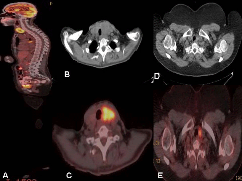
The most common primary malignancy was breast cancer in both groups. Of the 29 patients in group I, 20 patients had benign (69%) and 9 patients had malign (31%) histopathology results. Mean SUV max value in benign and malignant thyroid nodules were 5.3 (Range: 1-18) and 17.54 (Range: 7-35), respectively. Mean maximum standardized uptake value in malignant thyroid lesions was higher than that of benign lesions (P< 0.0001). Figure 1 illustrate two patients with hypermetabolic thyroid nodules. Table 1 demontrates TI RADS classification of benign and malignant nodules. Most of the benign thyroid nodules (85%) had TI RADS 1-3 classification while most malignant nodules had TI RADS 4-6 (87.5%). In one patient ultrasonography detected a lymph node instead of thyroid nodule with biopsy proven malignant lymphoma. One of the patients with malignant thyroid nodule had TI RADS 1 classification on ultrasound images.
Mean SUV max value in thyroid nodules were 5.35 (Range: 1-43) in Group II follow-up patients. Thyroid nodules were TI RADS 1-5 classification in this group, most of which were 1-3 as benign thyroid nodules in group II patients. This group consisted of patients who could not have a thyroid biopsy due to primary malignancy diagnoses and advanced stages. These patients were followed-up clinically and by ultrasound and PET-CT images for thyroid nodules. None of them were diagnosed for thyroid malignancy or progressed during follow-up. Four of the 24 patients in this group died during follow-up because of primary nonthyroid malignancies.
Discussion
Thyroid cancers are the most common endocrine organ malignancies. According to the ABDSEER (Surveillance, Epidemiology and End Results) program data, the probability of a diagnosis of thyroid cancer during a person’s lifetime (sex not observed) is 1.2 % [6]. Estimated of thyroid cancer in all new cancer cases in 2018 is 3.1 %. The prognosis in thyroid cancer is generally quite good. There is no increase in mortality in thyroid cancers despite the increase in incidence. In cancer screening study conducted in Japan, the highest rate of FDG PET positivity was found to be thyroid carcinoma in malignancies detected by screening program [7]. FDG positivity was detected 90.7% of the 353 patients with thyroid carcinoma. The mean sensitivity in the diagnosis of thyroid cancer for FDG PET-CT and FDG PET was calculated as 90.9%. This rate is superior to other methods and similar to ultrasound. F-18 FDG PET-CT sensitivity increases with ultrasonography [8]. The positive predictive value of FDG PET-CT in the diagnosis of thyroid cancer was found to be low as expected (29.5%). Differentiation of malignant from benign thyroid nodules is a critical issue in clinical practice in especially patients with other malignancies on FDG PET-CT imaging. Maximum standardized uptake value (SUVmax) is a semiquantitative parameter that reflect metabolic activity, but is not specific marker of malignancies. In one study, PET-CT imaging was performed in 15 patients with cytologically indetermined nodules. All patients underwent surgery and were conclusively evaluated. PET-CT was positive in 8 patients and 4 of them were diagnosed as thyroid ca. Thyroid ca was found in 3 of 7 patients with negative PET-CT. In this study, the positive predictive value was 50% and the negative predictive value was 57% [9]. In this study, it was concluded that FDG PET-CT did not provide additional contribution in suspicious nodules. Deandreis D, and colleagues [10] have found similar results in their study. A total of 56 nodules with suspicious FNAB results were evaluated in 55 patients and a positive predictive value of 57% and a negative predictive value of 81% results were found. In the literature, the incidence of thyroid incidentaloma in FDG PET-CT was reported as 1.1 % - 4.3 % [11,12]. Therefore, FDG PET-CT imaging is unlikely to detect thyroid incidentaloma. These rates are even lower than the possibility of detecting thyroid nodules by palpation [13,14]. FDG PET-CT imaging is important because the incidence of thyroid incidentaloma is rare but it is related to malignancy when it is detected to be high. In the literature, the prevalence of cancer has been reported between 14% and 63% in cases of focal incidentaloma. The multiple reported prevalence values are due to retrospective studies and the fact that patients who are already treated with another primary tumor have a definite diagnosis in a small number of patients. In 2 systematic reviews, the prevalence of malignancy was similar and 33.2% and 34.8%, respectively [15,16]. In our study, of the 29 patients in group I, 20 patients had benign (69%) and 9 patients had malign (31%) histopathology results. SUVmax values have been studied and it has been reported that the threshold values ranging from 3.8-6 can be used to differentiate benign from malignant lesions [17,18]. In our study, mean SUV max value in benign and malignant thyroid nodules were 5.3 (Range: 1-18) and 17.54 (Range: 7-35), respectively. Although, mean SUVmax value in malignant thyroid lesions was higher than that of benign lesions (P< 0.0001), it should be kept in mind that there is a significant overlap between the SUVmax values of benign and malignant lesions and malignancy can be seen even with low involvement. While evaluating the risk of malignancy in the nodule by ultrasound, central and lateral neck regions can be examined and lymph nodes with suspected metastasis can be detected. In one patient in group I, ultrasonography detected a lymph node instead of thyroid nodule with biopsy proven malignant lymphoma. In our study, group II patients were follow-up patients and this group included patients who couldn’t undergo thyroid biopsy due to advanced stage primary malignancy or other reasons. None of the patients in this group were diagnosed for thyroid malignancy or progressed during follow-up because of thyroid malignancy. Four of the 24 patients in this group died during follow-up for other reasons such as primary advanced staged malignancy or infections. Most of the benign thyroid nodules (85%) had TI RADS 1-3 classification while most malignant nodules had TI RADS 4-6 (87.5%) as expected. One of the patients with malignant thyroid nodule had TI RADS 1 classification on ultrasound images. Although diffuse FDG involvement of thyroid corresponds to thyroiditis or Graves disease, diffuse and focal FDG uptake can be detected in one patient and moreover malignancy can be detected such as in our patient. Thyroid nodules were TI RADS 1-5 classification in group II follow-up patients, most of which were TI RADS 1-3 as benign thyroid nodules. According to our results, TI RADS classification based on a score modified according to ultrasound criteria for malignancy of thyroid nodules determined on FDG PET-CT imaging can be used for oncological patients. Lower scored patients can be followed-up by ultrasound. TI RADS 4-5 patients with high score should be evaluated with FNAB (Fine needle aspiration biopsy).
Conclusions
The incidence of thyroid incidentaloma is rare but it is related to malignancy when it is detected to be high. Patients who are already treated with another primary tumor can not be searched for second thyroid malignancy each time. It should be known that approximately one-third of patients with focal incidentaloma with FDG PET-CT have a primary probability of thyroid cancer. Patients who do not have short expected duration of life due to other primary tumors should be evaluated with US. TI RADS classification based on a score modified according to ultrasound criteria for malignancy of thyroid nodules determined on FDG PET/CT imaging can be used for oncological patients.
- Hovarth E, Majlis S, Rossi R, Franco C, Niedmann JP, et al. (2009) An ultrasonogram reporting system for thyroid nodules stratifying cancer risk for clinical management. J Clin Endocrinol Metab 94: 1748-1751. Link: https://bit.ly/2IuZnLi
- Liberman L, Menell JH (2002) Breast imaging reporting and data system (BI-RADS). Radiol Clin North Am 40: 409-430. Link: https://bit.ly/31mozME
- Burnside ES, Sickles EA, Bassett LW, Rubin DL, Lee CH, et al. (2009) The ACR BI-RADS experience: learning from history. J Am Coll Radiol 6: 851-860. Link: https://bit.ly/2LiMtkp
- Kwak JY, Han KH, Yoon JH, Moon HJ, Son EJ, et al. (2011) Thyroid imaging reporting and data system for US features of nodules: a step in establishing better stratification of cancer risk. Radiology 260: 892-899. Link: https://bit.ly/2wLXGDw
- Sánchez JF (2014) TI-RADS classification of thyroid nodules based on a score modified according to ultrasound criteria for malignancy. Rev. Argent. Radiol 78: 138-148. Link: https://bit.ly/2WpymOd
- Cancer Stat Facts: Thyroid Cancer (2013) National Cancer Institute Link: https://bit.ly/2K46uNK
- Minamimoto R, Senda M, Jinnouchi S, Terauchi T, Yoshida T, et al. (2013) The current status of an FDG-PET cancer screening program in Japan, based on a 4-year (2006-2009) nationwide survey. Ann Nucl Med 27: 46-57. Link: https://bit.ly/2F0Vgpl
- Minamimoto R, Senda M, Jinnouchi S, Terauchi T, Yoshida T, et al. (2014) Detection of thyroid cancer by an FDG-PET cancer screening program: a Japanese nation-wide survey. Anticancer Res 34: 4439-4445. Link: https://bit.ly/2ZoWToD
- Hales NW, Krempl GA, Medina JE (2008) Is there a role for fluorodeoxyglucose positron emission tomography/computed tomography in cytologically indeterminate thyroid nodules? Am J Otolaryngol 29: 113-118. Link: https://bit.ly/2K9PlT0
- Deandreis D, Al Ghuzlan A, Auperin A, Vielh P, Caillou B, et al. (2012) Is (18)F-fluorodeoxyglucose-PET/CT useful for the presurgical characterization of thyroid nodules with indeterminate fine needle aspiration cytology? Thyroid 22: 165-172. Link: https://bit.ly/2WTmKY4
- Choi JY, Lee KS, Kim HJ, Shim YM, Kwon OJ, et al. (2006) Focal thyroid lesions incidentally identified by integrated 18F-FDG PET/CT: clinical significance and improved characterization. J Nucl Med 47: 609-615. Link: https://bit.ly/2WQU0zh
- Chun AR, Jo HM, Lee SH, Chun HW, Park JM, et al. (2015) Risk of malignancy in thyroid incidentalomas identified by fluorodeoxyglucose-positron emission tomography. Endocrinol Metab (Seoul) 30: 71-77. Link: https://bit.ly/31mpr3S
- Dean DS, Gharib H (2008) Epidemiology of thyroid nodules. Best Pract Res Clin Endocrinol Metab 22: 901-911. Link: https://bit.ly/2R0P1Xi
- Hegedüs L (2004) Clinical practice. The thyroid nodule. N Engl J Med 351: 1764-1771. Link: https://bit.ly/2MDKoE4
- Shie P, Cardarelli R, Sprawls K, Fulda KG, Taur A (2009) Systematic review: prevalence of malignant incidental thyroid nodules identified on fluorine-18 fluorodeoxyglucose positron emission tomography. Nucl Med Commun 30: 742-748. Link: https://bit.ly/2WC1cQf
- Soelberg KK, Bonnema SJ, Brix TH, Hegedüs L (2012) Risk of malignancy in thyroid incidentalomas detected by 18F-fluorodeoxyglucose positron emission tomography: a systematic review. Thyroid 22: 918-925. Link: https://bit.ly/2MCzmyG
- Bae JS, Chae BJ, Park WC, Kim JS, Kim SH, et al. (2009) Incidental thyroid lesions detected by FDG-PET/CT: prevalence and risk of thyroid cancer. World J Surg Oncol 7: 63. Link: https://bit.ly/2I6gR1u
- Are C, Hsu JF, Schoder H, Shah JP, Larson SM, et al. (2007) FDG-PET detected thyroid incidentalomas: need for further investigation? Ann Surg Oncol 14: 239-247. Link: https://bit.ly/2I6gV1e
Article Alerts
Subscribe to our articles alerts and stay tuned.
 This work is licensed under a Creative Commons Attribution 4.0 International License.


 Save to Mendeley
