Archives of Renal Diseases and Management
Open Repair of a Renal Artery Aneurysm with Hypogastric Artery Autograft and Hypothermic Perfusion Preservation
Vascular and Endovascular Surgery Division, São Paulo University Medical School, São Paulo - Brazil
Author and article information
Cite this as
De Luccia N, Queiroz AB, Mulatti GC, do Espirito Santo FRF, Rosa Schneidwind KPD (2015) Open Repair of a Renal Artery Aneurysm with Hypogastric Artery Autograft and Hypothermic Perfusion Preservation. Arch Renal Dis Manag. 2015; 1(1): 12-13. Available from: 10.17352/2455-5495.000004
Copyright License
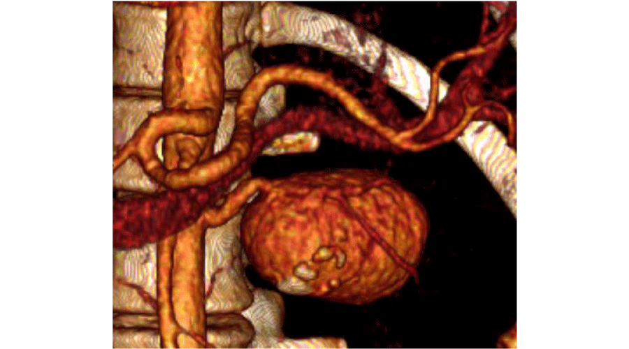
© 2015 De Luccia N, et al. This is an open-access article distributed under the terms of the Creative Commons Attribution License, which permits unrestricted use, distribution, and reproduction in any medium, provided the original author and source are credited.A 32-year-old woman presented with a one-year history of mild abdominal pain in the left upper quadrant and a palpable pulsatile abdominal mass on physical examination. The results of laboratory investigations, including serum urea and creatinine levels, were unremarkable. Contrast enhanced computed tomography (CT) showed a large left renal artery aneurysm, measuring 5,0 cm by 3,5 cm, but no evidence of renal perfusion alterations or other vascular abnormalities (Figure 1). She had been previously submitted to an unsuccessful endovascular approach with intention to treat the aneurysm and preserve left renal perfusion. Because she was young and in good health our purpose was to preserve left renal function and an open repair was adopted. The patient underwent a laparotomy with midline incision and the left kidney, left renal vein and artery were circumferentially mobilized from surrounding tissues. To permit a much better exposure, left renal vein and artery were clamped and transected while the ureter was left intact and the ex-situ reconstruction was performed on the body wall [1-3].
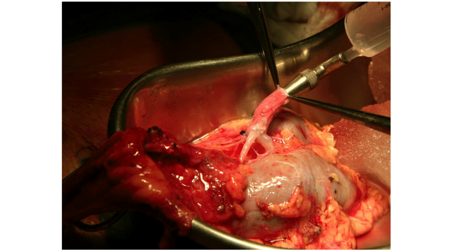
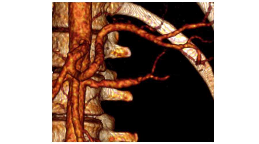
A 32-year-old woman presented with a one-year history of mild abdominal pain in the left upper quadrant and a palpable pulsatile abdominal mass on physical examination. The results of laboratory investigations, including serum urea and creatinine levels, were unremarkable. Contrast enhanced computed tomography (CT) showed a large left renal artery aneurysm, measuring 5,0 cm by 3,5 cm, but no evidence of renal perfusion alterations or other vascular abnormalities (Figure 1). She had been previously submitted to an unsuccessful endovascular approach with intention to treat the aneurysm and preserve left renal perfusion. Because she was young and in good health our purpose was to preserve left renal function and an open repair was adopted. The patient underwent a laparotomy with midline incision and the left kidney, left renal vein and artery were circumferentially mobilized from surrounding tissues. To permit a much better exposure, left renal vein and artery were clamped and transected while the ureter was left intact and the ex-situ reconstruction was performed on the body wall [1-3]. Surface cooling and hypothermic renal perfusion with Euro-Collins solution (4oC) was performed. To prevent the preservation solution (containing high levels of potassium) from entering the central circulation, it was washed out before finishing the anastomoses [1,2]. Other authors have suggested that when more than 40 minutes of warm ischemia are required, these measures to protect renal function should be instituted [1,4]. The aneurysm was resected leaving the stump of the distal renal artery, just proximal to its bifurcation. The left hypo gastric artery has been previously dissected and was used as auto graft for the renal artery reconstruction (Figure 2). Hypo gastric auto grafts has been used in larger series and have important role in complex branched Reno vascular lesions [5]. Left renal vein was primarily anastomosed and the kidney was returned to the original position. The patient was discharged six days later, renal function remained unchanged and symptoms resolved. Duplex scan and contrast enhanced CT surveillance demonstrated left renal artery and vein patency at six-month follow-up (Figure 3).
- Nelms JK, Benjamin ME (2013) Ex vivo renal repair: technical tips, when, and why. Semin Vasc Surg 26: 199-204 .
- Lacombe M (1994) Ex situ repair of complex renal artery lesions. Cardiovasc Surg 2: 767-771.
- English WP, Pearce JD, Craven TE, Wilson DB, Edwards MS, et al. (2004) Surgical management of renal artery aneurysms. J Vasc Surg 40: 53-60 .
- Crutchley TA, Pearce JD, Craven TE, Edwards MS, Dean RH, et al. (2007) Branch renal artery repair with cold perfusion protection. J Vasc Surg 46: 405-412 .
- Murray SP, Kent C, Salvatierra O, Stoney RJ (1994) Complex branch renovascular disease: management options and late results. J Vasc Surg 20: 338-45 .


 Save to Mendeley
Article Alerts
Subscribe to our articles alerts and stay tuned.
 This work is licensed under a Creative Commons Attribution 4.0 International License.

