International Journal of Oral and Craniofacial Science
An Easy Way of Identification and Protection of the Hypoglossal Nerve during Neck Dissection for Head and Neck Cancer
D Deskos* and N Papadogeorgakis
Department of Oral and Maxillofacial Surgery, Evgenideion Hospital, National and Kapodistrian University of Athens, Greece
Cite this as
Deskos D, Papadogeorgakis N. An Easy Way of Identification and Protection of the Hypoglossal Nerve during Neck Dissection for Head and Neck Cancer. Int J Oral Craniofac Sci. 2024;10(2):006-008. DOI: 10.17352/2455-4634.000063Copyright License
© 2024 Deskos D, et al. This is an open-access article distributed under the terms of the Creative Commons Attribution License, which permits unrestricted use, distribution, and reproduction in any medium, provided the original author and source are credited.Understanding the anatomical relationship between the Hypoglossal Nerve (HN) and the Facial Vein (FV) is crucial during neck dissections for head and neck cancer. The consistent placement of the HN beneath the FV provides a reliable anatomical landmark, aiding in the preservation of nerve function and reducing surgical complications. This paper reviews the anatomical pathway of the HN, its clinical implications, and surgical observations from 87 cases. Techniques for protecting the HN during neck dissection are also discussed, emphasizing the importance of precise surgical planning to avoid postoperative complications.
Introduction
The hypoglossal nerve (HN), or cranial nerve XII, is responsible for motor innervation of the tongue. Preserving this nerve during neck dissections for head and neck cancer is essential to prevent complications such as tongue paralysis. Accurate identification of the HN’s location, particularly concerning the facial vein (FV), is critical for nerve protection. This manuscript highlights the consistent anatomical relationship observed between the HN and FV, supported by clinical images from surgical dissections.
Detailed pathway of the hypoglossal nerve
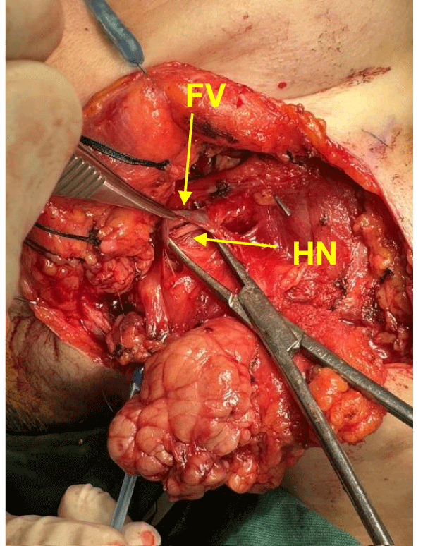
The HN originates from the hypoglossal nucleus in the medulla oblongata, exits the skull via the hypoglossal canal, and descends through the neck, passing between the internal carotid artery and internal jugular vein. It then travels medially, alongside the posterior belly of the digastric muscle and the stylohyoid muscle, eventually lying deep to the FV in the submandibular region. Understanding this anatomical course is crucial for avoiding nerve injury during surgical procedures [1,2].
Facial vein pathway
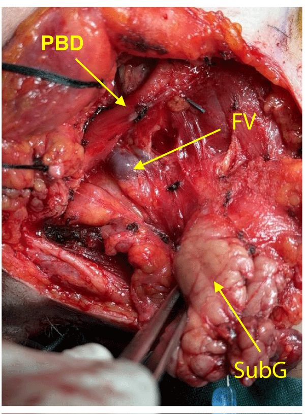
The FV begins near the medial canthus of the eye and descends along the lateral aspect of the face, passing superficially to the submandibular gland (SubG). As it travels inferiorly, it crosses over the HN before draining into the internal jugular vein. Its consistent superficial location relative to the HN makes it a useful surgical landmark during dissections [1,3].
Surgical observations
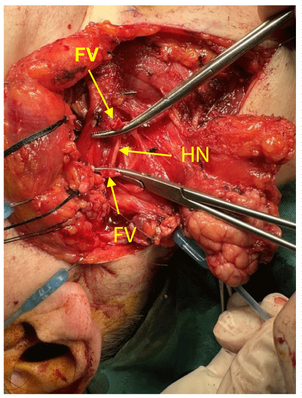
During neck dissections, especially in levels I and II, the HN is consistently located beneath the FV, as confirmed in 87 cases during neck dissection, where the nerve was always observed underneath the vein. Clinical images from our dissections vividly illustrate this relationship (Image 1-3). The FV, which often needs to be ligated or retracted for better exposure, crosses over the HN, making it a crucial landmark for identifying and preserving the nerve during surgery [4,5].
Demographic analysis
The demographic analysis of the 87 cases showed a slight predominance of female patients, with 55.17% (48 cases) being women and 44.83% (39 cases) being men (Table 1). The age range of the patients was between 32 and 93 years, with a mean age of 67 years, indicating that the anatomical consistency of the HN-FV relationship was observed across a wide spectrum of ages. All patients in this study were of Greek ethnicity, reflecting the population served by the Evgenideion Hospital in Athens. In all cases, the hypoglossal nerve was consistently located underneath the facial vein and never above it, confirming its reliability as an anatomical landmark for nerve preservation during surgery.
Regarding cancer types, the majority of cases involved squamous cell carcinoma (SCC) of the tongue, accounting for 60.92% (53 cases). Additionally, SCC of the mandible constituted 20.69% (18 cases), while SCC of the buccal mucosa and maxilla each accounted for 9.20% (8 cases each) (Table 2). This distribution underscores that the surgical technique and findings are applicable across various forms of head and neck malignancies, reinforcing the general relevance of the HN-FV relationship as a critical anatomical guide during neck dissections.
Statistical analysis
Clinical relevance: Damage to the HN during neck dissection can lead to significant post-operative issues, including tongue deviation, dysarthria, and dysphagia. Awareness of the nerve’s consistent location beneath the FV helps reduce the risk of such complications by guiding precise surgical techniques [6].
Discussion
The anatomical relationship between the hypoglossal nerve (HN) and the facial vein (FV) observed in this study holds significant clinical relevance for neck dissections involving head and neck cancer. In all 87 cases, the consistent placement of the HN beneath the FV provided a reliable and easily identifiable landmark that aided in the preservation of the nerve during surgical procedures. This consistency is vital because damage to the HN can result in severe postoperative complications, such as tongue deviation, dysarthria, and dysphagia. By utilizing the FV as a reference point for locating the HN, surgeons can minimize the risk of nerve injury, thereby improving patient outcomes.
The demographic analysis, showing a near-even distribution of male and female patients (55.17% women and 44.83% men) across a wide age range (32-93 years), with a mean age of 67 years, further supports the general applicability of these findings. The consistency of the HN-FV relationship across such diverse patient demographics underscores the reliability of using this anatomical landmark for nerve preservation during neck dissections. The findings are not limited to a specific age group or gender, making the surgical techniques described broadly applicable to head and neck cancer patients.
The distribution of cancer types among the cases also indicates the widespread applicability of the HN-FV relationship. With 60.92% of the cases involving squamous cell carcinoma of the tongue, 20.69% affecting the mandible, and 9.20% each involving cancers of the cheek mucosa and maxilla, the surgical approach proved useful across various malignancies in the head and neck region. This suggests that the HN-FV anatomical relationship is an essential consideration for surgeons performing neck dissections for different cancer types.
The consistent positioning of the HN relative to the FV was not only useful for nerve preservation but also facilitated better exposure and management of the surgical field. In cases where the FV needed to be ligated or retracted, the consistent anatomical relationship allowed surgeons to anticipate the location of the HN accurately, reducing the risk of inadvertent nerve damage. This approach is particularly valuable in complex cases where extensive dissection is required, as it helps guide surgeons in navigating the intricate anatomy of the neck.
In previous studies, similar findings have been reported, reinforcing the value of the FV as a surgical landmark for the HN. For example, Kariuki, et al. (2017) emphasized the importance of understanding the cervical part of the HN in surgical planning, highlighting the need for landmarks to avoid nerve injury [1]. Likewise, Cotofana, et al. (2017) discussed the implications of the anatomical course of the FV for reconstructive and aesthetic procedures, suggesting that knowledge of such relationships can optimize surgical strategies and reduce complications [2].
Furthermore, this study’s findings are consistent with Souza, et al. (2017), who noted the surgical significance of the HN in guiding head and neck procedures. By relying on predictable anatomical patterns, surgeons can refine their techniques to prioritize nerve preservation, thus preventing adverse outcomes associated with HN damage [4]. Moreover, the variations in superficial veins of the neck, as reviewed by Dalip, et al. (2018), highlight the complexity of the anatomical landscape surgeons must navigate during procedures [5].
Additionally, the recent study by Siwetz, et al. (2023) further underscores the variability in facial vein anatomy, emphasizing the importance of understanding these anatomical relationships to avoid complications during surgeries involving the head and neck [3]. Finally, the relationships among the submandibular duct, lingual nerve, and hypoglossal nerve explored by Beşer, et al. (2018) further elucidate the intricate anatomical configurations that require careful consideration to minimize the risk of nerve injury during surgical interventions [6]. Overall, the consistent findings across various studies support the critical role of anatomical knowledge in improving surgical outcomes in the head and neck region.
Ethical considerations
This observational study adhered to ethical standards, with all procedures conducted following the principles outlined in the Declaration of Helsinki. Informed consent was obtained from all patients for the publication of images and data collected during the neck dissections. Additionally, the study was reviewed and approved by the Institutional Review Board (IRB) of Evgenideion Hospital, ensuring compliance with ethical research practices.
Conclusion
The anatomical relationship between the HN and FV provides a reliable landmark for nerve preservation during neck dissections. Consistent placement of the HN beneath the FV in all 87 cases highlights the importance of understanding this relationship for successful surgical outcomes. Detailed knowledge of this anatomy reduces the risk of complications, improving the quality of life for patients postoperatively.
- Kariuki BN, Butt F, Mandela P, Odula P. Surgical anatomy of the cervical part of the hypoglossal nerve. Craniomaxillofac Trauma Reconstr. 2017;11(1):21-27. Available from: https://doi.org/10.1055/s-0037-1601863
- Cotofana S, Steinke H, Schlattau A, Schlager M, Sykes JM, Roth MZ, et al. The anatomy of the facial vein: implications for plastic, reconstructive, and aesthetic procedures. Plast Reconstr Surg. 2017;139(6):1346-1353. Available from: https://doi.org/10.1097/PRS.0000000000003382
- Siwetz M, Widni-Pajank H, Hammer N, Pilsl U, Bruneder S, Wree A, Antipova V. The course and variation of the facial vein in the face—known and unknown facts: an anatomical study. Medicina (Kaunas). 2023;59(8):1479. Available from: https://doi.org/10.3390/medicina59081479
- Souza AD, Hosapatna M, Naik KS, Souza AS, Ankolekar VH. Surgical anatomy of hypoglossal nerve as a guide for important head and neck surgeries. J Anat Soc India. 2017;66(1):54-57. Available from: https://doi.org/10.1016/j.jasi.2017.03.004
- Dalip D, Iwanaga J, Loukas M, Oskouian RJ, Tubbs RS. Review of the variations of the superficial veins of the neck. Cureus. 2018;10(6). Available from: https://doi.org/10.7759/cureus.2826
- Beşer C, Erçakmak B, Ilgaz HB, Vatansever A, Sargon MF. Revisiting the relationship between the submandibular duct, lingual nerve, and hypoglossal nerve. Folia Morphol. 2018;77(3):521-526. Available from: https://doi.org/10.5603/FM.a2018.0010
Article Alerts
Subscribe to our articles alerts and stay tuned.
 This work is licensed under a Creative Commons Attribution 4.0 International License.


 Save to Mendeley
