International Journal of Oral and Craniofacial Science
Evaluation of the Relationship between Gubernaculum Dentis and Tooth Development with Cone Beam Computed Tomography
Lale Rizeli and Ayşe Zeynep Zengin*
Oral and Maxillofacial Radiology, Ondokuz Mayis University, Faculty of Dentistry, Turkey
Cite this as
Rizeli L, Zengin AZ. Evaluation of the Relationship between Gubernaculum Dentis and Tooth Development with Cone Beam Computed Tomography. Int J Oral Craniofac Sci. 2024;10(2):009-016. DOI: 10.17352/2455-4634.000064Copyright License
© 2024 Rizeli L, et al. This is an open-access article distributed under the terms of the Creative Commons Attribution License, which permits unrestricted use, distribution, and reproduction in any medium, provided the original author and source are credited.Aim: The Gubernaculum Dentis (GD) is a structure that serves as the eruption path for permanent teeth, extending from the tooth follicle to the gingiva and including the gubernacular cord. It plays a crucial role in the normal eruption process by providing a defined path for the tooth to emerge through the bone. Radiographically, GD is visible as a radiolucent area, bordered by cortical bone, around the crown of an unerupted tooth on standard X-rays. On Cone Beam Computed Tomography (CBCT) scans, it appears as a hypodense cortical path adjacent to the dental follicle of the unerupted tooth.
This study aims to examine the prevalence, radiological appearance, and characteristics of GD in unerupted permanent teeth with CBCT and to investigate its characteristics according to the Demirjian dental calcification scale.
Materials and methods: Radiographic images of 75 pediatric patients whose CBCT images were taken for various reasons were analysed retrospectively. The prevalence of GD in the images, types of shapes, attachment sites to the teeth, and areas of openings in the alveolar crest were evaluated. GD angle and length measurements were made on CBCT images. Teeth were classified according to Demirjian tooth development method and the relationship between GD and tooth development was examined.
Results: The prevalence of GD was found as 73.5% in the 1055 unerupted teeth that were examined. While the prevalence of GD was 60% in the maxilla, it was found as 87.8% in the mandible. (p < 0.001) When the shapes of GDs were analyzed, the most common shape was flat with 54.8%, followed by rectangular with 38.7%. When the opening sites of teeth with GD were examined, the highest rate was found in palatinal and lingual with 57.8%, followed by the crest apex with 40.9%. The mean GD angle was found as 6.260, while the mean length was found as 3.93 mm. According to the Demirjian tooth calcification scale, the highest rate of GD was seen in stage C teeth with 92.7%, while the lowest rate of GD was seen in stage A teeth with 33.3% (p < 0.001).
Conclusion: According to the results of this study, it was found that GD was mostly in the maxilla and flat. GD length value was found to be higher in the mandibular teeth, Demirjian scale A and especially canine teeth.
Introduction
GD is an eruption pathway that extends from the follicle of permanent teeth to the gingiva and includes the gubernacular cord [1-4]. Since it represents the eruption pathway of the tooth in the bone, GD plays an important role in the normal eruption of permanent teeth [1,2,5,6]. Histologically, it is a bony canal that connects the pericoronal follicular tissue of successive teeth with the overlying gingiva, opens to the alveolar bone crest posterior to the deciduous teeth, and consists of a fibrous band [1]. It also produces many chemical mediators that induce osteoclastic bone resorption. Thus, an eruption pathway is formed on the lingual aspect of the deciduous teeth for permanent teeth, which guides these teeth to the eruption position [7].
GD has a small and thin anatomical structure with a mean diameter of 1 mm - 3 mm. Since this thin structure is in alveolar bone, it is extremely difficult to distinguish it from bone marrow cavities on conventional two-dimensional radiographs [8]. Radiologically, it is observed as a cortical limited radiolucent pathway around the crown of the tooth on direct radiographs [3]. However, GD may not always be observed, especially on panoramic radiographs [9]. Radiologically, it can be best seen in CBCT images [8]. It is observed as a hypodense cortical pathway adjacent to the tooth follicle of an unerupted tooth in CBCT and CT [3].
The method developed by Demirjian, et al. in 1973 assesses the dental morphological development of children using panoramic radiographs. It defines eight stages (A-H) of mineralization for the seven permanent teeth in the left mandible, and the numerical values corresponding to these stages are used to determine dental maturation. In 1976, Demirjian and Goldstein expanded the method by increasing the age group and sample size, making it possible to estimate age based on the development of four teeth. In this study, we aimed to explore the developmental stage of the tooth and its relationship with GD. To ensure a more objective and literature-supported evaluation of tooth development, we applied the Demirjian Tooth Development and Calcification Scale.
GD is mentioned in some anatomy textbooks, but there is limited research regarding its existence, appearance, and significance. Despite its importance, GD has not received considerable attention in dental fields, including pediatric dentistry and oral and maxillofacial radiology. It is crucial to analyze CBCT images from pediatric patients to better understand the radiological appearance and characteristics of GD, particularly during the mixed dentition phase. This is important for evaluating the prognosis and treatment planning of unerupted or impacted teeth, especially in cases involving orthodontic issues. This study aims to determine the prevalence of GD in permanent teeth, investigate its radiological features, and examine its characteristics based on the Demirjian tooth calcification scale.
Materials and methods
Ethical approval was obtained for this study from the Ondokuz Mayıs University Clinical Research Ethics Committee (OMÜ KAEK 2021/327).
This study was conducted as Lale RİZELİ’’s specialization thesis in 2023 at Ondokuzmayıs University, Faculty of Dentistry, Department of Oral and Maxillofacial Radiology.
Tomographic images of 75 pediatric patients who were admitted to Ondokuz Mayıs University, Faculty of Dentistry, Department of Oral and Maxillofacial Radiology between 2012 and 2019 and whose CBCT images were taken for various reasons were analyzed retrospectively.
Inclusion criteria
Permanent teeth that are expected to erupt normally in the tomographic images of pediatric patients between the ages of 5 and 12 who were in a mixed dentition period were examined.
Exclusion criteria
Teeth with eruption disturbances such as impacted teeth without normal eruption, supernumerary teeth, and transmigration, patients with systemic diseases that affect the bone (Rickets, hyperparathyroid, etc.), images that had artifacts for various reasons and diagnostically insufficient were excluded from the study.
The images were obtained with a dental volumetric imaging system (GALILEOS Comfort Plus, Sirona Dental Systems, Bensheim, Germany) operating at 98 kVp and 15-30 mAs, 15×15 cm FOV. CBCT images were created with 0.3/0.15 mm³ isotropic voxels, 14 sec scanning time, 2-6 sec irradiation time, and 204° rotation.
Simultaneous reconstruction was performed with SIRONA Sidexis XG 2.61 imaging software by using isotropic voxels of 12-bit grey scale depth and 0,25 mm³ size (measurement accuracy ± 0,15 mm).
All examinations and measurements were performed on a 3.7 MP, 68 cm, 2560 × 1440 resolution, 27-inch color LCD screen (The RadiForce MX270W, Eizo Nanao Corporation, Ishikawa, Japan).
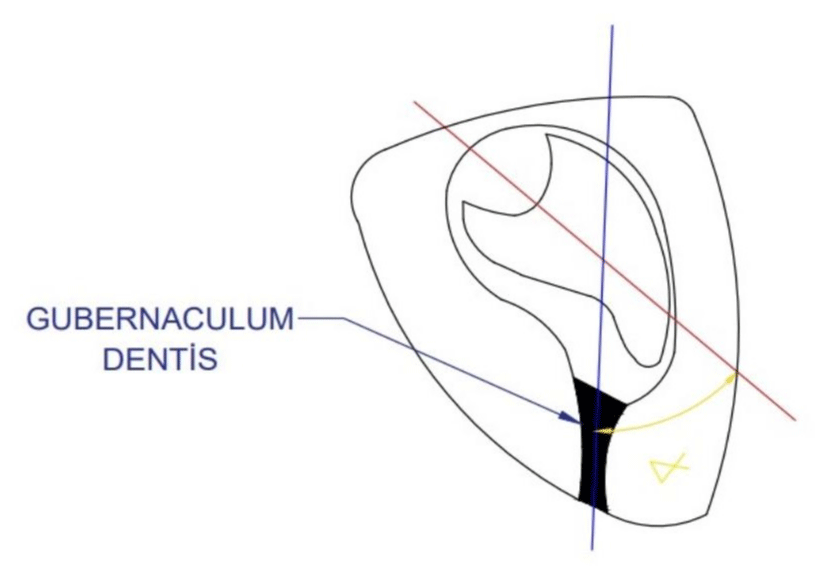
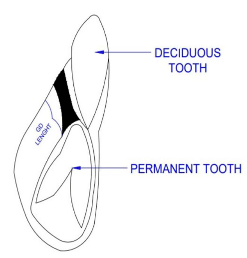
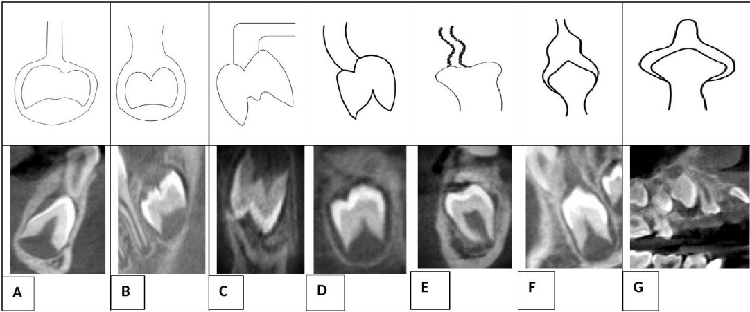
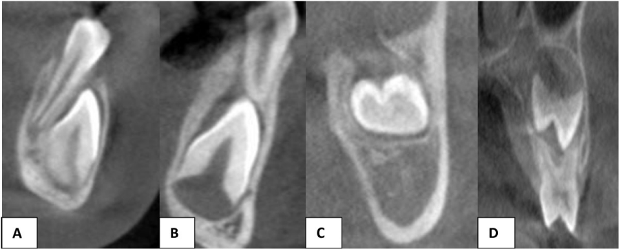
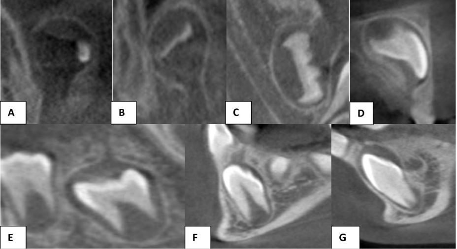
In the CBCT images, 1055 tooth germs in the unerupted alveolar jaws in the maxilla and mandible were examined in sagittal, coronal, and axial planes. Attachment properties of GD to the teeth and their opening to the alveolar crest were examined on CBCT images. The angular value between the long axis of teeth and the long axis of the gubernacular canal (Figure 1) and the length measurements of the gubernacular canal were measured on cross-sectional images (Figure 2). In the images, GD were classified as flat, rectangular, angular, curved, contracted, test tube and occluded according to their shapes (Figure 3). GD was classified according to the location of the opening in the alveolar crest. These are openings to palatinale in the maxilla and lingual in the mandible, to the buccal of the alveolar crest, to the apex of the alveolar crest, and between the roots of the deciduous teeth (Figure 4). Attachment of GD to tooth was classified as incisally and occlusally, crown lateral surface, cervical and root surface. The presence and absence of GD in each tooth were determined radiologically and the teeth were classified according to the tooth classification scale Demirjian method (Figure 5). Tooth development consisted of 8 stages viz., A, B, C, D, E, F, G, H according to Demirjian calcification scale [10].
Data analysis
The data were analyzed with IBM SPSS V23. Normality distribution was analyzed with the Shapiro-Wilk and Kolmogorov Smirnov Test. Mann Whitney U Test was used to compare non-normally distributed variables in paired groups. Kruskal Wallis Test was used to compare the non-normally distributed variables in groups of three or more and multiple comparisons were conducted with Dunn’s Test. Pearson Chi-square test was used to compare the categorical data. Spearman’s rho correlation coefficient was used to examine the correlations between non-normally distributed variables. Analysis results were shown as frequency (percentage) for categorical data and as mean± standard deviation and median (minimum-maximum) for quantitative variables. Intraobserver agreement was evaluated with the Intraclass Correlation Coefficient (ICC). The level of significance was taken as p < 0.050.
Results
In this study, GD was considered as a thin cortically circumscribed hypodense canal adjacent to the tooth follicle. A perfect fit was observed for all values between the measurements of the observer at different times. (ICC: 0,968-0,985) 68% of the participants were boys, while 32% were girls and the mean age of the participants was found as 8.51 ± 1.63.
GD was observed in 73.5% of the teeth analyzed. Distribution of the presence of GD in the maxilla was found to differ according to tooth groups (p < 0.001). GD was found with a rate of 89.6% in incisors, 73.3% in canines, 18.9% in premolars, and 95.5% in molars. Distribution of the presence of GD in the mandible was found to differ according to tooth groups (p = 0.009). GD was found with a rate of 92.7% in incisors, 90.5% in canines, 81.5% in premolars, and 92% in molars. It was found that the rate of the presence of GD differed according to the state of being in the mandible or maxilla (p < 0.001). While the rate of GD was 60% in the maxilla, it was found as 87.8% in the mandible.
In terms of the shapes of GDs, while the most common shape was flat with 54.8%, this was followed with the rectangle with 38.7%. A statistically significant difference was found between the distribution of shapes of GDs according to tooth groups (p < 0.001). Only flat shape was found in incisors. The most common type of GD in canines was flat with a rate of 89%, while it was also flat in premolars with a rate of 84.1%, and rectangular in molars with a rate of 96.8%.
When the attachment sites of GDs are examined, it can be seen that 100% are attached incisally and occlusally.
When the opening sites of teeth with GD are examined, it can be seen that the highest rate is palatinal and lingual with 57.8%, followed by the crest apex with 40.9%. The distribution of opening sites was found to differ according to tooth groups (p < 0.001). The opening site with the highest rate was palatinal and lingual in incisors with a rate of 94.9%, in canines with a rate of 99.4%, and in premolars with a rate of 91.7%, it was the apex of the crest in molars with a rate of 98.4%. A difference was found between the distribution of opening sites in terms of tooth shape (p < 0.001). The highest rate of opening was lingual and palatinal with a rate of 94.3% in flat shape and with a rate of 90.5% in curved shape. The highest rate of opening was the apex of crest with a rate of 98.7% in rectangular shape. In angular shape, the opening site was buccal with a rate of 50% and lingual and palatinal with a rate of 50%.
The mean GD angle was found as 6.260, while the mean length was found as 3.93 mm (Table 1).
A statistically significant difference was found between the median values of angles in terms of the shape of GD (p < 0.001). While the highest median value of angle was found in angular GDs with 15.850, the median value of angle was found as 110 in contracted GDs, as 10.40 in curved GDs, and as 5.10 in occluded GDs. No difference was found between the median values of the GD angle in terms of being in the maxilla or mandible (p = 0.882).
A difference was found between the median values of length in terms of GD being located in the maxilla or mandible (p < 0.001). While the median value of GD length in the maxilla was 3.24 mm, this value was found as 4.1 mm in the mandible. A statistically significant difference was found between the median values of GD angles in terms of tooth groups (p < 0.001). The highest median angle value was found in incisors at 7.20, while the median angle value was found at 6.30 in premolars.
A statistically significant difference was found between the median values of length in terms of the shape of GD (p < 0.001). While the highest length was found in a contracted shape with 8.79 mm, it was found as 6 mm in an angular shape, 5.29 mm in a test tube shape, and 5.64 mm in a curved shape. A statistically significant difference was found between the median values of GD length in terms of tooth groups (p < 0.001). The highest median length value was found in canines with 4.71 mm and this value was found to be different from other tooth groups.
Distribution of the presence of GD was found to differ in terms of calcification scale (p < 0,001). The lowest GD incidence was found in teeth with a calcification stage of A (33.3%), while the highest GD incidence was found in teeth with a calcification stage of C (92.7%) (Table 2). Median values of the GD angle were found to show statistically significant difference in terms of calcification scale (p < 0.001). While the highest angular median value was found in stage D with 5.90, this value was found at 5.80 in stage E, at 5.20 in stage F, and 4.550 in stage G. The Median value of GD was found to be statistically significantly different in terms of calcification scale (p < 0.001). The highest median length was found in stage A with 4.21 mm, while the lowest median length was found in stage G with 2.1 mm.
Discussion
GD is the eruption pathway that connects the enamel organ to the oral mucosa [11,12] and has an important role in the eruption of teeth [6,11]. However, in addition to the little interest in this structure in the field of dentistry, there are few studies in the literature on the presence and appearance of this structure [1,3].
GD is approximately 1 to 3 mm in diameter and cannot be seen clearly in two-dimensional imaging methods such as panoramic radiography. Besides, superpositions and magnifications in the images limit the evaluation of this structure. For this reason, GD can be accurately evaluated only with tomographic sections [3,12]. CBCT has a lower radiation dose than computed tomography and it is widely used in dentistry since it provides high-quality images [13,14].
Nishiado, et al. [3] reported that GD in normally erupted maxillary and mandibular central incisor, lateral incisor, canine and premolar teeth was observed as a hypodense elongated cortical canal adjacent to the tooth follicle in the lingual part of deciduous teeth in tomographic images. The same researchers reported GD in molars to be radiologically similar, but wider. In our study, GD was radiologically observed as a hypodense cortical canal adjacent to the tooth follicle in CBCT images.
Nishiada, et al. [3] examined normally erupted permanent teeth and 62 supernumerary teeth of 110 pediatric patients retrospectively from CBCT, MDCT, and panoramic radiographic images. While they reported the presence of GD less frequently in panoramic radiographs when compared with CBCT and MDCT, they reported that GD was found to be present at a lower rate in premolars compared to other teeth. Similar to this study, our study also had a pediatric patient population, 1055 teeth were analyzed in CBCT and 73.5% GD was found. Similar to this study, a lower rate of GD was found in premolars in the maxilla and mandible. We believe that the reason for this is the fact that the crowns and GDs of premolars are between the roots of temporary molars and they are superposed on each other in radiographs.
Oda, et al. [15] examined 725 normally erupted, late erupted, and mesiodens teeth of 205 patients between the ages of 4 and 81 in MDCT and CBCT images. While the rate of GD was higher than 90% in normally erupted teeth, in late erupted teeth, it was 83.3% in centrals, 83.3% in laterals, and 50% in canines. The rate of GD was found as 23.4% in mesiodens and this rate was found to be significantly low. In the present study, normally erupted teeth were examined, and the rate of GD was found as 73.3%. The fact that the presence of GD was high in both studies supports the view that GD has an important role in eruption.
Koç, et al. [16] used the CBCT images of 250 patients between the ages of 6 and 70 and analyzed 753 impacted and unerupted teeth of 250 patients between the ages of 6 and 70 by using CBCT images. Of all the teeth analyzed, GD prevalence was found to be relatively low in central, lateral, and first molar teeth. Koç, et al. [16] suggested that the reason for this was the relatively low number of CBCT images that belonged to patients younger than 9 years of age in the patient group. Since the patient group in our study was between the ages of 5 and 12 and since they had normally erupted teeth, the prevalence of GD was found to be different than this study, and higher statistical results were found. In addition, unlike this study, we found and radiologically defined GD in all teeth including third molars in the maxilla and mandible, although at different rates. In addition, in Koç, et al.’s [16] study, while GD was found with a rate of 91.8% in teeth that did not have eruption disturbances, it was found with a lower rate of 57.2% in teeth with eruption disturbances. While the rate of GD was low with a rate of 54.8% in teeth with a pathology, a negative significant correlation was found between the presence of pathology (follicle enlargement, root resorption, resorption in impacted teeth) and the presence of GD.
Araujo, et al. [17] examined GD in CBCT images of 159 patients between the ages of 5 and 36. The study included 423 normally erupted teeth (70%), 35 late erupted teeth (5.9%), and 140 impacted teeth (23.4%). While GD was found with a rate of 87.1% in normally erupted teeth, it was found with a rate of 62.9% in late erupted teeth and with a rate of 87.1% in impacted teeth. In addition, while the late eruption was found in 5.2% of the teeth with GD, a later eruption was found with a rate of 34.2% in teeth without GD. When the data of studies in the literature are reviewed, Oda, et al .[16], Koç, et al. [17] and Araujo, et al. [17] reported the presence of GD to be lower in teeth with late erupted, impacted teeth, and teeth with eruption anomaly when compared with normally erupted teeth. These three studies have parallel results and they support the importance of GD in normal eruption of teeth.
Zengin, et al. [9] found GD prevalence as 77.2% in supernumerary teeth. GD prevalence was found as 54.8% in the maxilla and 45.2% in the mandible and no significant difference was found between them. In the present study, GD was found with a rate of 60% in the maxilla and 87.8% in the mandible, and a significant difference was found between them. According to this result, the rate of GD was higher than the maxilla.
Araujo, et al. [17] classified teeth in terms of formation (crown formation, root formation, open apex, closed apex) and found higher GD in earlier stages of tooth formation. In our study, teeth were classified in terms of the Demirjian tooth calcification scale (A, B, C, D, E, F, G, H). In Demirjian classification, A, B, C, and D scales are the early formation stages of teeth and in stage D, crown formation is completed at the connection point of crown-cement [10]. While the highest GD was found in stage C (92.7%), this was followed by stage B (85%). The lowest GD was found in stage A (33.3%). In our study, except for stage A, a higher rate of GD was found in teeth in the early calcification stage, similar to Araujo, et al. [17] study. In teeth at E, F, G, and H Demirjian calcification stages where the beginning and development of root formation is observed, a lower rate of GD was found, in parallel with the results of this study. Unlike this study, we did not have a patient group in stage H where the root apex is closed and therefore it was not possible to evaluate GD at this stage.
Zengin, et al. [9] reported the highest rate of GD shape as flat with a rate of 46%. In our study, similar to the results of this study, flat GD was found with a rate of 54.8%, followed by rectangular shape with a rate of 38.7%. The lowest rate of GD shape was found as occluded GD (0.4%). Significant differences were found in our study between GD shapes and teeth. While only flat GD was found in incisors, rectangular GD was found in molars with a rate of 96.8%. Occluded and test tube GD were found only in canines. Angular and contracted GD were found only in premolars. In their study, Nishiado, et al. [3] reported wider GD shape (rectangular) in molars. Chaudhry, et al. [7] also described GD as a wider rectangle in the molars.
In our study, GD was attached to all teeth incisally and occlusally from the upper part of the crown with a rate of 100%. Cervical and root surface attachment were not observed. In Oda, et al.’[15] study, while the attachment site of GD to the tooth was the crown in all normally and late erupted maxillary teeth, the GD attachment site was mostly the cervical and root area in mesiodens. Unlike Oda, et al. [15], Zengin, et al. [9] reported that in supernumerary teeth, GD attached to teeth incisally and occlusally with a rate of 82.7%.
In the present study, no correlation was found between GD attachment in terms of GD shapes. In all shapes, GD is attached from the upper part of the crown, incisally and occlusally. Zengin, et al. [9] reported that in rectangular-shaped supernumerary teeth, GD is mostly attached to teeth from lateral surfaces.
In our study, GDs opened to alveolar crest in erupting teeth. GDs most frequently opened to palatine in the maxilla and the lingual crest in the mandible with a rate of 57.8%, followed by the apex of the crest with a rate of 40.9%. In addition, 1.2% GDs opened to the buccal of the crest and only one GD opened to the root of deciduous teeth. In Oda, et al.’ ‘s [15] study, in all maxillary central, lateral, and canine teeth with normal and late eruption, GDs opened to the alveolar crest. In the same study, while GDs opened to the alveolar crest in mesiodens teeth with normal orientation, inverted mesiodens teeth opened to the incisive canal at a rate of 90% and to the alveolar crest with a rate of 10%.
While the teeth analyzed in our study normally erupted, no abnormal angulation was found between the teeth and GD. When Oda, et al. [15] examined the angulation of GDs to teeth, they found that GD had higher angulation in late-erupted teeth when compared with normally erupted teeth. In this study, no difference was found between the jaws in terms of the median angulation values of GDs to the axis of teeth. In our study, when evaluated in terms of GD shape, the highest angulation value was found in angulated GD with (15.850), while when evaluated in terms of teeth, the highest GD angulation value (7.20) was found in incisors and Stage D. In their study with supernumerary teeth, Zengin, et al. [9] found mean angulation of GDs as 170 in the mandible and 430 in the maxilla and reported a difference between these.
In the present study, the median length value of GD was found to be higher in the mandible at 4.1mm. The values found in the study by Zengin, et al. [9] are different and there are differences between, with 2.8 mm in the maxilla and 1mm in the mandible. Zengin, et al. [9] reported that GD was shorter in rectangular type. In our study, GD was the longest in contracted type and canines. We believe that this is probably because canine teeth have longer eruption pathways than the other teeth [18].
In a 2024 study by Liu P, et al. [19], CBCT images of 50 patients were analyzed to reconstruct and evaluate the 3D models of the GD for mandibular canines. Various characteristics of the GD were assessed using a centerline fitting algorithm. The study found that among the 100 GDs examined, the length of the GD for mandibular canines decreased between the ages of 5 and 9 years, while the diameter increased until the age of seven.
When GD length was evaluated in terms of the Demirjian calcification scale, the highest GD median length was found in stage A teeth as 4.21 mm, while the shortest median length was found in stage G teeth as 2.1 mm. Demirjian Stage A is the earliest calcification stage and there are only cone-shaped calcification points on teeth, which have not yet fused. In Demirjian Stage G, root formation is completed to a large extent and the apex is open [10]. Carlson H. [20] reported that tooth eruption started after the formation of the crown of the tooth and eruption started in the crown after the formation of the root. No changes occur in the shape of GD in the pre-eruption stage of the tooth [21].
In the eruption stage, GD is enlarged with osteoclastic activity and the tooth moves to the oral mucosa [11]. Nishiado, et al. [3] reported that the length of GD became shorter and disappeared with the eruption of the tooth to the alveolar crest. In line with this information, we can see that while longer GD is seen in teeth at Stage A, GD is shorter in teeth at Stage G.
The present study has some limitations. The number of CBCT images is small and the sample size is also small. In addition, we do not have data about the clinical examination and results of patients. Since a control CBCT cannot be taken from the patients in terms of ALARA principles, it was not possible to follow up on the teeth that were expected to erupt normally. In addition, since there were no teeth at the Demirjian H stage among the teeth analyzed in our study, GD shapes and other radiological characteristics of teeth in this development stage were not evaluated.
GD plays a crucial role in tooth eruption, yet there remains a lack of sufficient information on this topic. For future studies, we suggest utilizing high-resolution CBCT devices with a small field of view (FOV) to gain more detailed insights into this subject.
Conclusion
In the present study, the radiological appearance and features of GD in unerupted permanent teeth were examined with CBCT, and the relationship between GD and tooth development was evaluated. According to our current knowledge, this is the first study to evaluate GD in terms of tooth development methods. It is very important to determine the radiological appearance and features of CBCT images taken from pediatric patients, especially in mixed dentition periods, and in the evaluation of the prognosis and treatment planning of unerupted/impacted teeth with orthodontic problems. Clinicians’ evaluation of the eruption process and prognosis in the light of this information can enable them to make more accurate decisions in treatment planning.
- Ide F, Mishima K, Kikuchi K, Horie N, Yamachika S, Satomura K, et al. Development and growth of adenomatoid odontogenic tumor related to formation and eruption of teeth. Head Neck Pathol. 2011;5:123-132. Available from: https://doi.org/10.1007/s12105-011-0253-3
- Nanci A, ed. Ten Cate’s Oral Histology. Development, Structure, and Function. 7th ed. St. Louis, MO: Mosby; 2008:79-289. Available from: https://books.google.co.in/books/about/Ten_Cate_s_Oral_Histology.html?id=nLW3Ts_TDpEC
- Nishida I, Oda M, Tanaka T, Kito S, Seta Y, Yada N, et al. Detection and imaging characteristics of the gubernaculums tract in children on cone beam and multi-detector CT. Oral Surg Oral Med Oral Pathol Oral Radiol. 2015;120:E109-E117.
Available from: https://doi.org/10.1016/j.oooo.2015.05.001 - Antonio N. Physiologic tooth movement: eruption and shedding. In: Nanci A, ed. Ten Cate’s Oral Histology: Development, Structure, and Function. 8th ed. St. Louis, MO: Elsevier; 2008:233-251.
- Wagner M, Katsaros C, Goldstein T. Spontaneous uprighting of permanent tooth germs after elimination of local eruption obstacles. J Orofac Orthop. 1999;60:279–285. Available from: https://doi.org/10.1007/bf01299786
- Oda M, Miyamoto I, Nishida I, Tanaka T, Kito S, Seta Y, et al. A spatial association between odontomas and the gubernaculum tracts. Oral Surg Oral Med Oral Pathol Oral Radiol. 2016;121:91-95.
Available from: https://doi.org/10.1016/j.oooo.2015.10.014 - Chaudhry A, Sobti G. Imaging characteristics of gubernacular tract on CBCT: a pictorial review. Oral Radiol. 2021;37:355–65.
Available from: https://doi.org/10.1007/s11282-020-00461-y - Koenig LJ. Gubernaculum dentis. In: Koenig LJ, editor. Diagnostic imaging: oral and maxillofacial. 2nd ed. Philadelphia: Elsevier; 2017; 276–7.
- Zengin AZ, Rizeli L, Sumer AP. Detection and characteristics of the gubernacular tract in supernumerary teeth on cone beam computed tomography. Oral Radiol. 2023 Apr;39(2):292-300.
Available from: https://doi.org/10.1007/s11282-022-00636-9 - Demirjian A, Goldstein H. New systems for dental maturity based on seven and four teeth. Ann Hum Biol. 1976;3:411–421.
Available from: https://doi.org/10.1080/03014467600001671 - Ferreira DCA, Fumes AC, Consolaro A, Nelson Filho P, de Queiroz AM, de Rossi A. Gubernacular cord and canal – do these anatomical structures play a role in dental eruption? RSBO. 2013;10:167-171. Available from: http://revodonto.bvsalud.org/scielo.php?script=sci_arttext&pid=S1984-56852013000200012
- Philipsen HP, Khongkhunthiang P, Reichart PA. The adenomatoid odontogenic tumour: an update of selected issues. J Oral Pathol Med. 2016;45:394-398. Available from: https://doi.org/10.1111/jop.12418
- Correia F, Salgado A. Cone beam computed tomography and its application in dentistry. Portuguese Journal of Stomatology, Dentistry and Maxillofacial Surgery. 2012;53:47-52. Available from: http://dx.doi.org/10.1016/j.rpemd.2011.11.010
- Rech AS, Toé KPD, Claus J, Junior BP, Freitas MPM, Thiesen G. Use of cone beam computed tomography in dental diagnosis. Full Dent Sci. 2015;6:261-275. Available from: https://www.researchgate.net/publication/281003801_Utilizacao_da_tomografia_computadorizada_de_feixe_conico_no_diagnostico_odontologico
- Oda M, Nishida I, Miyamoto I, Habu M, Yoshiga D, Kodama M, et al. Characteristics of the gubernaculum tracts in mesiodens and maxillary anterior teeth with delayed eruption on MDCT and CBCT. Oral Surg Oral Med Oral Pathol Oral Radiol. 2016;122:511-516.
Available from: https://doi.org/10.1016/j.oooo.2016.07.006 - Koc N, Boyacioglu Dogru H, Cagirankaya LB, Dural S, van der Stelt PF. CBCT assessment of gubernacular canals in relation to eruption disturbance and pathologic conditions associated with impacted/unerupted teeth. Oral Surg Oral Med Oral Pathol Oral Radiol. 2019;127:175-184.
Available from: https://doi.org/10.1016/j.oooo.2018.09.007 - Gaêta-Araujo H, da Silva MB, Tirapelli C, Freitas DQ, de Oliveira-Santos C, de Oliveira-Santos C. Detection of the gubernacular canal and its attachment to the dental follicle may indicate an abnormal eruption status. Angle Orthod. 2019;89:781-787. Available from: https://doi.org/10.2319/090518-651.1
- Ülgen M. Orthodonty Anomalies, Cephalometry, Etiology, Growth and Development. Dicle University, Faculty of Dentistry Publications; 5th ed. 205:343.
- Liu P, Li R, Cheng Y, Li B, Wei L, Li W, et al. Morphological variation of gubernacular tracts for permanent mandibular canines in eruption: a three-dimensional analysis. Dentomaxillofac Radiol. 2024;53(1):60-66. Available from: https://doi.org/10.1093/dmfr/twad008
- Carlson H. Studies on the rate and amount of eruption of certain human teeth. Am J Orthod Oral Surg. 1944;30:575-588.
- Cahill DR, Marks SC, Wise GE, Gorski JP. A review and comparison of tooth eruption systems used in experimentation – a new proposal on tooth eruption. In: Davidovitch Z, editor. Biological mechanisms of tooth eruption and root resorption. Alabama: EBSCO Media; 1988.
Article Alerts
Subscribe to our articles alerts and stay tuned.
 This work is licensed under a Creative Commons Attribution 4.0 International License.

 Save to Mendeley
