International Journal of Oral and Craniofacial Science
Beyond Pain Relief: Six-month Stability of LLLT Combined with Occlusal Splint in Temporomandibular Disorders: A Randomized Controlled Trial
Gnathology Department, San Paolo Dental Building, University of Milan, Via Beldiletto 1/3, Milan, Italy
Author and article information
Cite this as
Marchesi A, et al. Beyond Pain Relief: Six-month Stability of LLLT Combined with Occlusal Splint in Temporomandibular Disorders: A Randomized Controlled Trial. Int J Oral Craniofac Sci. 2026; 12(2): 16-22. Available from: 10.17352/ijocs.000068
Copyright License
© 2026 Marchesi A, et al. This is an open-access article distributed under the terms of the Creative Commons Attribution License, which permits unrestricted use, distribution, and reproduction in any medium, provided the original author and source are credited.Introduction: Low Level Laser Therapy (LLLT), or photobiomodulation, is a non-invasive technique employed for its anti-inflammatory, analgesic, and tissue healing-stimulating effects. This study evaluates the efficacy of LLLT, alone and in combination with a Michigan occlusal splint, in patients with temporomandibular disorders (TMD) of an arthritic or osteoarthritic nature, investigating whether their combination provides more stable long-term results through occluso-photobiomodulation synergy.
Methods: Forty patients were randomized into three groups: LLLT alone (n=13), LLLT + splint (n=14), and placebo + splint (n=13). Laser therapy (810 nm, 500 mW, 10 minutes per session) was administered in seven weekly sessions. Pain symptoms (NRS, GCPS 2.0) and mandibular kinematics (maximum opening, laterality, protrusion) were assessed before treatment (T0), after treatment (T1), and at six-month follow-up (T2). Repeated measures ANOVA was used for statistical analysis.
Results: All groups showed significant improvement in pain and mandibular function over time (p<0.05). Mean NRS scores decreased from 6.2 to 2.1 in group A, from 6.4 to 1.9 in group B, and from 6.1 to 2.4 in group C. Maximum mouth opening increased by approximately 5-7 mm in all groups. No statistically significant differences were found between groups at T1. At T2, however, group B (LLLT + splint) showed continued pain improvement (NRS 1.5), while group A (LLLT alone) exhibited partial recurrence (NRS 3.4). Group C showed stable results (NRS 2.6). CPI, Interference Score, and disability points improved in all groups without significant intergroup differences. However, the between-group differences at T2 did not reach statistical significance, and these findings should therefore be considered preliminary and hypothesis-generating.
Discussion: LLLT confirms its efficacy in reducing pain and improving function in arthritic/osteoarthritic TMD. The combination of LLLT with a Michigan occlusal splint, while not superior in the short term, showed a numerically more favorable pain trajectory at six months, suggesting a synergistic effect between photobiomodulation and occlusal stabilization. Given that between-group differences did not reach statistical significance, this potential synergy should be regarded as a hypothesis-generating observation rather than a confirmed effect. LLLT represents a safe, non-invasive therapeutic option. Larger samples and longer follow-ups are needed to establish standardized protocols.
The history of Low Level Laser Therapy (LLLT) began in 1967, when E. Mester observed that laser irradiation on shaved mouse skin did not induce neoplasms as expected, but rather accelerated hair growth and promoted faster wound healing [1]. This marked the first evidence of laser biostimulation, a technique now widely used for its anti-inflammatory, analgesic, and tissue healing effects [2,3]. Although the therapeutic mechanism is not fully understood, it is primarily attributed to the absorption of specific wavelengths (optimally 810 nm) by cytochrome c oxidase, which favors the detachment of nitric oxide (NO), thereby activating the enzyme and increasing ATP production [4,5]. Moreover, laser radiation generates reactive oxygen species (ROS): at low dosages, these enhance ATP production and regulate proteins involved in cell proliferation, while high dosages may cause cellular damage [6-8]. Studies have shown that increased ROS activates transcription factors that promote angiogenesis [9], and elevated VEGF levels have been measured in LLLT-treated areas [10]. Additionally, laser radiation interacts with photosensitive calcium channels, allowing calcium influx that increases DNA transcription activity and cell proliferation [11]. Through inhibition of cyclooxygenase-2 (COX-2), LLLT reduces pro-inflammatory cytokines while favoring anti-inflammatory cytokines and activating endogenous opioid production [12]. In the dental field, LLLT is used for temporomandibular disorders (TMD) of inflammatory nature. At the TMJ level, reactive oxygen species generated by mechanical trauma, hypoxia-reperfusion, and arachidonic acid catabolism induce oxidative stress in the synovium, leading to inflammation, movement limitation, and pain [13,14]. On this pathophysiological basis, a dual-mechanism therapeutic strategy emerges. LLLT acts at the molecular level by normalizing the oxidative stress environment within the synovium and downregulating pro-inflammatory cytokines. However, without addressing the underlying biomechanical instability, the inflammatory cascade may be reactivated. The Michigan occlusal splint provides continuous biomechanical protection by stabilizing the mandibular position, reducing excessive joint loading, and minimizing aberrant condylar movements. We propose that the combination (termed occluso-photobiomodulation synergy) operates through complementary mechanisms: LLLT resolves the inflammatory milieu, while the splint maintains mechanical stability, thereby preventing the reactivation of degenerative processes. This study evaluates whether this synergistic approach yields more stable long-term results compared to either modality alone. We explicitly acknowledge that demonstrating an additional benefit of combined therapy requires a direct comparison between LLLT + splint and splint alone, and that improvement within individual groups does not, by itself, establish such an additional laser effect.
Materials and methods
The study was conducted in the Gnathology department of the G. Vogel Dental Clinic in via Beldiletto in Milan. The research was conducted under the guidance of Prof. Andrea Sardella and Prof. Alessandro Marchesi. A total of 40 patients were selected following the application of specific inclusion and exclusion criteria. The inclusion criterion was: presence of clinical and instrumental diagnosis (MRI performed without contrast medium) of osteoarthritis or arthritis of the TMJ, accompanied by pain symptoms in the preauricular region. Subjects with TMD of a myofascial nature and those who did not present pain symptoms in the preauricular region were excluded from the study. All included subjects underwent a detailed medical history and an in-depth objective examination. The MRI examination was performed using dedicated acquisitions optimized for the study of the TMJ (T1 and T2 sequences, in opening and closing of the mandible), thus allowing the observation of the presence of both degenerative (osteoarthritis) and inflammatory (arthritis) processes. Although osteoarthritic degeneration is more easily identifiable by computed tomography, as it allows a detailed evaluation of the bone structures, in most cases, MRI is sufficient for the evaluation of the stage of progression of osteoarthritic degeneration, allowing the identification of characteristic signs such as: flattening of the condyles, formation of marginal osteophytes, irregularity of the condylar surface, indicators of initial erosive and degenerative phenomena affecting the articular heads. The presence of inflammatory infiltrate, better highlighted in T2-weighted MRI sequences, manifests as a hyperintense white area. This signal is usually located in the space between the mandibular condyle and the articular eminence, corresponding to the region of the glenoid fossa. In some cases, it is also possible to detect inflammatory infiltration at the retrodiscal level. Once the arthritic or osteoarthritic nature of the TMD was confirmed and the correlation between the pain symptoms and the articular condition was verified, we proceeded with the subdivision of the patients into 3 groups: A, B, and C. The subdivision of the participants into the 3 treatment groups was carried out by simple randomization. A subject external to the study, with no conflicts of interest, prepared opaque and sealed envelopes, each containing the assignment to one of the groups. Each patient independently selected an envelope, which was subsequently opened by a third operator, who was then responsible for initiating the assigned therapy. Allocation concealment was maintained until the moment of intervention assignment. Due to the nature of the interventions, complete blinding of participants and treating clinicians was not feasible across all treatment arms. Participants assigned to groups receiving the occlusal splint (Groups B and C) were necessarily aware of the mechanical device, whereas participants in Group A (LLLT alone) were aware that no splint was provided. However, blinding was successfully implemented for the laser component: in Group C (placebo + splint), the laser handpiece was positioned without being activated, and participants were informed that they would receive either active or inactive laser therapy. The operator administering laser therapy was not involved in outcome assessment. Outcome assessors were blinded to group allocation for all measurements (mandibular kinematics, NRS, and GCPS 2.0) at T0, T1, and T2.
A priori sample size calculation was performed to determine the minimum number of patients required to detect a clinically significant difference in pain reduction (NRS) between groups. Based on a previous study evaluating LLLT in TMD patients, assuming a moderate effect size (Cohen’s f = 0.35) for the primary outcome, with a significance level (α) of 0.05 and a desired statistical power (1-β) of 0.80, a total of 36 patients was estimated to be sufficient for the three-group repeated measures ANOVA design. Accounting for an anticipated dropout rate of approximately 10%, the enrollment target was set at 40 patients, which was achieved in the present study. This sample size ensures adequate power to detect clinically meaningful intergroup differences while minimizing the risk of Type II error.
Intervention protocol
Group A underwent LLLT exclusively, consisting of seven sessions, one week apart from each other. In the literature, there are few studies that have evaluated the effectiveness of LLLT on the TMJ using protocols based on weekly sessions. Among these, the study by Rady (2022) [15] is noted, which reported favorable results in the group treated with LLLT compared to anterior repositioning appliance. Each session involved the use of a defocused I-TECH 500 low-intensity laser (500 mW) with continuous emission and a wavelength of 810 nm. The therapy was delivered via a special handpiece held in direct contact with the preauricular region for a duration of 10 minutes. The irradiation was applied bilaterally at three predetermined points over the TMJ region, with a spot size of three cm2, an energy density of 10 J/cm2 per point. The device was operated in continuous-wave mode. The same operator performed all laser applications to ensure consistency.
Group B underwent LLLT with a protocol similar to Group A in association with a FLOS-type splint (flat occlusal splint or Michigan splint). This is a rigid splint featuring bilateral and balanced posterior contacts, and balanced canine guides. It is constructed in a physiological centric position, detected using the Intraoral Recording method. They were asked to wear the splint at night and possibly also during the day for a minimum of 8 hours and a maximum of 11 hours. Adherence to splint wear was assessed at each follow-up visit by means of a standardized interview; patients were also asked to report any days on which the splint was not worn. During the six-month follow-up, patients were instructed to avoid initiating any new analgesic, anti-inflammatory, or physical therapy for TMD. Any use of rescue medication (type, dose, and frequency) and any additional treatment sought outside the study were recorded at T1 and T2.
Group C is the placebo group: patients underwent seven laser therapy sessions in which the handpiece was positioned, but without being turned on. These patients were treated with the application of a FLOS-type splint.
The study was conducted in 3 phases: T0: before the start of LLLT, T1: at the end of LLLT, T2: six months after the end of LLLT. During the first visit (T0), the following measurements of mandibular kinematics were conducted: amplitude of the right laterality movement, amplitude of the left laterality movement, amplitude of the protrusion movement, and amplitude of the maximum opening movement. The measurements were performed with a caliper and were approximated to the nearest unit. Everything was recorded using a specific form.
Furthermore, evaluations were carried out on:
- pain symptoms using NRS (Numerical Rating Scale), a scale that goes from 0 (absence of pain) to 10 (worst pain describable by the patient)
- pain symptoms using the GCPS 2.0 questionnaire (Graded Chronic Pain Scale). Based on the patient’s responses, the following were calculated:
CPI (Characteristic Pain Intensity): it is obtained by multiplying by 10 the mean of responses 2-3-4.
Interference Score: representing the degree of functional limitation caused by the gnathological condition. It is obtained by multiplying by 10 the mean of responses 6-7-8.
Disability points: points given by the sum of “points for disability days” (points assigned based on the number of days in which there was a functional limitation) and “points for pain related interference score” (points assigned based on the degree of functional limitation related to pain). Points are assigned according to the methodology shown in Table 1.
The grade of disease is therefore determined based on: functional limitation, pain, CPI, and disability points, as shown in Table 2.
Concurrently with the first visit, once it was established that the patients met the inclusion criteria of our study, a subsequent appointment was scheduled in which the bite was applied to patients in groups B and C, and the first of the seven LLLT sessions was performed for patients in groups A and B and for group C as placebo. Some patients reported, during the LLLT sessions, mild heat in the affected area, but no discomfort or annoyance. Once the LLLT sessions were completed (T1), concurrently with the last session, the opening, laterality and protrusion movements and pain symptoms were re-evaluated using the same NRS scale and the GCPS. Six months after the end of therapy (T2), a further evaluation of the same parameters was performed.
Results
At the end of the observation period, the means of the values obtained in the different groups at times T0, T1 and T2 were calculated. The analysis population comprised all 40 randomized patients (13 in Group A, 14 in Group B, 13 in Group C); no patient was lost to follow-up, and no data were missing, so no imputation was required. The number of participants analyzed at each time point is therefore 40 (13/14/13). Degrees of freedom for the repeated measures ANOVA reflect this sample size (between-group df = 2; error df = 27 for the linear and quadratic contrasts), as shown in Tables 3 and 4. From a statistical point of view, the analysis of the results was conducted using repeated measures analysis of variance (ANOVA), since multiple measurements were performed on the same experimental units. This type of design requires a repeated measures model, as the measurements performed on the same individual are not independent, but correlated with each other. Since the data observed on the same subject are correlated, it is necessary to make an assumption of sphericity: it is assumed that all experimental conditions (for example, the monthly measurements) are correlated with each other with the same intensity, with the effect of these conditions constant. To verify the validity of ANOVA in this context, Mauchly’s test of sphericity was used. In case of violation of the sphericity assumption, corrections are applied, such as the Huynh-Feldt correction. Following the repeated measures ANOVA, post-hoc pairwise comparisons were performed to identify specific differences between time points (T0 vs. T1, T0 vs. T2, T1 vs. T2) within each group and between treatment groups at each time point. To account for multiple comparisons and control the family-wise error rate, the Bonferroni correction was applied. Adjusted significance levels were set at p < 0.017 for within-group comparisons across three time points (0.05/3) and p < 0.0083 for between-group comparisons across six pairwise contrasts (0.05/6). This approach minimizes the risk of Type I errors while maintaining adequate statistical power. All analyses were performed on the complete dataset, and the original analysis output was rechecked for internal consistency with the reported sample sizes and degrees of freedom.
About pain, Mauchly’s test indicated that sphericity could be assumed, and the repeated measures ANOVA showed a significant Time effect for both the linear (F = 199.412; p < 0.001; observed power = 1.000) and quadratic (F = 40.188; p < 0.001; observed power = 1.000) contrasts, indicating a significant change in pain over time across all groups. The Time × Group interaction was significant for the linear contrast (F = 6.413; p = .005; observed power = .867) and for the quadratic contrast (F = 12.937; p < .001; observed power = .994). Applying the pre-specified Bonferroni threshold of p < 0.0083, the linear Time × Group effect (p = .005) does not survive correction, whereas the quadratic Time × Group effect (p < .001) does. This indicates that the shape of the pain trajectory over time differed between groups, although the pairwise between-group differences at T2 must be interpreted with caution given the small sample size.
The estimated marginal means for pain at T2 were: Group A (LLLT alone) = 4.200 (SE = 0.585; 95% CI 3.000–5.400); Group B (LLLT + splint) = 1.400 (SE = 0.585; 95% CI 0.200–2.600); Group C (placebo + splint) = 1.600 (SE = 0.585; 95% CI 0.400–2.800). The corresponding pairwise differences at T2 were: Group B vs. Group C = −0.200; Group A vs. Group C = +2.600; Group B vs. Group A = −2.800. These differences are consistent with the descriptive trajectories shown in Figure 1, in which Group B shows a continued improvement at T2, Group A shows partial recurrence, and Group C remains stable.
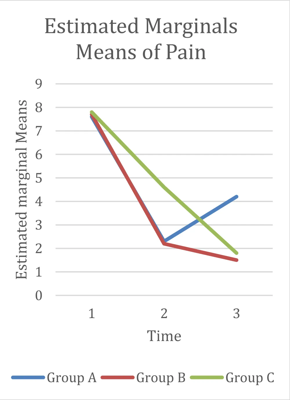
It should be noted that the pairwise between-group comparisons at T2 did not reach the pre-specified Bonferroni-adjusted significance threshold (p < 0.0083). The numerical advantage of Group B over Group C at T2 (−0.200 points) is therefore small and not statistically significant, and the observed pattern should be regarded as hypothesis-generating rather than as evidence of a confirmed additional benefit of combined LLLT + splint therapy. The significant quadratic Time × Group interaction (p < .001) indicates that the shape of the pain trajectory differed between groups, but this does not equate to a statistically significant between-group difference at T2.
In summary, the data show a statistically significant effect of time on pain in all groups, and a statistically significant quadratic Time × Group interaction indicating different trajectory shapes. However, the pairwise between-group differences at T2 do not reach the pre-specified Bonferroni-adjusted significance threshold, and the observed numerical advantage of Group B over Group C at T2 is small and not statistically significant.
Figure 1 shows the trend of pain in the three groups. The graph thus shows that:
- Group A: exhibits a rapid reduction of pain symptoms between 1 (T0) and 2 (T1). Between 2 and 3 (T2), however, there is a worsening of pain symptoms.
- Group B: shows a reduction of pain symptoms comparable to group A between 1 and 2, followed by an improvement of pain symptoms between 2 and 3.
- Group C: demonstrates an almost uniform reduction of pain symptoms between 1 and 3.
In all three groups, there is an evident improvement in pain symptoms; however, the differences between these groups are not statistically significant. It should be emphasized that the values plotted in Figure 1 are model-estimated marginal means derived from the repeated measures ANOVA, not raw observed means. The observed means at each time point are reported in the supplementary data table, and any discrepancy between observed and estimated values is attributable to the model’s adjustment for within-subject correlation.
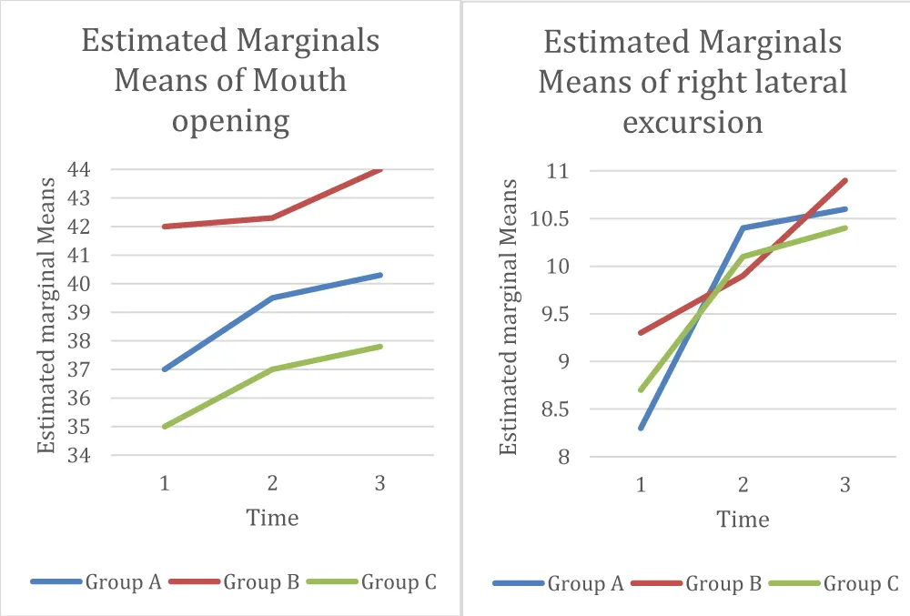
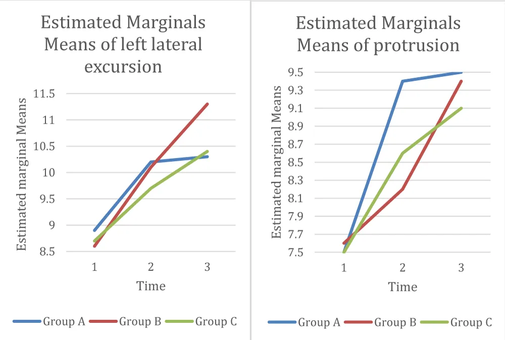
Regarding mandibular kinematics, the statistical analysis is visible in Table 4: the significance is close to 0.000, and this indicates an improvement in mandibular mobility and mandibular function in all groups. It is also true that in the “Time * GROUP” row there is a significance greater than the cutoff of 0.05, which translates into an absence of difference between the groups on this aspect. It follows that there is a statistically similar improvement in the three groups; Figure 2 shows opening and right laterality and Figure 3 shows left laterality and protrusion. As with Figure 1, the values presented in Figures 2 and 3 are model-estimated marginal means; observed values are provided in the supplementary data table.
The difference between the CPI of the various groups is not significant (the p-value is close to 0.05, but still higher), as is that of the Interference Score. The trend of the means of the collected data, both for the CPI and for the Interference Score, follows the trend of the pain means, demonstrating that there has been a general improvement in the grade of the pathology in all three groups. There was only one patient, belonging to group A, who experienced no improvement from time T0 to time T2. Similarly to the CPI and the Interference Score, for disability points, no statistically significant difference was found between the three groups: in each of the three groups, three subjects did not have an improvement in disability points between T0 and T2 even though they underwent therapy, regardless of whether the subject belonged to the group undergoing LLLT, splint + LLLT, or splint + placebo. No patient reported the use of rescue medication or the initiation of additional TMD treatments during the six-month follow-up period.
Discussion
The results of the present study demonstrate that all three therapeutic modalities improved pain symptoms and mandibular kinematics in patients with arthritic or osteoarthritic TMD. However, the critical finding lies not in short-term efficacy but in the divergent long-term trajectories observed across groups, which may suggest the mechanistic synergy between photobiomodulation and occlusal stabilization (a concept we term occluso-photobiomodulation synergy). It must be stressed, however, that the between-group differences at T2 did not reach the pre-specified adjusted significance level; the following interpretation is therefore hypothesis-generating and requires confirmation in adequately powered studies. These data align with a growing body of literature supporting the efficacy of photobiomodulation and occlusal splints in TMD management [16]. The pain reduction observed in Group A confirms LLLT’s role as a modulator of inflammatory processes, consistent with preclinical studies demonstrating reduced pro-inflammatory cytokines and inhibition of matrix metalloproteinases. Recent randomized controlled trials have further confirmed the effectiveness of photobiomodulation in reducing pain in TMD patients [17], while prospective studies have demonstrated its efficacy to be comparable to other therapeutic modalities such as therapeutic ultrasound [18]. A systematic review has highlighted differential effectiveness of photobiomodulation in muscular versus articular TMD, supporting the need for targeted therapeutic approaches [19]. The efficacy of the splint in group C is also well documented, with the Michigan splint acting as an occlusal stabilizer that reduces biomechanical load on the joint [20]. The most relevant finding is the divergent symptom trend at six-month follow-up: group B (LLLT + splint) was the only one showing maintained or further improved pain symptoms at T2, while group A exhibited partial recurrence. This may suggest a synergistic effect: LLLT acts on the acute inflammatory component, whereas the splint provides continuous biomechanical protection, preventing microtrauma that reactivates inflammation and degeneration. However, because the direct comparison between Group B and Group C did not reach statistical significance, the possibility that the observed difference is due to chance or to the small sample size cannot be excluded. A formal claim of synergy would require a statistically significant interaction and a larger sample. This combination might be particularly valuable in osteoarthritic patients, in whom the degenerative component cannot be resolved but can be stabilized. The literature emphasizes that LLLT efficacy is dose-dependent, and the systematic review by Díaz et al. [21] confirms that optimal protocols use 810-940 nm wavelengths and energy densities of 3-12 J/cm2, thereby validating our methodological choice. However, recurrence in group A suggests that even seven sessions of LLLT alone may not guarantee long-term stability without mechanical support. Regarding comparison with other therapies, the 2025 review suggests LLLT shows results comparable or superior to NSAIDs and TENS, with an excellent safety profile. While some studies show no significant differences between LLLT and occlusal splints, others demonstrate LLLT is as effective in improving mandibular movement [22]. Our study raises the possibility that the question is not which therapy is superior, but that their combination may provide superior medium-term outcomes. We refrain from claiming prevention of degenerative progression, as this was not assessed by instrumental follow-up. Study limitations include the sample size of 40 patients, insufficient for subgroup analyses, and the six-month follow-up, adequate for short-term stability but not for evaluating effects on degenerative disease progression. The integration of instrumental follow-up (e.g., T2 MRI) or salivary biomarkers could have further objectified the biological benefits of combined therapy. The researchers are currently expanding the sample and prolonging observation times to consolidate the role of photobiomodulation as a first-line therapy in arthrogenous TMD, ideally in association with occlusal therapy.
Conclusion
In conclusion, this study demonstrates that LLLT effectively reduces pain and improves mandibular function in arthritic and osteoarthritic TMD. The combination of LLLT with a Michigan occlusal splint showed a numerically more favorable pain trajectory at six months compared to LLLT alone; however, this difference did not reach statistical significance, and the finding should therefore be considered preliminary and hypothesis-generating. These findings support further investigation of a paradigm shift in TMD management: from isolated symptom relief toward integrated mechano-biological stabilization. The association of photobiomodulation with occlusal rebalancing may not only achieve short-term improvement but also potentially ensure durable results, addressing the chronic, recurrent nature of degenerative TMJ conditions. Given its non-invasive nature and excellent safety profile, LLLT represents an ideal adjunct to occlusal therapy, offering patients sustained benefit beyond the active treatment phase. Future studies, based on larger samples, long-term follow-ups, and standardized laser protocols, will be necessary to define evidence-based clinical guidelines and consolidate the role of photobiomodulation as a first-choice treatment in TMD.
Acknowledgments
We would like to extend our sincere gratitude to the University of Milan and San Paolo Hospital for the support and collaboration.
Authors contributions
AM and AS have contributed to the analysis and interpretation of the statistical data and the conception of the manuscript; PA, SB, CB and GN have given substantial contributions to the acquisition of the data. All authors have contributed to the drafting of the manuscript, and they have read and approved the final version of the manuscript.
Ethical policy and institutional review board statement
The study was carried out following the Declaration of Helsinki on Human Clinical Research. The study protocol was submitted to the Ethics Committee of the University of Milan. The Committee determined that formal approval was not required because the clinical procedures employed were not experimental in nature and fell within routine clinical practice. Recruitment took place between November 2024 and March 2025. The study was not registered in a public trial registry, as it was classified as a non-experimental clinical audit. All participants received detailed oral and written information about the study procedures, the use of their clinical data, and their right to withdraw at any time, and provided written informed consent before enrollment. The consent form explicitly included permission to use and publish anonymized data from their medical records. No participant was included without documented consent.
Patient declaration of consent
All participants provided written informed consent prior to enrollment, including explicit consent for the publication of anonymized data derived from their medical records.
Data availability statement
The datasets generated and analyzed during the current study are available from the corresponding author on reasonable request.
- Mester E, Szende B, Gartner P. The effect of laser beams on the growth of hair in mice, Radiobiol Radiother (Berl) 9 1968: 621-6.
- Boaviagem A, Nóbrega Júnior JC, da Silva RCB, Lemos A. Effectiveness of photobiomodulation for pain management and wound healing in the immediate postoperative period after cesarean section: A systematic review. Complement Ther Med. 2026 Jul 15;101:103403. Available from: https://dx.doi.org/10.1016/j.ctim.2026.103403.
- Hashim PK, Shaji AT, Amrutha AS, Ahmad S. Conceptual expansion of photomedicine for spatiotemporal treatment methods. RSC Med Chem. 2025 Mar 11;16(6):2360-2372. Available from: https://dx.doi.org/10.1039/d4md01005a.
- Da Silva D, Crous A, Abrahamse H. Enhancing osteogenic differentiation in adipose-derived mesenchymal stem cells with near-infrared and Green Photobiomodulation. Regen Ther. 2023 Nov 10;24:602-616. Available from: https://dx.doi.org/10.1016/j.reth.2023.11.003.
- Zamani ARN, Saberianpour S, Geranmayeh MH, Bani F, Haghighi L, Rahbarghazi R. Modulatory effect of photobiomodulation on stem cell epigenetic memory: A highlight on differentiation capacity. Lasers Med. Sci. 2019;35:299–306. Available from: https://dx.doi.org/10.1007/s10103-019-02873-7.
- Garzón J, Baldion PA, Grajales M, Escobar LM. Response of osteoblastic cells to low-level laser treatment: a systematic review. Lasers Med Sci. 2022 Oct;37(8):3031-3049. Available from: https://dx.doi.org/10.1007/s10103-022-03587-z.
- Dos Santos Soares F, de Souza Pinto M, Kruger A, Coracini CA, Bertolini GRF. Photobiomodulation therapy on skeletal muscles exposed to diabetes mellitus: a systematic review of animal studies. Lasers Med Sci. 2023 Aug 14;38(1):185. Available from: https://dx.doi.org/10.1007/s10103-023-03853-8.
- Garzón J, Baldion PA, Grajales M, Escobar LM. Response of osteoblastic cells to low-level laser treatment: a systematic review. Lasers Med Sci. 2022 Oct;37(8):3031-3049. Available from: https://dx.doi.org/10.1007/s10103-022-03587-z.
- Dragojević IM, Kisić B, Mirić D, Ilić A, Todić JT, Kostić M, et al. Oxidative Stress Biomarkers in Oral Mucosal Wound Healing and Photobiomodulation: Biochemical Pathways, Experimental Models, and Translational Perspectives. Int J Mol Sci. 2026 Jun 26;27(13):5763. Available from: https://dx.doi.org/10.3390/ijms27135763.
- Hanna R, Dalvi S, Benedicenti S, Amaroli A, Saălaăgean T, Pop ID, et al. Photobiomodulation Therapy in Oral Mucositis and Potentially Malignant Oral Lesions: A Therapy Towards the Future. Cancers. 2020;12:1949. Available from: https://dx.doi.org/10.3390/cancers12071949.
- Shivappa P, Basha S, Biswas S, Prabhu V, Prabhu SS, Pai AR, et al. From light to healing: photobiomodulation therapy in medical disciplines. J Transl Med. 2025 Dec 29;23(1):1430. Available from: https://dx.doi.org/10.1186/s12967-025-07466-3.
- Wade-Vallance A, Ng M, Kuo K, Luo C, Adams S, Dhaliwal G, et al. A central role for reactive oxygen species (ROS) in the pathogenesis of temporomandibular joint disorders: All roads lead to ROS. Catal. Facet. Biochem. Biomed. Sci. 2017;2:1–7.
- Eslami H, Katebi K, Ghaffaripour Saleh S, Mirizadeh L, Hashemi M. The relationship between oxidative stress markers and temporomandibular disorders: A systematic review and meta-analysis. J Res Med Sci. 2024 Jul 12;29:33. Available from: https://dx.doi.org/10.4103/jrms.jrms_660_23.
- Farshidfar N, Farzinnia G, Samiraninezhad N, Assar S, Firoozi P, Rezazadeh F, et al. The effect of photobiomodulation on temporomandibular pain and functions in patients with temporomandibular disorders: an updated systematic review of the current randomized controlled trials. J Lasers Med Sci. 2023;14:e24. Available from: https://dx.doi.org/10.34172/jlms.2023.24.
- Rady NA, Bahgat MM, Abdel-Hamid AM. Promising minimally invasive treatment modalities for symptomatic temporomandibular joint disc displacement with reduction: a randomized controlled clinical trial. BMC Oral Health. 2022 Dec 1;22(1):547. Available from: https://dx.doi.org/10.1186/s12903-022-02579-3.
- Ahmad SA, Hasan S, Saeed S, Khan A, Khan M. Low-level laser therapy in temporomandibular joint disorders: a systematic review. J Med Life. 2021 Mar-Apr;14(2):148-164. Available from: https://dx.doi.org/10.25122/jml-2020-0169.
- Trindade LP, Urruty MJ, Bruno LH, Todeschini F, Moya LEP, Lestido V, et al. LED Photobiomodulation for Pain Reduction in Temporomandibular Disorder: A Randomized, Controlled, and Blinded Clinical Trial. J Biophotonics. 2026 Mar;19(3):e70243. Available from: https://dx.doi.org/10.1002/jbio.70243.
- Budakoti A, Misra D, Vyas T, Vihan A, Singh R, Adarshi N. Efficacy of Photobiomodulation Therapy and Therapeutic Ultrasound in Temporomandibular Joint Disorder Patients: A Prospective Study. Ann Maxillofac Surg. 2025 Jul-Dec;15(2):175-179. Available from: https://dx.doi.org/10.4103/ams.ams_61_25.
- Candido-do-Prado LG, Ribeiro-Silva VHA, Simões-Barbosa AF, Mazzi-Chaves JF, Magri LV. Differential effectiveness of photobiomodulation in muscular and articular temporomandibular disorders: a systematic review and critical appraisal. Lasers Med Sci. 2025 Nov 18;40(1):487. Available from: https://dx.doi.org/10.1007/s10103-025-04751-x.
- Maracci LM, Stasiak G, de Oliveira Chami V, Franciscatto GJ, Milanesi J, Figueiró C, et al. Treatment of myofascial pain with a rapid laser therapy protocol compared to occlusal splint: A double-blind, randomized clinical trial. Cranio. 2022 Sep;40(5):433-439. Available from: https://dx.doi.org/10.1080/08869634.2020.1773661.
- Díaz L, Restelli L, Valencia E, Atalay DI, Abarca JM, Gil AC, et al. Effectiveness of low-level laser therapy on temporomandibular disorders. A systematic review of randomized clinical trials. Photodiagnosis Photodyn Ther. 2025 Jun;53:104558. Available from: https://dx.doi.org/10.1016/j.pdpdt.2025.104558.
- Shukla S, Srivastava A, Batra S, Pal S, Darjee S, Shekhar A. Adjunctive 810 nm photobiomodulation with pharmacotherapy for trigeminal neuralgia: A randomized controlled trial in a tertiary care centre. J Photochem Photobiol B. 2025 Nov;272:113249. Available from: https://dx.doi.org/10.1016/j.jphotobiol.2025.113249.


 Save to Mendeley
Article Alerts
Subscribe to our articles alerts and stay tuned.
 This work is licensed under a Creative Commons Attribution 4.0 International License.