International Journal of Oral and Craniofacial Science
Can human amniotic membrane be used as an Ideal Suture or Filling Material? An Experimental study
Fatma Bilgen1*, Alper Ural1, M Nuri Karatoprak2 and Mehmet Bekerecioğlu1
2MNK Aesthetic Clinic, Turkey
Cite this as
Bilgen F, Ural A, Karatoprak MN, Bekerecioglu M (2019) Can human amniotic membrane be used as an Ideal Suture or Filling Material? An Experimental study. Int J Oral Craniofac Sci 5(1): 005-009. DOI: 10.17352/2455-4634.000037Background: The use of the patient’s own tissues in surgery increases the morbidity rate, whereas the use of injected or implanted materials can cause allergic reactions in the body mostly via immunogenic pathways. In addition, absorbable suture materials and some fillers used in almost all surgical applications can cause reactions in the body.
Objective: The use of human amniotic membrane (HAM) as a suture and/or filling material has been successfully investigated in many areas to solve or minimize these problems.
Material and Method: In this study, 40 rats were divided into four groups (10 rats per group). The HAM was removed with the surrounding tissue at the first week from group 1, the second week from group 2, the first month from group 3, and the third month from group 4, and underwent clinical and histopathological examination.
Results: When the rats that had HAM placed under the skin and intramuscle were evaluated statistically using a points-based scoring system, there was a statistically significant difference among all groups (p<0.001).
Conclusion: In our study, we found that HAM progressively biodegraded by macrophages within 3 months could be used as an absorbable suture material; therefore, its use as a filling material was limited over time. In addition, it is also likely to be used as an absorbable suture material, as it can accelerate wound healing due to the growth factors secreted. However, further studies are needed to prevent adhesions of factors isolated from amniotic fluid or the HAM and to investigate its use in wound healing.
Introduction
Correction of congenital or acquired tissue defects and scars has been one of the most controversial issues since the beginning of plastic surgery. Despite advances in reconstructive and aesthetic surgery, the prevention of aging signs and the elimination of tissue defects continue to be major issues. Due to age, smoking, sun exposure, and gravity, collagen in the skin decreases and wrinkles become apparent. The history of soft tissue fillings applied to correct these wrinkles is based on the first use of Neuber’s fat grafts in the face in the 1800s [1,2]. Since the use of the first filling material, the search for the ideal material is ongoing.
Suture materials are one of the fundamental issues that concern every surgeon. The main goals of the use of suture are to complete the natural wound-healing process and to ensure that the wound edges are properly conjoined until the old tension is restored. Suture materials are foreign bodies implanted into the tissues and are likely to cause foreign body reactions[2].
In the literature, it has been shown that human amniotic membrane (HAM) has several functions: it accelerates epithelization [2], prevents protein and fluid loss of the wound surface [3], reduces adhesion formation [4], has antibacterial and non-immunological effects [5], contributes to collagen synthesis by increasing the fibroblastic activity [6], and reduces angiogenesis, scar formation, pain and inflammation [7]. However, there is no study in the literature using HAM as a suture and/or filling material.
Based on these properties of the amnion membrane, in the present study we aimed to evaluate the usability of HAM as ideal suture and/or filling material considering the fact that it does not contain tissue antigens.
Materials and Methods
This study protocol was approved by the (03.2010-24) Ethics Committee. The study was prepared in accordance with Animals in Research: Reporting in vivo Experiments (ARRIVE) guidelines
Preparation
The study was conducted on 40 male Wistar species, male albino rats (weighing 250-350 g and aged 105-120 d). The rats were randomly selected and divided into four groups of 10 rats in each group.
Surgical technique
All rats were anesthetized with 30 mg/kg intramuscular ketamine hydrochloride (Ketalar®, Eczacıbası, Turkey) and 10 mg/kg intramuscular xylazine hydrochloride (Rompun®, Bayer, Turkey).
After receiving the consent forms, the HAMs were obtained following cesarean operations, when the patient was known to have been previously seronegative for infection. The blood was removed by washing with sterile 0.9% NaCl solution and washing with 0.025% sodium hypochlorite for sterilization. Strips 2 × 1 in size of HAM were prepared as 500,000 to 2,000,000 IU/L as described in the literature. Penicillin and 0.5 to 2 g streptomycin were used after soaking in sterile saline for 12 to 24 hours at +4°C.
After surgical site shaving, povidone-iodine (Betadin®, Merkez İlaç, Turkey) was used for sterilization. The HAM was placed into the pouch opening in the left lumbar area and into the right quadriceps femoris muscle and fixed with 5/0 non-absorbable suture to prevent prolapsing when placed in the muscle. Skin suturing was done with 5/0 non-absorbable sutures. The laboratory temperature was set between 25°C and 30°C. The wound location of each rat was dressed daily with povidone-iodine (Betadine®). All groups received a single dose of subcutaneous ampicillin 150 mg/kg daily for 5 days for infection prophylaxis.
The HAM was removed with the surrounding tissue at the first week from group 1, the second week from group 2, the first month from group 3, and the third month from group 4, and underwent clinical and histopathological examination.
Measurements
In the clinical and histopathological examinations, muscle and subcutaneous tissue samples were initially taken from all groups. These tissue samples were then fixed in 10% formalin and embedded in paraffin blocks. The sections were stained with hematoxylin and eosin (H&E) and examined under the light microscope. In the pathological examination of the tissue specimens, parameters such as polymorphonuclear neutrophil leukocytes (PMNL), lymphocytes, fibroblast proliferation, eosinophil density, and foreign body granulation tissue were examined as was the presence of HAM and indicators of an inflammatory response in the specimens. The pathological degree of inflammation was scored for the numerical transfer of the means of the data (Table 1).
Statistical analysis
Statistical analysis was performed using the Statistical Package for Social Sciences (SPSS) version 15.0 software. Histopathological scores obtained from each specimen included in the study were statistically analyzed using the chi-square test for baseline variables among the groups. The chi-square test was used to analyze statistically significant differences between first- and second-week and first- and third-month values of intramuscular and subcutaneous applications. A p value of < 0.001 was considered statistically significant in all groups.
Results
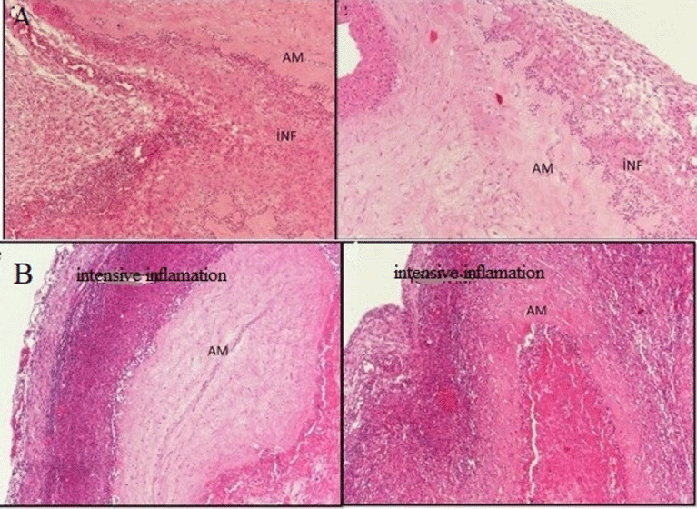
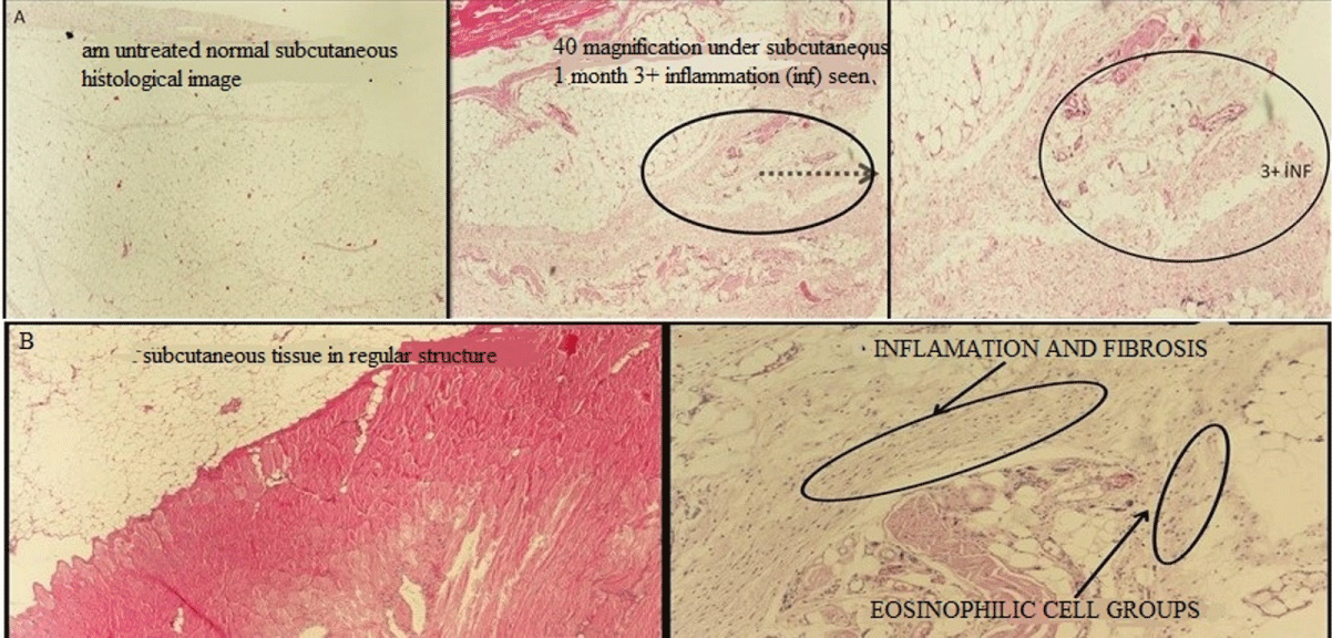
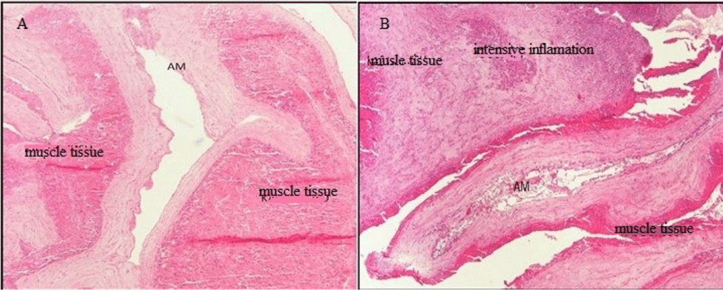
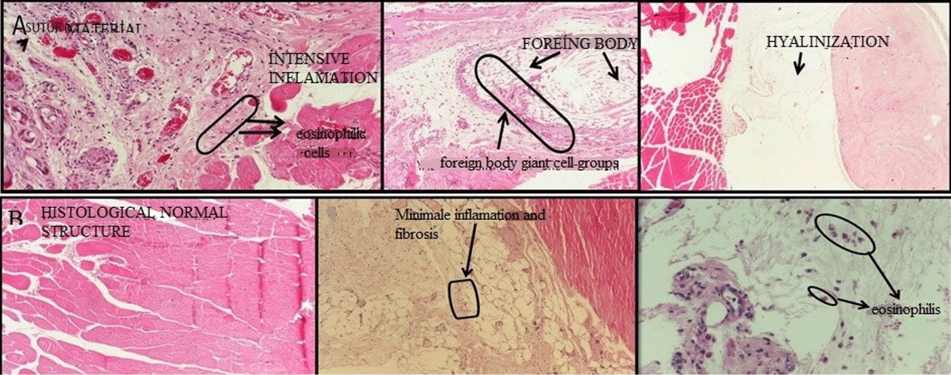
Following surgical interventions, the adhesions in the tissues gradually decreased and the fibrotic adhesions at the first and second week gradually decreased toward the first month and were nearly undetectable at the third month. The HAM placed under the skin and into the muscle in the first week could be obviously selected, whereas it was still in sight macroscopically in the second week. During the first and third months following surgery, however, the HAM was unable to be visualized even at the microscopic level.
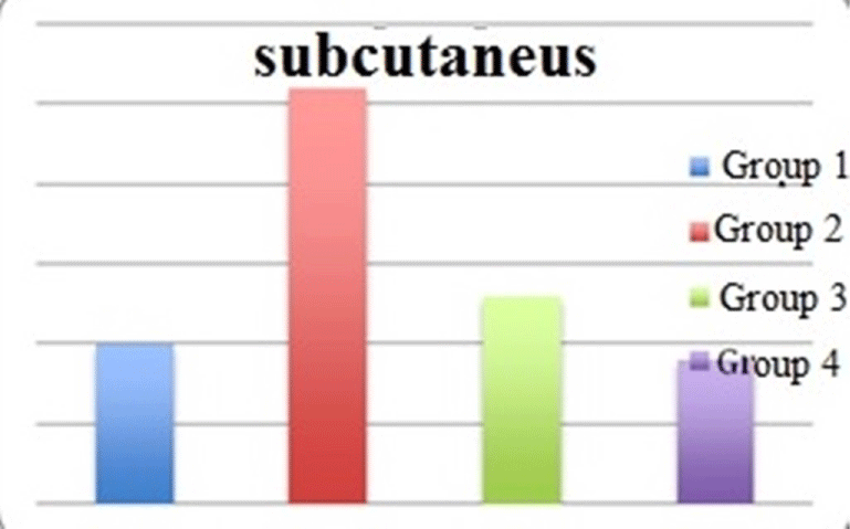
When the rats in which the HAM was placed under the skin were evaluated statistically, there was a statistically significant difference among all groups (p<0.001). The reaction against the HAM increased in the first week, reaching the maximum values in the second week, although it progressively reached values close to normal at the end of the third month (Figures 1,2, Graphic 1).
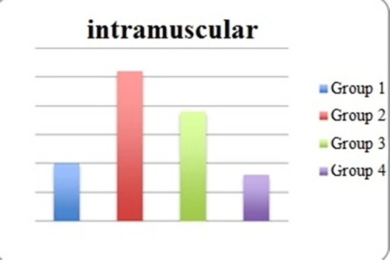
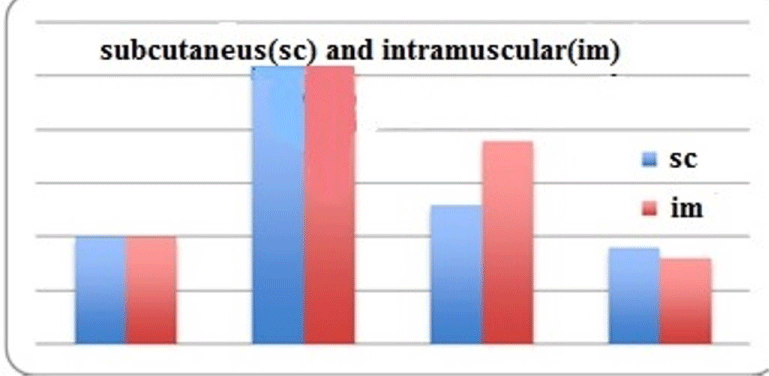
When the HAM was placed into the muscle and the scores of the rats were statistically analyzed, there was a statistically significant difference among all groups (p<0.001). The reaction to the HAM increased in the first week, reaching the maximum values in the second week, although it progressively reached values close to normal at the end of the third month. This statistically significant difference was related to the reduced reaction after the first month (Figures 3,4, Table 2, Graphics 2,3).
Discussion
Currently, the allergic potentials of the suture materials and all organic or inorganic filling materials and the body’s reactions against these materials are not yet fully understood. Owing to technological developments, materials that are minimally allergic are preferred with an aim at improved results with minimal morbidity for the patient [1,6,7].
HAM contains many growth factors such as epidermal growth factor (EGF), keratocyte growth factor (KGF), hepatocyte growth factor (HGF), transforming growth factor (TGF), steroid hormones (estrogen, progesterone), hydrolytic enzymes, oxidation-reduction enzymes, secondary enzymes, and is called the “living enzyme museum” [8-10].
HAM was first used by Davis in 1910 as a graft material in the closure of skin defects [11-14]. In the following years, it was used successfully in several cases as follows: the repair of symblepharon, the treatment of acute ocular burns, for accelerating wound healing, the repair of abdominal defects, unhealed skin ulcers, urinary bladder reconstruction, artificial vaginal reconstruction, the prevention of abdominal chirurgical adhesions, the treatment of corneal surface defects and ulcers, repair of urethral defects, myelomeningoceles repair in vestibuloplasty cases, prevention of tendon adhesions, peripheral nerve anastomosis, and complicated scleritis [12-15].
HAM, a rapidly growing biological cover with increasing value, is an easy and convenient way to accelerate epithelialization, to prevent protein and fluid loss from the wound surface, to reduce adhesion formation, to have antibacterial and non-immunological effects, to contribute to collagen synthesis by increasing fibroblastic activity, and to reduce angiogenesis, scar formation, pain, and inflammation [3,4,16].
The mechanisms of HAM are as follows: acting as a new substrate suitable for epithelization, acceleration of epithelial cell migration, reinforcement of adhesion of basal epithelial cells, inducing epithelial differentiation, prevention of epithelial apoptosis, blocking tissue metalloprotease inhibitors and tissue destruction, increasing fibroblastic activity and contributing to collagen synthesis, and accelerating wound healing through many growth factors and some enzymes [16-19].
HAM fills the wound floor as mass in the area where it is applied and prevents loss of protein and fluid from the wound surface. Morever, it reduces pain by closing the open nerve endings. Due to non-allergic and non-immunologic features, graft excretion is not a concern. In addition, it has a reducing effect on wound contracture for moderate defects [17-19]. Epithelial cells in the stromal matrix of HAM exhibit anti-inflammatory effects by depressing interleukin-1 α and β, which play a role in inflammation by transforming lipopolysaccharides. Again, due to the presence of progesterone and other factors, it shows bacteriostatic effect against gram-positive microorganisms [18,19].
It was reported that HAM acquires the feature of the body region in which it is applied over time; thus, a new surgical procedure is not required [18,19]. Studies have shown that in the tendon incision model, HAM completely disappeared over time and that it transformed into connective tissue composed of cells originating from the epitenon [19,20]. In an effort to identify the incidence and characterization of infection after HAM transplantation, Marangon [20], suggested that infection occurred in 3.4% of 326 (11 cases) transplantations. In addition, the most isolated microorganisms in cultures were gram-positive ones.
In the present study, we found that the highest level of inflammatory reactions in the early postoperative period, particularly in the second week, may be due to inflammatory response after surgical trauma, due to the absence of Human Leucoyte Antigene in HAM. The fact that the statistically significant difference in this study can be attributed to the reduced reaction after the first month and to the fact that that these reactions completely disappeared in the third month also showed once again that HAM had no allergenic potential.
In conclusion, in our study, we found that HAM progressively underwent biodegradation by macrophages over a period of 3 months. Therefore, it was concluded that the use of HAM as a filling material was limited over time. However, it can be used as an absorbable suture material, as it accelerates wound healing due to the growth factors secreted and does not have allergic potential. Furthermore, to prevent adhesions of the factors to be isolated from the amniotic fluid or HAM and to enhance their potential for the use in wound healing, further immunohistochemical studies are required.
All of the authors declare that they have all participated in the design, execution, and analysis of the paper, and that they have approved the final version. Additionally, there are no conflicts of interest in connection with this paper, and the material described is not under publication or consideration for publication elsewhere.
- Thorne CH (2010) Grabb and Smith’s Plastic Surgery. 6. Baskı. Güneş Kitapevler, No 7/2. Esentepe, İstanbul 468-474.
- Mostaque AK, Rahman KB (2011) Comparisons of the effects of biological membrane (amnion) and silver sulfadiazine in the management of burn wounds in children. J Burn Care Res 32: 200–209. Link: https://tinyurl.com/y5cw7hrt
- Eskandarlou M, Azimi M, Rabiee S, Seif Rabiee MA (2016) The healing effect of amniotic membrane in burn patients. World J Plast Surg 5: 39-44. Link: https://tinyurl.com/y5g8wfyk
- Rouher N, Pilon F, Dalens H, FauquertJl, KemenyJl, et al. (2004) Implantation of preserved human amniotic membrane for the treatment of shield ulcers and persistent corneal epithelial defects in chronic allergic keratoconjunctivitis. J Fr Ophthalmol 27: 1091-1097. Link: https://tinyurl.com/y3x29r3l
- Mamede AC, Carvalho MJ, Abrantes AM, Laranjo M, Maia CJ, et al. (2012) Amniotic membrane: from structure and functions to clinical applications. Cell Tissue Res 349: 447-458. Link: https://tinyurl.com/y4mol24k
- Lo V, Pope E (2009) Amniotic membrane use in dermatology. Int J Dermatol 48: 935-940. Link: https://tinyurl.com/y4xwmcrx
- Sippel KC, Ma JJ, Foster CS (2001) Amniotic membrane surgery. Curr Opin Ophtalmol 12: 269-281. Link: https://tinyurl.com/yxvmbdum
- Koob TJ, Rennert R, Zabek N, Massee M, Lim JJ, et al. (2013) Biological properties of dehydrated human amnion/chorion composite graft: implications for chronic wound healing. Int Wound J 10: 493–500. Link: https://tinyurl.com/yxvowvzp
- Yazdanpanah G, Paeini-Vayghan G, Asadi S, Niknejad H (2015) The effects of cryopreservation on angiogenesis modulation activity of human amniotic membrane. Cryobiology 71: 413-418. Link: https://tinyurl.com/y4modpds
- Mc Carthy JG, May JW, Littler JW (1990) General Principles and Implantation materials. In: Mc Carthy’s Plastic Surgery. W.B. Saunders Company Phiedelphia 186-207.
- Tosi GM, Massaro-Giordano M, Caporossi A, Toti P (2005) Amniotic membrane transplantation in ocular surface disorders. J Cellular Physiology 202: 849-851. Link: https://tinyurl.com/yxs52g86
- Hanada K, Nishikawa N, Miyokawa N, Yoshida A (2017) Long-term outcome of amniotic membrane transplantation combined with mitomycin C for conjunctival reconstruction after ocular surface squamous neoplasia excision. Int Ophthalmol 37: 71-78. Link: https://tinyurl.com/y6pgmz64
- Klama-Baryła A, Łabuś W, Kitala D, Kraut M, Nowak M, et al. (2018) Experience in Using Fetal Membranes: The Present and New Perspectives. Transplant Proc 50: 2188-2194. Link: https://tinyurl.com/y25s63yk
- Singh R, Chouhan US, Purohit S, Gupta P, Kumar P, et al. (2004) Radiation processed amniotic membranes in treatment of non-healing ulcers of different etiologies. Cell Tiss Bank 5: 129-134. Link: https://tinyurl.com/y2o7oahd
- Taylan Sekeroglu H, Erdem E, Dogan NC, Yagmur M, Ersoz R, et al. (2011) Sutureless amniotic membrane transplantation combined with narrow-strip conjunctival autograft for pterygium. Int Ophthalmol 31: 433-438. Link: https://tinyurl.com/y589t455
- John A, Oommen J (2010) Use of amniotic membrane in dermatology. Indian J Dermatol Venereol Leprol 76: 196-197. Link: https://tinyurl.com/y598nhr8
- Dorazehi F, Nabiuni M, Jalali H (2018) Potential Use of Amniotic Membrane-Derived Scaffold for Cerebrospinal Fluid Applications. Int J Mol Cell Med 7: 91-101. Link: https://tinyurl.com/y3qpbyzd
- Maan ZN, Rennert RC, Koob TJ, Januszyk M, Li WW, et al. (2014) Cell recruitment by amnion chorion grafts promotes neovascularization. J Surg Res 193: 953-962. Link: https://tinyurl.com/y6ntqav4
- Ozgenel GY, Samli B, Ozcan M (2001) Effects of human amniotic fluid on peritendinous adhesion formation and tendon healing after flexor tendon surgery in rabbits. J Hand Surg 26: 332-339. Link: https://tinyurl.com/y2spatob
- Marangon FB, Alfonso EC, Miller D, Remonda NM, Muallem MS, et al. (2004) Incidence of microbial infection after amniotic membrane transplantation. Cornea 23: 264-269. Link: https://tinyurl.com/y2k4khfj
Article Alerts
Subscribe to our articles alerts and stay tuned.
 This work is licensed under a Creative Commons Attribution 4.0 International License.
 Save to Mendeley
