International Journal of Oral and Craniofacial Science
Prolotherapy with 12.5% dextrose to treat temporomandibular joint dysfunction (TMD)
Ehab Shehata1,2*
2Assistant Professor, Oral and Maxillofacial Surgery Department, College of Dentistry, University of Kentucky, USA
Albert B. Chandler Hospital, D542, Lexington, Kentucky 40536-0297, Tel: +1 859 489 4803, +1 859 323 8520; Fax: +1 859 323 3679; Email: [email protected]
Associate Professor, Maxillofacial and Plastic Surgery Department, Faculty of Dentistry, University of Alexandria, Egypt
Cite this as
Shehata E (2019) Prolotherapy with 12.5% dextrose to treat temporomandibular joint dysfunction (TMD). Int J Oral Craniofac Sci 5(1): 015-019. DOI: 10.17352/2455-4634.000039Introduction: Temporomandibular joint dysfunction (TMD) is a collective term used to describe a complex and multifactorial disorders of the orofacial region. Symptoms commonly associated with TMD include TMJ pain, limited mandibular movement or locking and painful clicking or popping sounds. Most of patients diagnosed with TMD are initially treated conservatively. Failure of such conservatism poses a great challenge for the treating physician. Prolotherapy has been used successfully in many joints in the body by orthopedics and spinal surgeons. Injection prolotherapy has also been used in management of weakening tendons or ligaments in head and neck.
Aim of the study: To evaluate the efficacy of dextrose prolotherapy with 12.5% concentration in the treatment of intractable temporomandibular joint dysfunction.
Material & Methods: A prospective clinical study with 33 patients with the diagnosis of TMD were included in this study during the period from Jan. 2012 to Jan.2015. Inclusion criteria were; adult patients above 18 years old, with TMD symptoms for more than 6 months, had failed conservative treatment and have no pathological findings on dynamic magnetic resonance imaging. Prolotherapy was achieved by six sessions of 12.5% dextrose (3ml) injection per joint with one month interval. A Semi-structured questionnaire developed by the author was utilized to gather all demographic data. All variables were collected, tabulated and analyzed. Follow up appointments were booked after the last session, every 3 months for up to 24 month.
Results: I used maximum mouth opening, clicking and visual analogue pain score to judge the efficacy of my protocol. There was statistical significant improvement of all previous variables outcomes.
Conclusion: Dextrose prolotherapy can be used in patients diagnosed with TMD whom had failure of conservative treatment to control their symptoms. More than four prolotherapy sessions is not recommended as per results of this study. It is safe and efficient technique.
Introduction
Temporomandibular joint disorder, commonly known as TMJD or TMD, was defined by the American Dental Association (ADA) as “A group of orofacial disorders characterized by pain in preauricular region, TMJ or muscles of mastication, limitation or deviation of mandibular range of motion, TMJ sounds during mandibular function” [1].
National institute of Dental and Craniofacial research has reported an incidence of 10.8 million people in USA at any given time [2]. TMJD occurs predominantly in women with the female to male ratio ranging from 2:1 to 6:1 with 90% of those seeking treatment being women in their child-bearing years [3,4].
Symptoms commonly associated with TMD include pain localized to the joint, generalized oro-facial pain, chronic headache and jaw dysfunction which includes hyper and hypo-mobility, limited movement or locking of the jaw, painful clicking and or popping sounds [5]. Additional symptoms may include otalgia, decreased hearing, dizziness and visual problems [6].
Causes of TMD are often unclear and are usually considered to be multifactorial. TMJ capsular damage or inflammation and muscle pain / spasm can be caused by malocclusion, para-functional habits, stress, anxiety or abnormalities of the intra-articular disc (meniscus) [7]. It can be due to hormonal or psychological problems [8,9]. The common way to manage patients experiencing TMD but without pathological findings is with a conservative (non-surgical) approach. This includes diet modifications, medications, occlusal appliances and physical therapy. Medications used could be non-steroidal anti-inflammatories, steroids, muscle relaxants, antidepressant or anxiolytics. A relatively new technique directed at those with a disordered TMJ system is prolotherapy.
Prolotherapy means rehabilitation of an incompetent structure such as a ligament or tendon by induction of cellular proliferation. Prolo comes from the word proliferate. Prolotherapy injections stimulate growth of new, normal ligament and tendon tissues by stimulation of low grade inflammation [10]. Monocytes, granulocytes, macrophages migrate to injured tissue by prolotherapy with activation of fibroblasts to produce matrix and new collagen fibrils. The temporary cellular stress causes release of cytokines and increased growth factors activity. Unlike repair after injury, disruption of the architecture of the tissue does not occur from injury and new cells and matrix are deposited in an organized fashion, with maturation of new tissue for 6-8 weeks [11]. Different concentration of dextrose solution have been used as a therapeutic injecting material. Prolotherapy has been used successfully to treat many painful joints in the body [12].
The aim of this study was to evaluate the efficacy of injecting dextrose solution (12.5%), a prolotherapy technique, in the treatment of resistant tempro-mandibular joint dysfunction.
Material & Methods
In the period from January 2012 to January 2015, 33 consecutive patients diagnosed with TMD were treated by prolotherapy. Inclusion criteria were adult patients above 18 years old, with TMD symptoms for more than 6 months, or who had failed conservative treatment and showing no pathological findings on dynamic magnetic resonance imaging (MRI). Exclusion criteria were patients who have corn allergy (Dextrose is a corn product), previous history of open joint surgery, presence of abnormal pathological findings on MRI that need surgical intervention and patients who did not agree with the injections protocol. The study was approved by the research & ethics committee.
Patients were interviewed for detailed history and examination by the author. Their ages ranged from 23-55 years with the mean of 37.7 years. They were 25 female and 8 male. Patients who matched the inclusion criteria were scheduled for 6 prolotherapy sessions with one month intervals. All Patients in this study had basic panoramic x- rays as well as magnetic resonance image (MRI) in open and closed position (Dynamic) to assess condylar- disc unit for osseous or soft tissue pathologies. A semi-structured questionnaire was developed to gather the following variables: age, sex, duration of complain, number of previous physicians, current/ previous medications, treatment plan by other physicians, pain score (pre session and during the follow up appointments), inter-incisal opening with overtime measurement changes, clicking (0:absent 1:subjective/audible by stethoscope, 2: very audible; without stethoscope), pain medications before ,during and after prolotherapy sessions, and during the follow up period. Data were tabulated and statistically analyzed. The procedure of prolotherapy was carried out as an outpatient procedure under local anesthesia.
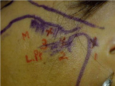
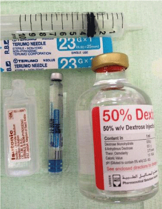
Prolotherapy technique: With some modifications to Hakala’s original technique [13], a one inch long 23-gauge needle was used (instead of the 30-gauge of Hakala). Three anatomical points are targeted (Figure 1). 12.5% dextrose (3 ml) is prepared by diluting 50% dextrose (one part: 0.75 ml) with 1% lidocaiene (two parts: 1.5 ml) and bacteriostatic water (one part: 0.75 ml) (Figure 2). Before the injection, a bite block was inserted between patient’s anterior teeth to induce forward translation of the condyle leaving a space for the needle. 1ml of the prolotherapy solution was injected into the posterior compartment aiming for the retro-discal tissue (10 mm anterior to mid-tragal point then 2 mm below) - point 1. The second point is the anterior disc attachment where it connects to the lateral pterygoid muscle. Point 2 is marked before injecting point 1. It is identified by a slight depression just anterior to the condyle when the mouth is closed. For purposes of injecting point 2, the bite block is removed and patient was asked to close her/his mouth to direct the condyle back into the fossa. The needle is inserted in a medial and slightly anterior direction. The third point of injection is identified as the most tender point of masseter muscle attachment to the inferior border of zygomatic arch. This point is determined before injecting points 1&2.
After completion of the procedure patients were instructed to eat soft diet, prescribed analgesics as required but prohibited from using any anti-inflammatory medications. The remaining 5 injections sessions were scheduled at one month intervals on an outpatient basis. Outcomes of treatment were measured at the follow up appointments every 3 months for up to 24 months.
Success was defined as absence or reduction of pain (at least 75% on visual analogue score) or absence of the need to take analgesics, improvement of maximum incisal distances and the absence or reduction of clicking. The data were analyzed to assess the efficacy of the prolotherapy technique. Quantitative data are presented as mean (SD). Wilcoxon signed rank test was used to identify significant changes in outcomes. Qualitative data are presented as numbers (%). Probabilities (P- values) of ≤ 0.05 were accepted as significant.
Results
This study involved 33 patients. A total of 62 joints (bilateral n=29 and unilateral n=4). Female to male ratio was 3:1 with a mean age of 37.7 years (range 23-55 years). Mean duration of complains was 24 months (range 8-48 m). Two third of patients (n=22) were secondarily referred for consultation after primary failure of conservative approaches. Management options offered by other physicians included no treatment option in 36% (n=12), continue conservative management in 48.5% (n=16) and surgery in 15.2% (n=5). A total of 25 patients (75%) were taking analgesic medication on referral. At subsequent radiological assessment, all the 33 patients had no soft tissue or osseous pathological abnormalities detected on MRI. The mean maximum mouth opening (MMO) for the study group was 33.7 ± 10.6 mm.
Maximum incisal opening (MIO), clicking and pain scores were used to judge the efficacy of the protocol. A Wilcoxon signed- rank test was used to identify significant changes in outcomes pre and post treatment. There was statistically border-line significant improvement in MIO (P = 0.05), while statistically significant difference in clicking (P=0.00) and pain reduction (P= 0.00) (Table 1). Using Wilcoxon signed-rank test to evaluate pain reduction during prolotherapy sessions, there was a significant pain reduction only after first four sessions (P=0.002) while became non-significant during the last two sessions (P= 0.25 & P= 0.18) table 2. Only 28 patients (84.8%) reported an improvement in the quality of life at the end of follow up period.
All patients tolerated the technique without serious complications. 3 patients developed temporary frontal branch nerve palsy; all resolved within three hours following the procedure. 2 patients did not comply with the appointment protocol and were excluded from the study.
Discussion
The exact cause of chronic TMJ dysfunction is obscure. The mechanism of dextrose prolotherapy is to improve the stability of TMJ by enhancing capsular and ligament strength [14]. Weakening of the TMJ capsule and ligaments would explain joint subluxation, disc displacement as well as muscle spasms and myofacial pain patterns. The most common cause of TMJ pain is myofacial pain dysfunction syndrome and primarily involves the muscles of mastication [15]. In general, prolotherapy agents are four general types: osmotic agents, inflammatory mimetics, chemical and physical irritants. In this study, osmotic agent was used. 12.5% dextrose produces a hypertonic extracellular environment causing lysis of the adjacent cell walls. Release of cellular proteins, inflammatory breakdown products and debris cause localized inflammation and fibrous healing.
It was the aim of this study to assess the efficiency of dextrose prolotherapy for treatment of TMD. Dextrose 12.5% was used as the active gradient in the solution as it is the most common proliferant agent in prolotherapy management. It is readily available, inexpensive and has satisfactory safety profile [16,17]. Dextrose concentration more than 10% partially works by stimulation of inflammatory reaction as do phenol and sodium morrhuate [18]. Hakala et al advocate for the use of dextrose in a concentration greater than 10%, which is enough to initiate adequate cell wall lysis to attract fibroblasts and begin the required proliferation/regeneration process [12]. The exact concentration of dextrose proliferant agent may not be critical as long as it is above 10%. Different concentration have been published in recent articles suggesting 12.5%, 15%, 25% of dextrose solution for prolotherapy management of TMD [13,16-18].
Refai, et. al., reported usage of 10% dextrose as sub-inflammatory concentration in tightening loose ligaments for treatment of TMJ hypermobility [16]. The effects of 10% dextrose concentration also showed statistically significant benefit upon injecting the prolotherapy agents in small joints [19-21]. In the current study, the effectiveness of prolotherapy technique in treatment of TMD was judged based on clinical parameters of pain, maximum incisal opening (MIO) and clicking. Except for the duration of complaints, the subjects’ characteristics would match with most of the reported publications. In my study, the duration of symptoms was maximumally 48 month (Mean of 24 m ± 11.4) while patients in other studies reported longer period of symptoms [17,22,23].
In the current study, patients reported a pain intensity that ranged between 5-10 on visual analogue scale. This range dropped to 2 or less after the fourth session of prolotherapy with statistically significant pain reduction (P value of 0.002) except in 8 patients. After completion of the injections protocol, 5 of them were referred to facial pain clinic for other conservative modalities while 3 patient denied the referral. Among all cases, the mean percentage of pain reduction was 87.3%. That would give better outcome as compared to Hauser, et. al., who showed that 71% of their 14 patients study sample had pain improvement of at least 75% [17]. Hakala’ s protocol was modified as 23-gauge needle was used instead of 30-gauge needle for easier injection. 6 prolotherapy sessions with one month intervals instead of the 2, 4 and 6 weeks intervals in Hakala’s protocol. No statistically significant improvement in pain reduction was identified after the 5th and 6th sessions with P values of 0.257 and 0.180 respectively. It appears that four sessions of dextrose injection with 12.5% concentration is optimum number of treatments to gain the maximum improvement. This would coincide with the original Hakala’s technique where four sessions of prolotherapy were recommended [13]. There was no significant correlation between the number of sessions the subjects received and the persistence of TMJ pain-free period during the follow up. Hauser, et. al., reported, that 91% of their patients retained at least 50% of the improvement in pain during 18 month follow up [17]. In the current study, the subjects’ reported TMJ pain-free period post-prolotherapy ranged between 4 – 24 m (mean: 16.5 ± 4.3).
Borderline significant improvement in mouth opening was demonstrated between pre and post prolotherapy (P= 0.05), however that was satisfactory to 28 patients (84.8%) who reported as well an improvement in the quality of life at the end of follow up period. This positive outcome can be attributed to the significant reduction of pain intensity. Kim, et. al., reported in his study that at 12 week recall, 43 of TMJ’s in his 30 patients sample had substantial improvement in clicking ( no longer detected by clinical palpation) while 32 patients eventually had completely quit clicking [13]. In my study, the significant clinical improvement in clicking would support the therapeutic benefits of dextrose (12.5%) prolotherapy. Many of the recent literatures studied the efficacy of prolotherapy with different dextrose concentrations as a tissue tightening technique in treatment of temporomandibular joint hypermobility disorders [24]. In my clinical study, none of my patients complained of hypermobility symptoms and seems that dextrose 12.5 % concentration is optimum to treat patients with symptoms of temporomandibular joint dysfunction (TMD).
Conclusion
Prolotherapy with dextrose 12.5% concentration can be used in patients diagnosed with TMD whom had failure of conservative treatment to control their symptoms. It is apparently an efficient and safe technique. The encapsulation of the TMJs and the small amount of injected proliferant enhance the safety profile of the injections. The technique gives promising results regarding improvement of pain, clicking and mouth opening and hence the quality of life. Also it seems that more than four prolotherapy sessions is not recommended as per results of this study. More number of patients and more solid studies as prospective randomized controlled design will be eventually needed to evaluate the true efficacy of this technique in treating patients with TMD.
- Al-Riyami S (2010) Temporomandibular joint disorders in patients with skeletal discrepancies. Dental Institute for Oral Health Sciences. Link: https://tinyurl.com/y4oayg5l
- Van Korff M, Dworkin SF, Le Resche L, Kruger A (1988) An epidemiologic comparison of pain complaints. Pain 32: 173-183. Link: https://tinyurl.com/yypk9sp5
- Ta LE, Dionne RA (2004) Treatment of painful tempromandibular joints with cyclooxygenase-2 inhibitor: a randomized comparison of celeoxib to naprosyn. Pain 111: 13-21. Link: https://tinyurl.com/y66g4pvp
- Helland MM (1980) Anatomy and function of tempromandibular joint. JOSPT 1: 145-152. Link: https://tinyurl.com/yxzzpemg
- The TMJ Association (2007) Link: https://tinyurl.com/y273w6p6
- Malone TP, McPoil T, Nitz AJ (1997) Orthopedic and sports physiotherapy. 3rd edition. Mosby. Philadelphia, PA.
- Tabassum N, Trang D, Gihan H (2005) Relaxin’s induction of metalloproteinases is associated with the loss of collagen and glycosamineglycans in synovial joint fibrocartilagenous explants. Arthritis Res Ther 7: 1-11. Link: https://tinyurl.com/y3scxdoy
- Meldolesi G, Picardi A, Accivile E, Toraldo di Francia R, Biondi M (2000) Personality and psycopathology in patients with tempromandibular joint dysfunction syndrome. A controlled investigation. Psychother Psychosom 69: 322-328. Link: https://tinyurl.com/y4wl4z3v
- Dorman T (1991) Treatment for spinal pain arising in ligaments using prolotherapy: A retrospective study. Journal of Orthopedic Medicine 13: 13-19.
- Hackett G, Henderson D (1955) Joint stabilization: an experimental histologic study with comments on the clinical application in ligament proliferation. Am J Surg 89: 968-973. Link: https://tinyurl.com/y695ba7y
- Hackett G, Hemwall G, Montegomery G (1993) Ligament and tendon relaxation treated by prolotherapy. Oak Park IL; Gustav A Hemwall MD. 51-78.
- Hakala R, Ledermann K (2010) The use of prolotherapy for tempromandibular joint dysfunction. J Prloloth 3: 441-447.
- Hagberg C, Korpe L, Berglund B (2004) Tempromandibular joint problems and self-registration of mandibular opening capacity among adults with Ehlers-Danlos syndrome. A questionnaire study. Orthod Craniofc Res 7: 40-46. Link: https://tinyurl.com/yxt95jlu
- Darnell M (1983) A proposed chronology of events for forward head posture. Journal of Craniomndibular Practice 1: 49-54. Link: https://tinyurl.com/yy5ms2h6
- Refai H, Altahhan O, Elsharkawy R (2011) The Efficacy of Dextrose Prolotherapy for Temporomandibular Joint Hypermobility: A Preliminary Prospective, Randomized, Double-Blind, Placebo-Controlled Clinical Trial. J Oral Maxillofac Surg 69: 2962-2970. Link: https://tinyurl.com/y4j5myhd
- Hauser R, Hauser M, Blakemore K (2007) Dextrose prolotherapy and pain of chronic TMJ dysfunction. Practical Pain Management 49-55. Link: https://tinyurl.com/y6o2m2m3
- Reeves KD (2000) Prolotherapy: Basic science, clinical studies and technique, in lennard TA (ed): Pain procedures in clinical practice (2nd), Philadelphia, PA. Hanley and Belfus 172-190. Link: https://tinyurl.com/y6cjbl93
- Reeves KD, Hassanein K (2000) Randomized prospective placebo-controlled double blind study of dextrose prolotherapy for osteoarthritis thumb and finger (DIP, PIP, and trapezio-metacarpal) joints: Evidence of clinical efficiency. J Altern Complement Med 6: 311-320. Link: https://tinyurl.com/y4wzwe3a
- Reeves KD, Hassanein K (2000) Randomized prospective double blind placebo-controlled study of dextrose prolotherapy for knee osteoarthritis with or without ACL laxity. Altern Thera Health Med 6: 68-74. Link: https://tinyurl.com/y2r22t5e
- Reeves KD, Hassanein KM (2003) Long-term effects of dextrose prolotherapy for anterior cruciate ligament laxity. Altern Ther Health Med 9: 58-62. Link: https://tinyurl.com/y3qve3x4
- Rao V, Ferule A, Karasick D (1990) Temporomandibular joint dysfunction: correlation of MR imaging, arthrography and arthroscopy. Radiology 174: 663-667. Link: https://tinyurl.com/y64abt5g
- Hall L (1984) Physiotherapy treatment results for 178 patients with tempromandibular joint syndrome. Am J Itol 5: 183-196.
- Kajumder SK, Krishna S, Chatterjee A, Amsari N (2017) Single injection technique prolotherapy for hypermobility disorders of TMJ using 25% dextrose: A clinical study. J Maxillofac Oral Surg 16: 226–230. Link: https://tinyurl.com/y6l3rbxd
- Mustafa R, Gungormus M, Mollaoglu N (2018) Evaluation of the efficacy of different concentrations of dextrose Prolotherapy in temporomandibular joint hypermobility treatment. J Craniofacial surg 29: 461-465. Link: https://tinyurl.com/y3fu7drz
Article Alerts
Subscribe to our articles alerts and stay tuned.
 This work is licensed under a Creative Commons Attribution 4.0 International License.


 Save to Mendeley
