International Journal of Oral and Craniofacial Science
Bone Quality Obtained in Sinus Lifting with Anorganic Bovine Bone. A CBCT Study
Martínez Soledad Melisa1, Ibañez Maria Constanza1* and Ibañez Juan Carlos2
2Doctor of Dentistry, Director and Professor in the Specialization in Oral Implantology, School of Medicine, Catholic University of Córdoba, Córdoba, Argentina
Cite this as
Martinez SM, Ibañez MC, Ibañez JC (2020) Bone Quality Obtained in Sinus Lifting with Anorganic Bovine Bone. A CBCT Study. Int J Oral Craniofac Sci 6(1): 016-020. DOI: 10.17352/2455-4634.000045Objective: Quantitatively assess bone density in maxillary sinus lifting with ABB (Bio-Oss®), using Cone-Beam computed tomography 6 months after surgery.
Material and methods: A retrospective observational study was conducted between February 2018 and February 2019 on 33 Cone-Beam tomographic studies of 29 adult patients of both genders, with maxillary sinus floor lift, using Bio-Oss® small particles ( 0.25-1um), large particles (1-2um) or a 50-50 % mix of both, always using Bio-Gide® membranes, in order to measure bone density through Hounsfield Units (UH) after 6 months of healing.
Results: The average density of the grafted sinuses was 586 ± 238 HU, corresponding to Type II-III bone. Gender (p = 0.079) and Residual bone (p = 0.681) showed no significant differences, while Age (p = 0.015) and Particles Size (p = 0.002; p <0.05), were significant. The highest tomographic density was obtained in patients older than 65 years; and in mixed particles size grafts.
Conclusion: The bone density obtained after maxillary sinus lifting with Bio-Oss shows quantitative values similar to those of type II-III bone, providing a high degree of predictability for implant placement 6 months after the intervention.
Introduction
Sinus lifting technique and implant placement have been successfully used when vertical height available in the posterior maxilla is reduced [1,2].
One of the most documented osteoconductive materials used for bone grafts, is anorganic bovine bone (ABB) hydroxyapatite: Bio-Oss® (Geistlich Pharma Wolhusen Switzerland) [3-7].
Although ABB particle size indicated for use in sinus floor elevation is “large” [8], it would also be possible to use small particles, and even mix both sizes. These modifications in the selection of particle size could produce different results in the final density of the graft.
On the other hand, it is important to note that the area where the highest density of the graft is needed would be the most occlusal part of it, because that is where the implants will be placed. Furthermore, due to the improvements in ultra-microtopography, currently shorter fixations are used, and there is no need for long implants [9-16].
One of the possible ways to assess the effect of particle size on the final graft could be by measuring the bone density obtained at 6-8 months after the healing of the bone filling, at which time the implants could also be placed [17,18].
Currently, to determine the available bone density, measuring Hounsfield Units (HU) by Computed Tomography (CT) scan is the most objective assessment method [19]. Soardi, et al. [19], confirmed that Cone-Beam Computed Tomography (CBCT) and medical imaging software used to visualize images are reliable tools to study the behavior of biomaterials after sinus augmentation procedures. Several authors achieved similar conclusions in relation to the effective of CBCT for sinus lifting evaluation [17,20,21].
The present research was proposed to determine the bone quality obtained 6 months after performing maxillary sinus lifting with lateral window open using piezo surgery and filled with Bio-Oss® with different particle sizes, especially in the middle inferior portion of the graft, through measure density in CBCT scans.
Material and methods
A retrospective observational study was carried out between February 2018 to February 2019. CBCT of 29 patients of the Career of Specialization in Oral Implantology were analyzed; all of them adults (54 years old average) and both genders (f = 13, M = 20) who received sinus lifting filled with ABB (Bio-Oss®) and using native collagen membrane (Bio-Gide®), in order to measure after 6 months the bone density obtained using software based in HU measurements. Sample size (33 sinuses) was established based on similar research papers. Cordaro, et al. [22], compared 48 sinuses regenerated with Bio-Oss® in 37 patients; Seiler, et al. [18], studied 26 maxillary sinuses grafted with ABB and measured HU in CBCT.
Successive non-probabilistic sampling was performed until the sample size was completed according to the established inclusion criteria.
Inclusion Criteria: CBCT of patients of both genders aged from 41 to 70 who had received unilateral or bilateral maxillary sinus lifting performed with piezo-surgery and filled with ABB (Bio-Oss®) and native collagen membrane (Bio-Gide®) for closing the lateral window.
All the patients signed an informed consent form and the study was carried out in accordance with the International Ethical Guidelines for the Research and Biomedical Experimentation on Human Beings (Declaration of Helsinki 2008), ensuring the protection and confidentiality of patient data.
Surgical protocol
The surgeries were carried out during the regular courses of the Career of Specialization in Oral Implantology of the Catholic University of Cordoba, Argentine. Each patient was treated by different operators, although they were instructed exactly under the same protocol.
Lateral window sinus elevation technique [23], using piezo surgery [24], was used in all cases. The graft material was ABB (Bio-Oss®) in its two sizes, small particles (0.25-1um) n= 11, large particles (1-2um) n=19 or a 50-50 % mix of both, n=3, always using native collagen membranes (Bio-Gide®) hydrated with saline solution [25].
Measurement of bone density in tomographic studies.
CBCT were studied before and after 6 months of sinus surgery. DICOM images were taken by a Kodak 9000 tomographer. (Kodak-Carestream Health, CS 9300, NY, USA) in a digital dental diagnostic center (Córdoba, Argentina) and were analyzed with Blue Sky Plan 3 software (Blue Sky Bio, USA).
All the measurements were performed by the same operator.
In pre-surgical tomographies, the measurement of the maxillary residual bone was taken at the point of lowest height in the affected areas in a sagittal section (Figure 1a,b).
In CBCTs taken 6 months after sinus lifting, the area with the greatest volume of the graft was located in a sagittal section in the center of the ridge. Measurements of bone density (HU) were taken in three points:
anterior point: 1 mm from the (horizontal) junction of the alveolar crest or floor of the maxillary sinus with the lateral wall of the nostrils (vertical).
midpoint: 1 mm from the sinus floor in the lowest portion of the residual bone.
posterior point: 1 mm from the junction of the sinus floor with the posterior wall of the maxillary sinus.
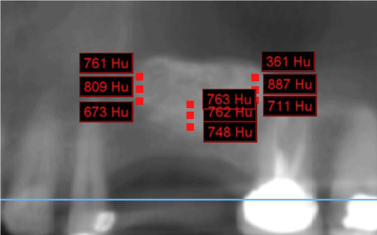
Three HU measurements were taken for each sector; that is to say, a total of 9 measurements were made moving the measurements apically 1.2 mm from the first measurement and 1.2mm apically again (Figure 2).
Finally, the residual bone height was measured again in three different points.
All these measurements were transcribed in a spreadsheet taking the average values of each group.
The variables analyzed were the following:
- Particle sizes, thickness of remnant bone, age, gender, and distance to remnant bone.
Statistical analysis of the data
Tomographic density contrasts according to the categories were carried out using parametric tests (Student test, one-way ANOVA and repeated measures ANOVA and Tukey post hoc test) and non-parametric tests (Mann-Whitney) according to distribution type for each evaluated factor: particle size, remnant bone thickness, age and gender. The Pearson correlation test was used to analyze the correlation between radiographic density and distance to remnant bone. For all tests the level of statistical significance was set at 0.05.
Results
The average tomographic density of the grafted sinuses was 586 ± 238 HU (mean ± standard deviation). This value corresponds to Type II - III bone considering the classification proposed by Lekholm and Zarb [26], (500-850 HU) and Type II considering Norton’s and Gamble’s [27], classification.
Radiographic density according to the particle size of the graft material
Table 1 shows the results in relation to particle sizes. The cases grafted with mixed particles of both sizes (small and large) recorded the higher radiographic density. The differences obtained were significant.
Tomographic density according to the residual bone
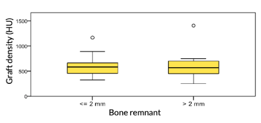
Residual bone was dived into two groups: <= 2mm and > 2mm. Values were similar in both groups. As seen in the box diagram of Figure 3, there were two outliers values, one for each category, which correspond to cases of grafts performed with mixed particles (small and large). The results did not show significant differences.
Tomographic density according to patient’s age
When comparing the density values considering only the age factor, the differences were significant (Mann-Whitney test: p = 0.015; p <0.05) Table 2.
Tomographic density according to patient’s gender
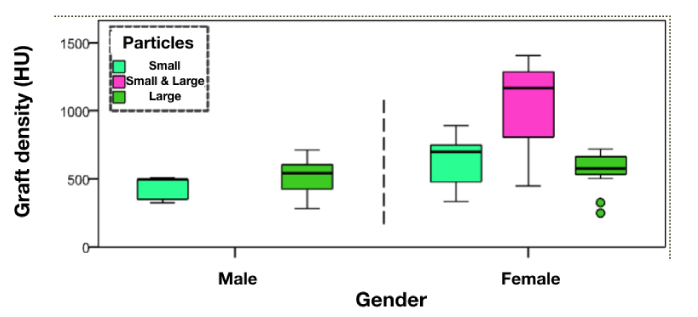
The results were considered without taking into account the influence of mixed particle cases, since the 3 mixed particle cases corresponded to women. No significant differences were obtained Figure 4.
Tomographic density according to distance to residual bone
Distance of the measure to the remnant bone was divided into three groups as Table 3 and Figure 2 show. The distributions were very similar, with no significant differences between the three groups (ANOVA: p = 0.879; p> 0.05).
Discussion
The average radiographic density of the grafted sinus was 586 ± 238 UH (mean ± standard deviation), a value that corresponds to Type II-III bone considering the classification proposed by Lekholm and Zarb [26], (500-850 UH) and Type II according to Norton and Gamble [27]. Seiler, et al. [18], after comparing two filling materials (Osteodens vs Bio-Oss) at 6-8 months after regeneration, obtained an average bone density value of 625.0 UH for Bio-Oss®, result which is similar to the one obtained in this study. Soardi, et al. [19], found similar values but using different biomaterials (Puros and Biomend) and they confirm the results using biopsies. Besides in another study [28], they performed CBCT scans for each patient in the maxillary region following this sequence: before surgery, after sinus augmentation, immediately after implant insertion (6 months), and consecutively after 10 and 18 months and reported similar results to the present study.
In relation to the variable particle size, it was observed that in cases where mixed particles were used, radiographic density was higher, (1007.7 HU), like a Type I bone. It was statistically significant compared to the average of 547 HU and 541 HU that groups of small and large particles had. Seiler, et al. [18]. did not register statistically significant differences in their comparative study between small and large particles within the Bio-Oss group, (650.74 HU and 595 HU respectively.) However, they did not study the combination of both particles. Similar results were obtained by Cassini, et al. [3], in a clinical study in which they study the bone density in sinus grafting using Resonance Frequency Analysis (RFA).
Regarding thickness of bone remnant, no statistically significant results were observed. In their study, Seiler, et al. [18], obtained similar results within the Bio-Oss group, with no statistically significant differences registered. However, Torres J, et al. [29], did find a correlation between insufficient residual bone and sinus grafting density
In relation to age of the patient, an increase in density was observed in patients > 65 years. In contrast, Seiler, et al. [18], didn’t have significant results in relation to age. Similar findings show Uzbeck, et al. [30], in their CBCT study.
With respect to the gender of the patients, there was a slight increase in women being not statistically significant. Similar results were obtained by Seiler, et al. [18] and Uzbeck, et al. [30].
Regarding the distance to the bone remnant, no significant differences were found. Similar results obtained Soardi C, et al. when they measure the density at 6, 8 and 10mm from the residual ridge [19,28].
As it can be seen in the present study, most of the measurements were performed in the lower portion of the grafted sinus, not higher than 8 mm. The reason for this decision was that most of the implants used nowadays are not too long due the improvements in their microtopography [9-13]. The use of short implants is as effective as using longer implants [12,15,16,31-34]. Moreover, Shi, et al. [35] showed similar results between 6 and 8mm long implants and 10mm implants in combination with sinus lifting. In addition, the 1 to 9 mm lower portion of a grafted sinus would have greater vascularization, as shown in the work done by Wong, et al. [36], The results of these last two investigations may be related to the findings of the present research.
Conclusion
The bone density obtained after grafting maxillary sinuses using lateral window technique with ABB as a bone graft and collagen membrane as barrier , and measuring Hounsfield Units in CBCTs, shows quantitative values similar to those of a bone type II - III, providing a high degree of predictability for implant placement 6 months after the sinus intervention.
- Valentini P, Abensur D, Wenz B, Peetz M, Schenk R (2000) Sinus grafting with porous bone mineral (Bio-Oss®) for implant placement: A 5-year study on 15 patients. Int J Periodontics Restorative Dent 20: 245-253. Link: https://bit.ly/2KISebz
- Chanavaz M (2000) Sinus Graft procedures and implant Dentistry: A review of 21 years of surgical experience (1979-2000) Implant Dent 9: 197-206. Link: https://bit.ly/2Sl1Spc
- Cassini M, Villavicencio H, Ibañez MC, Juaneda MA, Ibañez JC (2014) Does the size of bone graft particles in maxillary sinuses have differential effects on implant stability? Keys of Dentistry 73: 9-22.
- Pasquali PJ, Lucchesi Teixeira M, Altro de Oliveira T, Macedo Scavone LG, et al. (2015) Maxillary Sinus Augmentation Combining Bio-Oss with the Bone Marrow Aspirate Concentrate: A Histomorphometric Study in Humans. Int J Biomater 2015: 121286. Link: https://bit.ly/2xgShbu
- Smith M, Duncan WJ, Coates DE (2017) Attributes of Bio-Oss® and Moa-Bone® graft materials in a pilot study using the sheep maxillary sinus model. J Periodont Res 1-11. https://bit.ly/2SjFX1o
- Akbarzadeh Baghban A, Dehghani A, Ghanavati F, Zayeri F, Ghanavati F (2009) Comparing alveolar bone regeneration using Bio-Oss and autogenous bone grafts in humans: a systematic review and meta-analysis. Iran Endod J 4: 125-130. Link: https://bit.ly/3bQ8kMG
- Froum SJ, Wallace S, Cho SC, Rosenburg E, Froum S, et al. (2013) A histomorphometric comparison of Bio Oss alone versus Bio Oss and platelet-derived growth factor for sinus augmentation: a post-surgical assessment. Int J Periodontic Restorative 33: 269-279. Link: https://bit.ly/3aLs4j7
- Geistlich-pharma.com [internet] Geistlich FAQ Geistlich Bio-Oss Link: https://bit.ly/2YfeoKp
- Ibañez JC, Jalbout ZN (2002) Immediate loading of Osseotite Implants: two-year results. Implant Dent 11: 128-136. Link: https://bit.ly/3aM2lHh
- Ibañez JC, Tahhan MJ, Zamar JA (2003) Performance of double acid-etched surface external hex titanium implants in relation to one- and two-stage surgical procedures. J Periodontol 74: 1575-1581. Link: https://bit.ly/2YekKK5
- Ibañez JC, Tahhan MJ, Zamar JA, Menendez AB, Juaneda AM, et al. (2005) Immediate occlusal loading of double acid-etched surface titanium implants in 41 consecutive full-arch cases in the mandible and maxilla: 6- to 74-month results. J Periodontol 76: 1972-1981. Link: https://bit.ly/35m7zbK
- Rodriguez MR, Ibanez MC, Juaneda MA, Marengo H, Ibanez JC (2014) Supervivencia de implantes cortos <- 8mm de superficie arenada y grabada con ácido” Claves de Odontología 73: 49-66. Link: https://bit.ly/2W8V5Qn
- Bustos Malberti S, Correa Patino D, Crespo I, Juaneda MA, Ibanez MC, et al. (2016) Evaluation of removal torque in 3i, B&W and Tree-Oss dental implants. An experimental study in rabbits. Revista de la Asociación Odontológica Argentina 104: 150-159. Link: https://bit.ly/2W6GtkB
- Sezin M, Croharé L, Ibanez JC (2016) Microscopic Study of Surface Microtopographic Characteristic of Dental Implants. Open Dent J 10: 139-147. Link: https://bit.ly/35eXJIn
- Ibanez JC et al. (2020)Long-Term Evaluation of Dental Implants in the Elderly Population. Scientific Archives of Dental Sciences 3: 01-09.
- Ibanez JC, Juaneda MA, Ibanez MC, Juaneda M (2009) Comportamiento de implantes cortos de superficie microtexturada obtenida por doble grabado ácido: resultados de 1 a 9 años. Rev la Acad Nac Odontol 7: 26-33. Link: https://bit.ly/35eXU6v
- Soardi C, Spinato S, Zaffe D, Wang HL (2011) Atrophic maxillary floor augmentation by mineralized human bone allograft in sinuses of different size: an histologic and histomorphometric analysis. Clin Oral Impl Res 560-566. Link: https://bit.ly/3cWemLF
- Seiler E, Ibañez C, Ibañez JC (2019) Bone density evaluation measured at Cone-Beam Computed Tomography by means of Hounsfield Units in Maxillary Sinus Floor Augmentation. Link: https://bit.ly/3cZXNyi
- Soardi CM, Zaffe D, Motroni A, Wang H (2014) Quantitative Comparison of Cone Beam Computed Tomography and Microradiography in the Evaluation of Bone Density after Maxillary Sinus Augmentation: A Preliminary Study. Clin Implant Dent Relat Res 16 Link: https://bit.ly/3cWE4zD
- Kumar SM, Al Hobeira H, Aljanakh MD, Shaikh S, Ponnuse K, et al. (2018) Cone-beam computed tomography versus orthopantomography in sinus lift procedures: Two-dimensional versus three-dimensional imaging. Saudi Surg J 6: 113-121. Link: https://bit.ly/35fsBZm
- Jaju P, Jaju S (2014) Clinical Utility of Dental Cone-beam computed Tomography: Current perspectives. Clin Cosmet Investig Dent 6: 29-43. Link: https://bit.ly/2KHmljH
- Cordaro L, Bosshardt D, Palattella P, Rao W, Serino G, et al. (2008) Maxillary sinus grafting with Bio-Oss or Straumann Bone Ceramic: histomorphometric results from a randomized controlled multicenter clinical trial. Clin Oral Implants Res 19: 796-803. Link: https://bit.ly/2VMSih2
- Boyne PJ, James RA (1980) Grafting of the maxillary sinus floor with autogenous marrow and bone. J Oral Surg 38: 613-616. Link: https://bit.ly/2YdN0fR
- Agarwal E, Surendra Masamatti S, Kumar A (2018) Escalating Role of Piezosurgery in Dental Therapeutics. J Clin Diagn Res. Link: https://bit.ly/2ShFw7W
- Geistlich-Bio-Oss.com Geistlich Biomaterial. Link: https://bit.ly/3cUZsW2
- Lekholm U, Zarb GA (1985) Patient selection and preparation. Tissue integrated prostheses; osseointegration in clinical dentistry. Edited by Branemark PI, Zarb GA, Albrektsson T. Quintessence Publishing Company 199-209. Link: https://bit.ly/3cWRDz1
- Norton MR, Gamble C (2001) Bone classification: an objective scale of bone density using the computerized tomography scan. Clin Oral Impl Res 12: 79-84. Link: https://bit.ly/35cnRDZ
- Soardi CM, Suárez-López del Amo F, Galindo-Moreno P, Catena A, Zaffe D, et al. (2016) Reliability of Cone Beam Computed Tomography in Determining Mineralized Tissue in Augmented Sinuses. Int J Oral Maxillofac Implants 31: 352-358. Link: https://bit.ly/2VJgP6u
- Torres J, Tamimi F, Martinez PP, Alkhraisat MH, Linares R, et al. (2009) Effect of platelet-rich plasma on sinus lifting: a randomized-controlled clinical trial. J Clin Periodontol 36: 677-687. Link: https://bit.ly/2VMK2gJ
- Uzbek UH, Rahman SA, Alam MK, Gillani SW (2014) Bone Forming Potential of An-Organic Bovine Bone Graft: A Cone Beam CT study. J Clin Diagn Res 8: ZC73-ZC76. Link: https://bit.ly/2xm7N66
- Nedir R1, Bischof M, Briaux JM, Beyer S, Szmukler-Moncler S (2004) A 7 year life table analysis from a prospective study on ITI implants with special emphasis on the use of short implants. Clin Oral Impl Res 150-157. Link: https://bit.ly/2SgPfLO
- Fugazzotto PA (2008) Shorter implants in clinical practice: Rationale and treatment results. Int J Oral Maxillofac Implants 23: 487-469. Link: https://bit.ly/2VLKuvT
- Lombardo G, Pighi J, Marincola M, Corrocher G, Simancas-Pallares M, et al. (2017) Cumulative Success Rate of Short and Ultrashort Implants Supporting Single Crowns in the Posterior Maxilla: A 3-Year Retrospective Study. Int J Dent 2017: 8434281. Link: https://bit.ly/3f0Md8g
- Fan T, Li Y, Deng WW, Wu T, Zhang W (2017) Short Implants (5 to 8 mm) Versus Longer Implants (>8 mm) with Sinus Lifting in Atrophic Posterior Maxilla: A Meta-Analysis of RCTs. Clin Implant Dent Relat Res19: 207-215. Link: https://bit.ly/35cqzt6
- Shi JY, Gu YX, Qiao SC, Zhuang LF, Zhang XM, et a. (2015) Clinical evaluation of short 6-mm implants alone, short 8-mm implants combined with osteotome sinus floor elevation and standard 10-mm implants combined with osteotome sinus floor elevation in posterior maxillae: study protocol for a randomized controlled trial. Trials 16: 324. Link: https://bit.ly/35fgvPX
- Wong K (2000) Laser Doppler flowmetry for clinical Detection of blood flow as a measure of vitality in sinus bone crafts. Implant Dent 9: 133-42 Link: https://bit.ly/3eVjLEJ
Article Alerts
Subscribe to our articles alerts and stay tuned.
 This work is licensed under a Creative Commons Attribution 4.0 International License.


 Save to Mendeley
