Journal of Dental Problems and Solutions
Immediate Implant Placement in the Esthetic Zone Utilizing a Fully Digital Workflow for Crown Delivery
Tiago Diomar Claudino1*, Matheus dos Santos Bonfim1, Catherine Leticia Costa Rodrigues1, Fabiana Rodrigues Monteiro Santos1, Murilo Jacinto Santiago1, Leandro Henrique da Silva2, Marcus Vinicius Alves Fonseca3, Marcelo Benedito Ruffini Penteado4 and Rafael Alves de Camargo5
1Postgraduate Program in Implantology, Senac University Center, São Paulo, Brazil
2Coordinador of the Digital Dentistry Program, Senac University Center, São Paulo, Brazil
3Associate Professor of Digital Dentistry, Senac University Center, São Paulo, Brazil
4Associate Professor of Implantology Specialty, Senac University Center, São Paulo, Brazil
5Program Director of Implantology Specialty, Senac University Center, São Paulo, Brazil
Cite this as
Claudino TD, Santos Bonfim MD, Costa Rodrigues CL, Monteiro Santos FR, Santiago MJ, Da Silva LH, et al. Immediate Implant Placement in the Esthetic Zone Utilizing a Fully Digital Workflow for Crown Delivery. J Dent Probl Solut. 2025;12(1):001-003. Available from: 10.17352/2394-8418.000130Copyright License
© 2025 Claudino TD, et al. This is an open-access article distributed under the terms of the Creative Commons Attribution License, which permits unrestricted use, distribution, and reproduction in any medium, provided the original author and source are credited.The installation of immediate implants in fresh alveoli is a valuable technique for preserving surrounding structures. In this case, a male patient presented with trauma leading to a root fracture of tooth 21. Atraumatic extraction and immediate implant placement were performed, with biomaterial used to maintain periodontal tissues. A provisional restoration was placed due to torque exceeding 45N, and monthly follow-ups were conducted for six months. Prosthetic rehabilitation was completed through a digital workflow using CAD design and 3D milling technology.
Introduction
Immediate implant placement, introduced in 1976, offers advantages such as reduced treatment time and fewer surgical interventions, ensuring greater patient comfort. Studies indicate a high success rate for immediate implants in fresh sockets compared to healed ridges. Primary stability (40-60 N/cm²) is crucial, achieved through mechanical fixation within the residual socket. Proper implant positioning, especially with a palatal approach, minimizes bone remodeling and enhances stability. Biomaterials mixed with autogenous grafts help compensate for natural bone resorption. Maintaining the gingival contour and papillae is essential for aesthetics, facilitated by temporary crowns with an appropriate emergence profile. Atraumatic extraction techniques are critical in anterior regions to preserve soft and hard tissue architecture. Digital workflows, including intraoral scanning and 3D printing, optimize implant planning and execution. These advancements improve long-term functional and aesthetic outcomes in implant rehabilitation.
Clinical case report
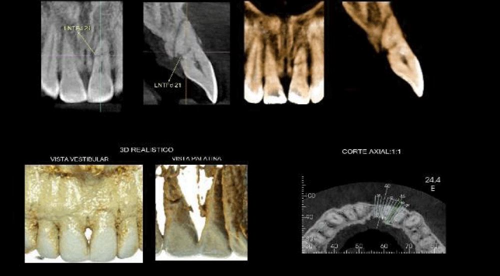
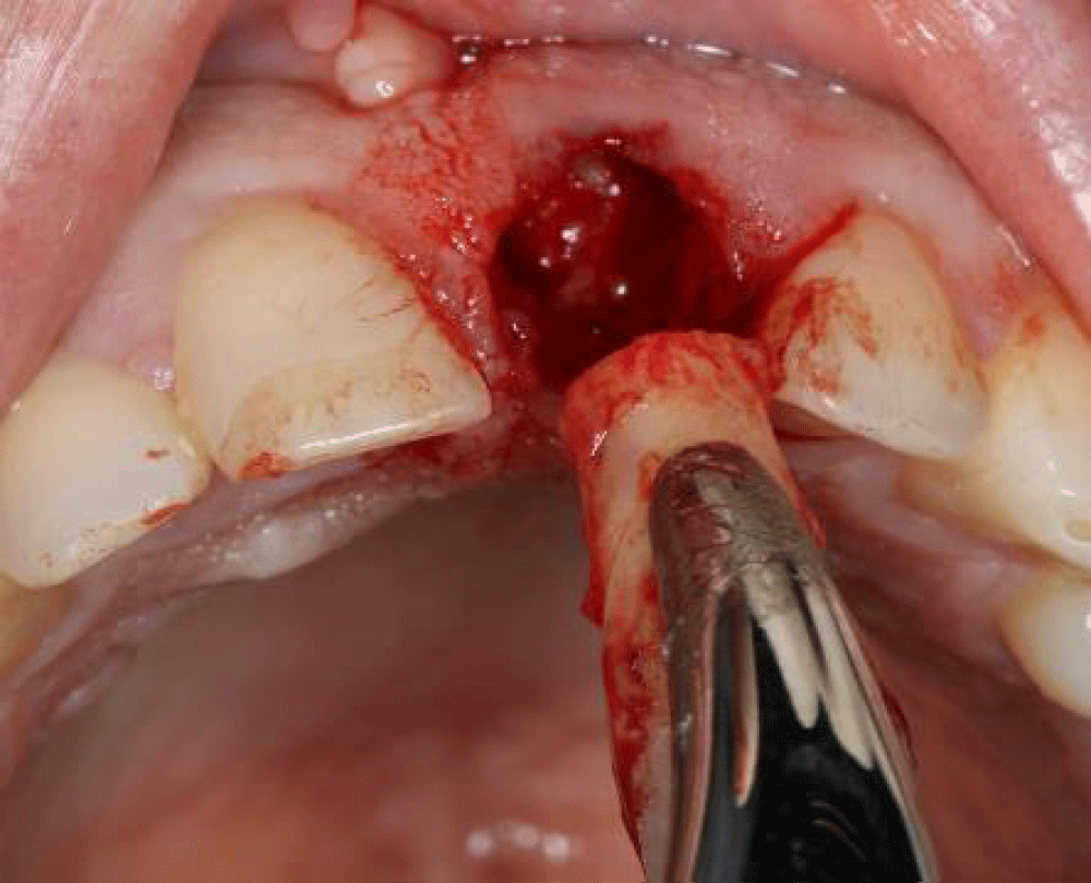
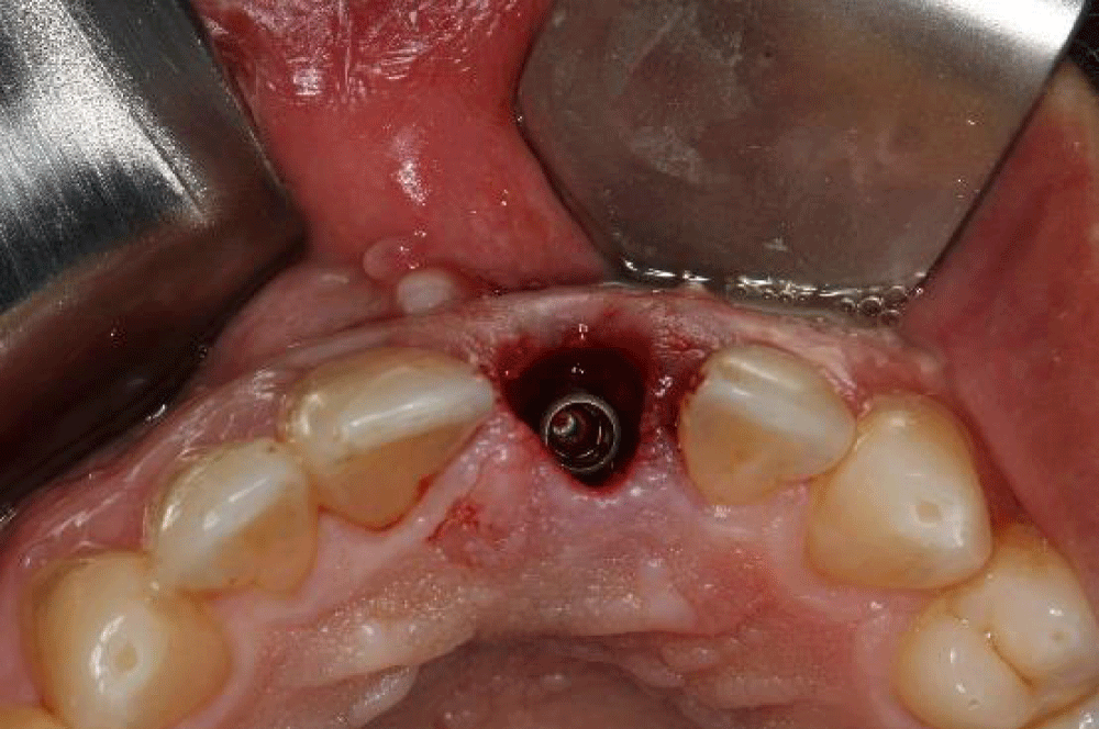
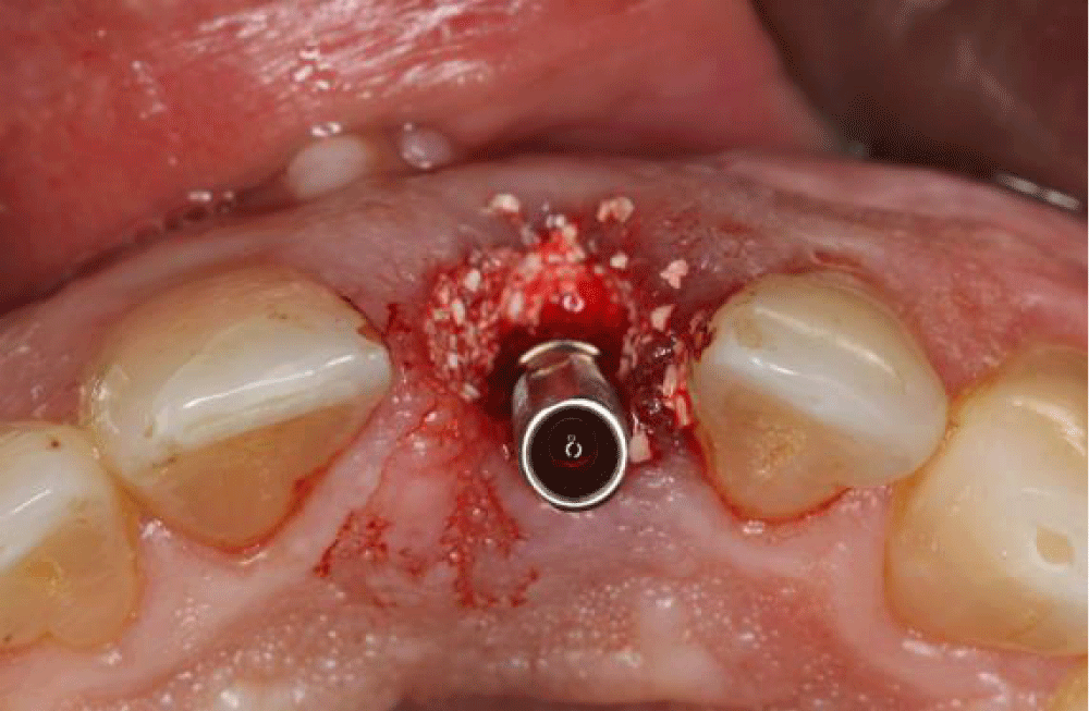
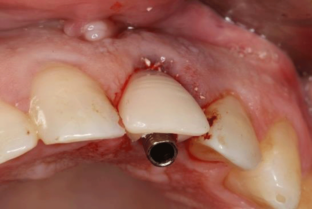
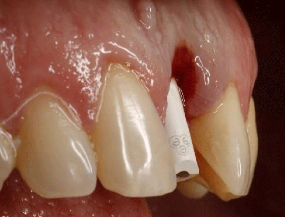
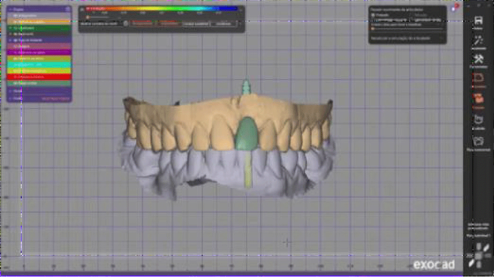
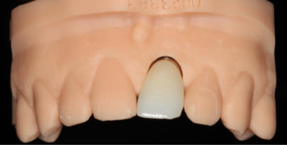
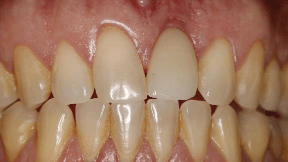
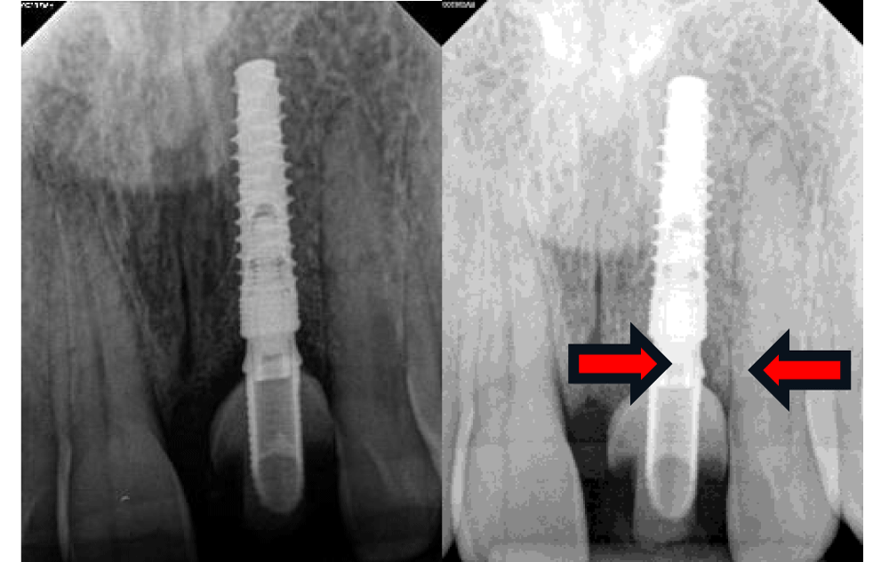
A 46-year-old male patient sought treatment at the Senac University Center in Brazil after experiencing trauma to the anterior region of his teeth. The CBCT examination revealed a root fracture in tooth 21 (Figure 1). Consequently, the patient underwent an atraumatic extraction of tooth 21 (Figure 2), followed by the immediate placement of a Bionnovation CM 3.5 × 15 mm implant, during the surgical procedure, the decision was made to initiate the osteotomy for implant placement using a lance drill, engaging the palatal wall of the socket, 6 mm above the apex of the extracted root. The drilling was centered on the bisector of the socket of the recently extracted tooth (Figure 3). To preserve the bone and gingival tissue architecture, Bionnovation material was used: small, dense fine xenograft particles of Bonefill material, with a volume of 0.50 cc was applied to fill the peri-implant gaps. (Figure 4) provisional restoration was installed without occlusal contact to support the osseointegration process. The patient was instructed to follow a soft diet consisting of cold or room temperature liquid and pasty foods for the first three days. During the six-month bone healing period, the patient was advised to avoid biting or applying functional load with the anterior region. Oral hygiene routines were to be maintained as usual, with special care to avoid pressure on the surgical site (Figure 5). The prosthetic rehabilitation was performed through a fully digital workflow, including intraoral scanning, CAD design, and 3D printing, ensuring the precise fabrication of the final restoration (Figures 6-9). Periapical x-rays were taken at the time of implant placement and after 2 months with the zirconia crown (Figure 10).
Discussion
The upper anterior teeth are highly prone to trauma, cavities, periodontal disease, and failed endodontic treatments, often leading to tooth loss. Minimally invasive extractions help preserve the fragile bone structure, preventing additional resorption [1]. Immediate implant placement following extraction has proven to be a predictable treatment, requiring careful planning and specific conditions such as absence of infection, sufficient apical bone, and good vestibular cortical bone support [2].
This approach offers functional, aesthetic, and psychological benefits by reducing surgical interventions and overall treatment time. Proper three-dimensional implant positioning, particularly using a palatal approach, ensures better primary stability and space for bone regeneration [3]. Bone grafting in the jumping gap minimizes ridge resorption and enhances long-term stability [4].
Studies show that up to 30% of horizontal bone volume may be lost within three months after extraction. Thus, immediate implantation with grafting is preferred to avoid additional regenerative procedures. Additionally, atraumatic surgical techniques that preserve soft tissues contribute to better outcomes [5].
Achieving aesthetic success in the anterior maxilla is challenging, as it depends on bone volume and interdental papilla preservation. Immediate loading implants support soft tissues and facilitate hygiene, while provisional restorations help shape the gingival architecture. Various techniques, such as gingivoplasty and customized impressions, enhance tissue adaptation [6].
Finally, early implant placement maintains the socket anatomy, reducing alveolar ridge resorption, which can reach 50% post-extraction. Strict adherence to clinical criteria is essential for osseointegration and achieving both functional and aesthetic success in rehabilitation [7].
Final considerations
In conclusion, immediate-load implants represent a major advancement in dentistry, offering a fast and efficient solution for tooth replacement. This technique reduces treatment time and enhances quality of life by enabling prosthesis placement on the same day as the implant. However, careful planning and execution are essential to prevent complications such as micromovements and implant instability.
Advancements in implant technology and clinical expertise continue to improve the success of immediate-load procedures, making them a reliable option in modern dentistry. Maintaining gingival contour is crucial for aesthetic outcomes, requiring precise implant positioning to preserve tissue integrity and ensure seamless integration with adjacent teeth.
The immediate placement of implants in fresh extraction sockets represents a predictable and efficient approach for preserving hard and soft tissues, especially in esthetically demanding regions. In the present case, atraumatic extraction, careful implant positioning, and the use of biomaterials contributed to favorable clinical and esthetic outcomes. However, as this is a single case, clinical variability must be considered. Future studies involving multiple cases would provide a broader understanding of the overall success rate and limitations of immediate implant placement.
Ethical considerations
Written informed consent was obtained from the patient for the treatment and the publication of this case report, including accompanying images.
- Vasconcelos WL, Hiramatsu AD, Paleckis PGL, Francischone EC, Vasconcelos BCR, Chaves GT. Immediate implant and alveolar preservation with Bio-Oss Collagen in the esthetic area. Int J Oral Maxillofac Surg (Portuguese edition). 2016;1.
- Oliveira CA, Souza RJ, Thomé G, Melo MCA, Satori MAI. Immediate single tooth implant in immediate loading – case report. RFO. 2008;13(1):70-74.
- Passoni B, Dalago HR, Cid R. Immediate implant with immediate aesthetics, definitive restoration, and tomographic follow-up of the vestibular bone plate – case report. Full Dent Sci. 2015;6(23):183-190.
- Naiem NS, Nawas AIB, Tawfik KO, Nahass EIH. Jumping gap in immediate implant placement in the esthetic zone: a virtual implant planning using cone-beam computed tomography. J Prosthodont Res. 2024;68(2):347-353.
- Andreiuolo R, Vasconcelos F, Andrade A, Groisman M, Junior VMG. Immediate Implant in the Anterior Region: surgical and prosthetic aspects. Rev Bras Odontol. 2016;73(1):84-88. Available from: http://revodonto.bvsalud.org/scielo.php?script=sci_abstract&pid=S0034-72722016000100016&lng=p&nrm=iso&tlng=en
- Fernandes G, Mysore A, Shetye O, Aras M, Chitre V. Customizing the Emergence Profile Around an Immediately Loaded Single Implant in the Esthetic Zone: A Case Report. Cureus. 2024;16(4):e58279. Available from: https://doi.org/10.7759/cureus.58279
- Martins SHL, Vieira GHA, Bezerra FJB, Ghiraldini
Article Alerts
Subscribe to our articles alerts and stay tuned.
 This work is licensed under a Creative Commons Attribution 4.0 International License.

 Save to Mendeley
