Journal of Dental Problems and Solutions
Er, Cr: YSGG Laser Excision of an Ulcerated Pyogenic Granuloma of the Upper Lip Vermilion in a Child Initially Diagnosed as Hemangioma: A Case Report
1Faculty of Dental Medicine, International Balkan University, Skopje, North Macedonia
2Private Dental Clinic “Dr. Bashkim Ismaili”, Gostivar, North Macedonia
3Daci Dent, North Macedonia
4Faculty of Dental Medicine, University of East Sarajevo, Foča, Bosnia and Herzegovina
5Department for Pediatric and Preventive Dentistry, Faculty of Dental Medicine, University Ss.Cyril & Methodius, Skopje, North Macedonia
Author and article information
Cite this as
Ismaili B, et al. Er, Cr: YSGG Laser Excision of an Ulcerated Pyogenic Granuloma of the Upper Lip Vermilion in a Child Initially Diagnosed as Hemangioma: A Case Report. J Dent Probl Solut. 2026; 13(2): 13-21. Available from: 10.17352/2394-8418.000136
Copyright License
© 2026 Ismaili B, et al. This is an open-access article distributed under the terms of the Creative Commons Attribution License, which permits unrestricted use, distribution, and reproduction in any medium, provided the original author and source are credited.Background: Pyogenic granuloma is a benign reactive vascular lesion frequently encountered in the oral cavity, most commonly on the gingiva. Localization on the vermilion border of the lip is uncommon, particularly in pediatric patients, and may present a diagnostic challenge because of its clinical resemblance to hemangiomas and other vascular lesions. Laser-assisted surgery has been introduced as a minimally invasive alternative to conventional surgical excision.
Case presentation: A 10-year-old girl presented with a vascular lesion located on the left lateral aspect of the upper lip vermilion. The lesion had been present for approximately one year and was initially diagnosed as a hemangioma after consultations with several specialists. During the two months preceding treatment, recurrent episodes of spontaneous bleeding were reported. Complete excision was performed on 07 May 2026 using an Er, Cr: YAG laser (Waterlase iPlus®, BIOLASE, Irvine, CA, USA). Histopathological examination confirmed an ulcerated pyogenic granuloma (Granuloma pyogenicum ulceratum) with negative peripheral and deep surgical margins.
Results: The procedure was completed under minimal local anesthesia with satisfactory intraoperative hemostasis and without the need for suturing. Healing progressed uneventfully, with progressive epithelialization and no signs of infection, prolonged bleeding, functional impairment, or recurrence. Follow-up demonstrated preservation of the normal lip contour and a highly satisfactory esthetic outcome.
Conclusion: Er, Cr: YSGG laser excision may represent a safe and effective minimally invasive treatment option for pyogenic granuloma of the upper lip vermilion in pediatric patients. The technique provides precise tissue removal, satisfactory hemostasis, favorable healing, and highly satisfactory esthetic results.
Pyogenic granuloma (granuloma pyogenicum) is a benign reactive vascular proliferation of the skin and mucous membranes that develops in response to various local irritative factors, trauma, or hormonal influences. Pyogenic granuloma (lobular capillary hemangioma) is a common vascular lesion seen in children and adolescents. Despite the widespread use of this term in clinical practice, the designation “pyogenic granuloma” is considered inappropriate, since the lesion is neither associated with a pyogenic infection nor histologically a true granuloma. Therefore, other terms, such as lobular capillary hemangioma [1], have also been used in the literature to describe more accurately the histomorphological characteristics of this lesion.
Pyogenic granuloma may occur at any age, but it is more frequently described in children, adolescents, and young adults. In the oral cavity, the gingiva is the most common site and accounts for approximately 75% of oral cases, whereas other locations, including the lips, tongue, buccal mucosa, and palate, are considerably less common. Lesions localized on the vermilion of the lip represent a diagnostic and therapeutic challenge because of their rarity and the esthetic importance of this anatomical region [2,3].
The etiopathogenesis of pyogenic granuloma has not been fully clarified. It is believed that the development of the lesion is associated with an exaggerated angiogenic tissue response to chronic irritation, local trauma, dental plaque and calculus, hormonal changes, or certain medications. In children, mechanical injuries, lip-biting habits, and repeated microtrauma may represent important predisposing factors [4].
According to Epivatianos A and all, there are two histological types of pyogenic granuloma (PG) in the oral cavity: the lobular capillary hemangioma (LCH) type and the non-LCH type. Their study aimed to compare their clinical, etiological, and immunohistochemical features to determine whether they represent distinct entities. Thirty cases of LCH and 26 cases of non-LCH were analyzed. Clinically, LCH lesions more often appeared as sessile masses, while non-LCH lesions were more frequently pedunculated. The non-LCH type was more commonly associated with identifiable etiological factors. Histologically, LCH showed a higher number of blood vessels with small lumens, whereas the non-LCH type had more vessels containing perivascular mesenchymal cells that were negative for certain muscle markers. Overall, their study concluded that these differences suggest that LCH and non-LCH pyogenic granulomas may be two separate histological entities [5].
The 2003 study by Toida et al. examines 43 cases of oral lobular capillary hemangioma (LCH), commonly known as pyogenic granuloma, detailing its clinical, histopathological, and immunohistochemical characteristics. The condition affects slightly more females than males (ratio of 1:1.5), with an average patient age of 52.7 years. The most common sites were the gingiva (15 cases), followed by the tongue (13 cases) and the labial mucosa (10 cases). Lesions typically presented as pedunculated (stalked) masses up to 15 mm in size, frequently accompanied by ulceration [6].
The study by Gordón-Núñez et al aimed to retrospectively analyze the clinical, demographic, and pathological characteristics of oral pyogenic granuloma in a Brazilian population. A total of 293 cases diagnosed between 1970 and 2008 were reviewed. Data collected included gender, age, race, lesion site, predisposing factors, clinical features, diagnosis, treatment, and recurrence. The results showed a female predominance (female-to-male ratio of 2.38:1), with a mean age of 27 years, and most cases occurring in the second decade of life. White patients were most commonly affected (44.7%). The gingiva (83%), especially in the maxilla, was the most frequently involved site. Clinically, lesions were typically symptomatic, bleeding nodules with a soft consistency, red surface, and a pedunculated base. The average size was about 1.3 cm, and the recurrence rate was 8.2%. They concluded that the clinical, demographic, and pathological features of oral pyogenic granuloma in the Brazilian population were similar to those reported in other populations worldwide [7].
A retrospective study by Patrice SJ and all of 178 patients aged 17 years or younger (mean age 6.7 years) showed that 42% of cases occurred in the first five years of life, while 12% were found in infants under one year. The male-to-female ratio was 3:2. In most cases (74.2%), no trauma or predisposing condition was identified. The average lesion size was 6.5 mm, with a mean duration of 3.8 months before diagnosis. Lesions were most frequently located on the head and neck (62.4%), followed by the trunk, upper extremities, and lower extremities. Most lesions (88.2%) involved the skin, while a smaller number affected the oral cavity and conjunctiva. Histology showed normal mast cell numbers, unlike proliferative hemangiomas. Surgical excision with linear closure resulted in no recurrences, whereas shave excision or cautery had a higher recurrence rate (43.5%) [8].
Clinically, pyogenic granuloma usually manifests as an exophytic, pedunculated or sessile lesion with a smooth or lobulated surface and a bright red to dark red coloration. Because of its marked vascularity, the lesion is prone to spontaneous or provoked bleeding, especially during mastication, speech, or minimal trauma. In some cases, surface ulceration occurs, further complicating the establishment of an accurate clinical diagnosis.
The differential diagnosis of pyogenic granuloma includes numerous benign and malignant soft-tissue lesions of the oral cavity. Vascular lesions such as hemangiomas and vascular malformations are particularly important because of their similar clinical appearance and pronounced tendency to bleed. Peripheral giant cell granuloma, fibroma, peripheral ossifying fibroma, and selected malignant tumors with a vascular component should also be considered. Therefore, histopathological analysis of the excised tissue remains the gold standard for establishing the definitive diagnosis.
Conventional surgical excision is considered the treatment of choice for pyogenic granuloma. However, scalpel surgery in esthetically sensitive regions may be associated with more pronounced intraoperative bleeding, the need for suturing, and potential scar formation. The development of laser technologies in recent decades has enabled the introduction of minimally invasive procedures that offer several advantages over conventional surgical techniques.
The Er, Cr: YSGG laser (erbium, chromium: yttrium-scandium-gallium-garnet) emits light at a wavelength of 2780 nm and shows a high affinity for water, allowing efficient ablation of soft and hard tissues through a hydrokinetic effect. Minimal thermal damage to adjacent tissues, satisfactory hemostasis, reduced need for local anesthesia, absence or reduced need for suturing, less postoperative pain and edema, and a favorable esthetic outcome represent important advantages of this technology. These characteristics make the Er, Cr: YSGG laser particularly suitable for pediatric dentistry and for the management of lesions localized in esthetically significant regions of the orofacial complex [9,10].
Although the use of lasers in oral surgery is increasing, data regarding the application of the Er, Cr: YSGG laser in the treatment of pyogenic granuloma localized on the vermilion of the lip in children remain limited. Therefore, reporting such cases may contribute to a better understanding of the possibilities and limitations of contemporary minimally invasive therapeutic approaches.
This paper aimed to present the successful treatment of an ulcerated pyogenic granuloma of the upper lip in a child using an Er, Cr: YSGG laser, with special emphasis on differential diagnostic challenges, histopathological verification of the diagnosis, postoperative healing, and the achieved esthetic result.
Case report
A 10-year-old female patient presented to our private dental clinic with a referral diagnosis of upper lip hemangioma, established after previous examinations by several specialists. According to the parents, the lesion first appeared approximately one year before the intervention as a small pinpoint red lesion located on the left lateral aspect of the upper lip vermilion. Over time, the lesion gradually increased in size.
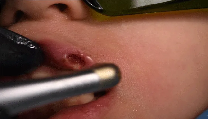
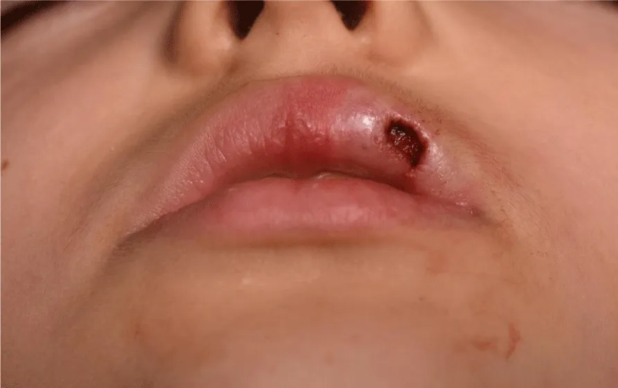
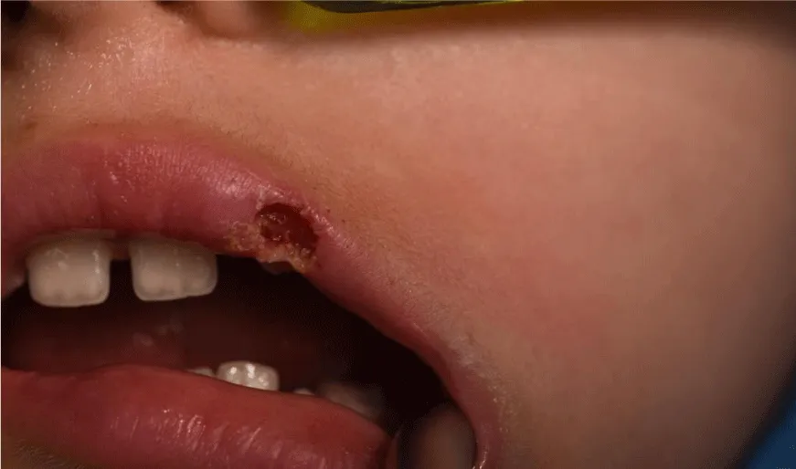
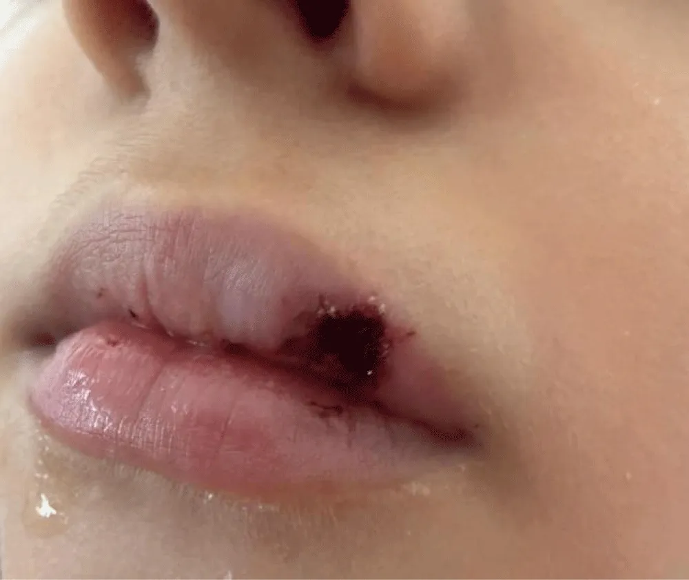
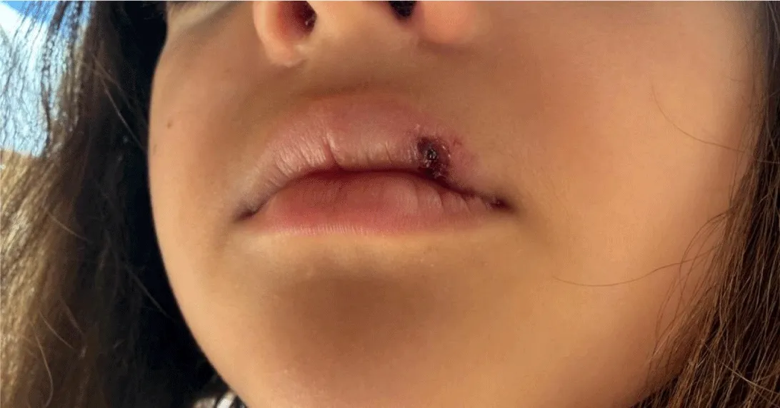
Approximately two months before presentation to our clinic, the parents noticed intermittent episodes of spontaneous bleeding from the lesion, particularly after minimal trauma during everyday activities (Figure 1). Because of these symptoms, the patient had previously been examined by a pediatrician, dermatologist, and plastic surgeon. Various topical preparations had been prescribed on several occasions, without clinical improvement or regression of the lesion. After failure of conservative management, surgical removal was recommended.
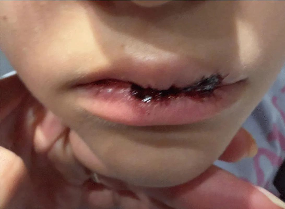
Ultrasound examination has an additional, but not primary, role in the diagnosis of pyogenic granuloma. Clinical examination and histopathological analysis after excision remain the gold standard, but ultrasound can provide important information before surgical treatment. Ultrasound examination, especially high-frequency color Doppler ultrasound, allows for noninvasive assessment of soft tissue lesions in the oral cavity. Pyogenic granuloma is most commonly seen as a well-circumscribed hypoechoic or heterogeneous soft tissue mass, with marked vascularization on color Doppler, which is characteristic due to the rich capillary network of the lesion. Assessment includes size, depth, and relationship to surrounding structures, as well as determination of whether there is infiltration into deeper tissues or involvement of adjacent structures. Ultrasound is particularly useful for differential diagnosis between pyogenic granuloma and other vascular or reactive lesions (hemangioma, peripheral giant cell granuloma, peripheral ossifying fibroma), planning surgical excision by assessing vascularity and possible risk of bleeding, monitoring the lesion after treatment, and early detection of possible recurrence. Although ultrasound provides valuable information about the structure and blood supply of the lesion, it cannot confirm the diagnosis with certainty. The final diagnosis is made by histopathological examination of the excised tissue [11].
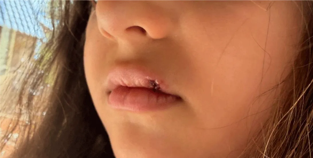
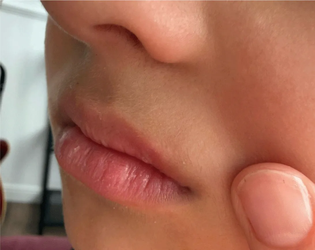
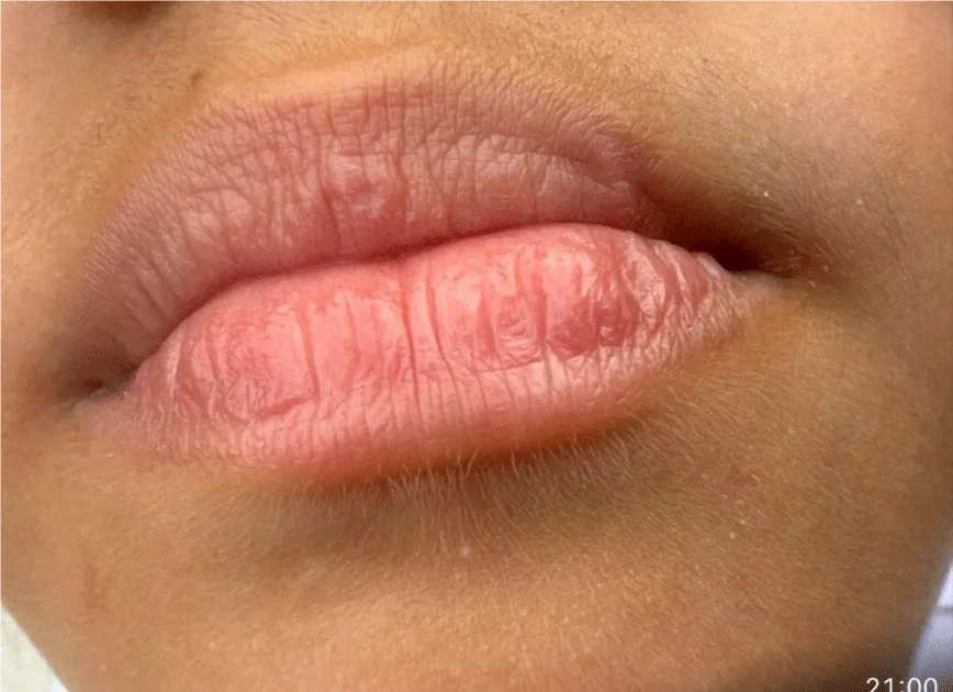
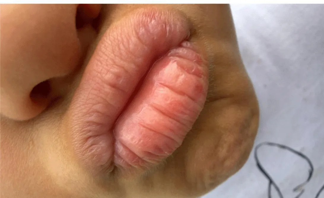
Clinical examination revealed a well-defined exophytic reddish lesion located on the left lateral aspect of the upper lip vermilion (Figures 2-4). The lesion was soft in consistency, had a smooth surface, and tended to bleed upon manipulation. Based on the medical history and clinical findings, the differential diagnosis included hemangioma, pyogenic granuloma, and other vascular soft-tissue lesions.
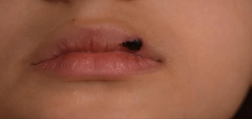
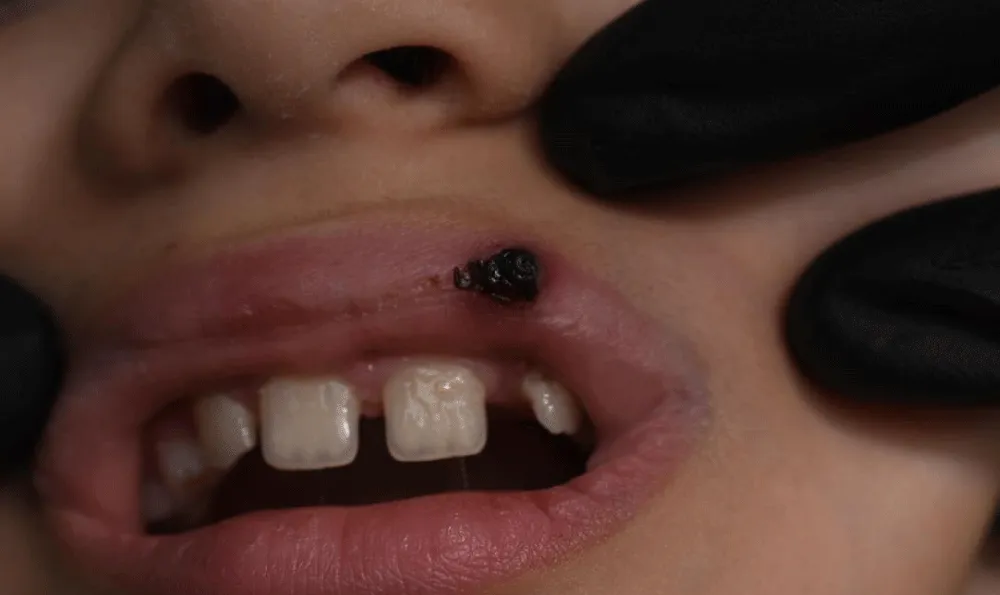
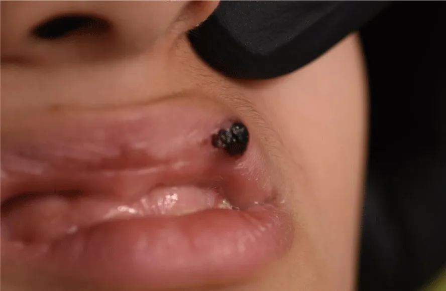
Considering the patient’s age, the esthetic importance of the affected region, and the advantages of laser surgery compared with conventional scalpel excision, the decision was made to remove the lesion using an Er, Cr: YAG laser (Waterlase iPlus®, BIOLASE, Irvine, CA, USA).
Before the intervention, a topical local anesthetic was applied to the operative field to reduce discomfort during infiltration anesthesia. Infiltration anesthesia was then performed using articaine with epinephrine (Artibsa®), with a minimal amount of anesthetic solution. Several small anesthetic deposits were applied along the periphery of the lesion, and the total administered amount did not exceed 1 mL.
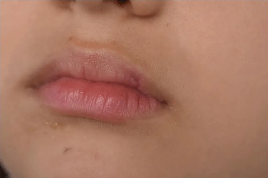
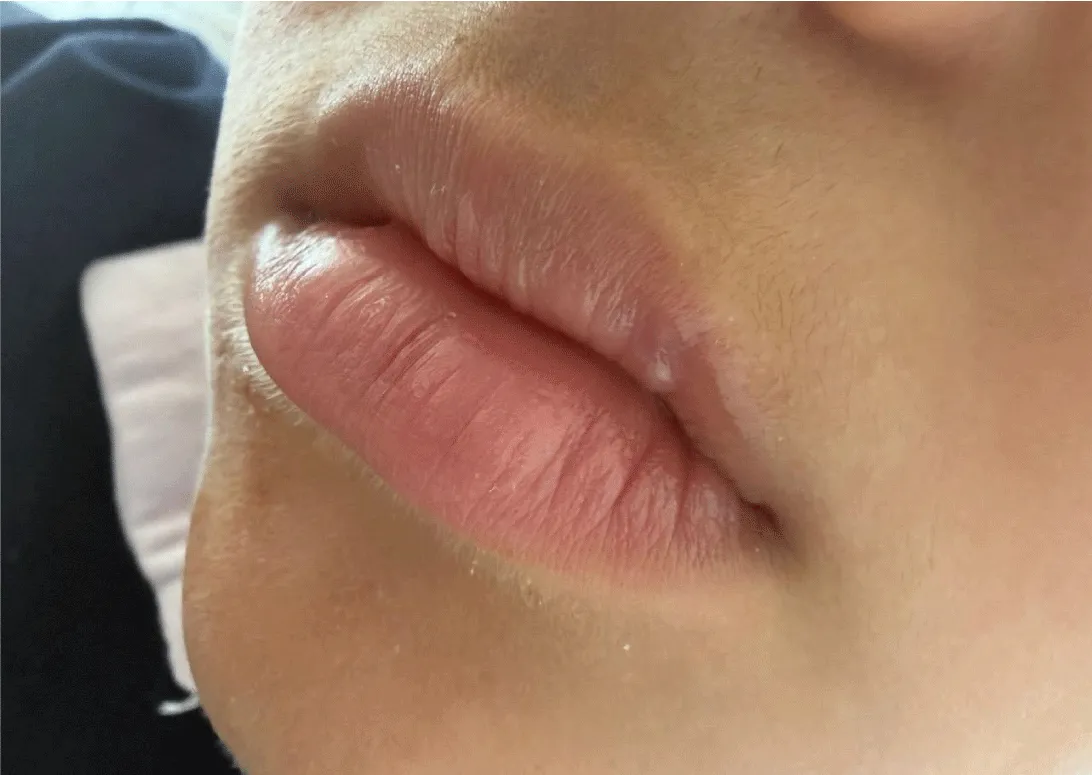
After adequate anesthesia had been achieved, complete excision of the lesion was performed using the Er, Cr: YSGG laser. The total duration of the procedure, including anesthesia administration and the surgical intervention itself, was approximately 20 minutes. During the procedure, satisfactory hemostasis was achieved without significant intraoperative bleeding, despite previous spontaneous bleeding episodes from the lesion (Figure 5). Because of the favorable intraoperative findings, sutures were not required, and the surgical wound was left to heal by secondary epithelialization (Figures 6-7 right after the intervention and Figures 8-15 one day, three days, ten days, 3 weeks, and 1 month after the intervention).
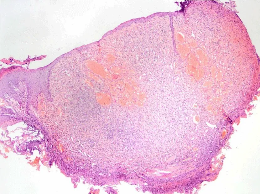
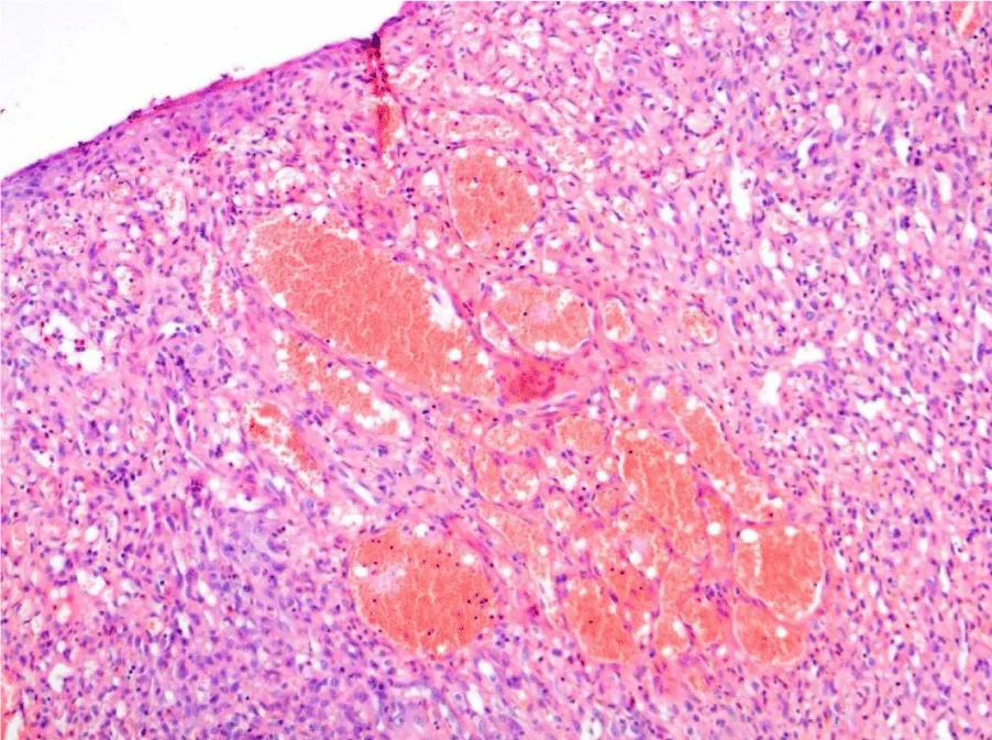
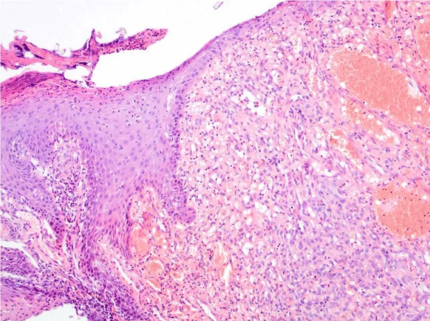
The excised specimen was submitted for histopathological analysis. Macroscopically, the submitted soft-tissue specimen measured 0.5 × 0.5 × 0.4 cm and contained a hemispherical tumefaction approximately 4 mm in diameter. Histopathological examination revealed a surface ulceration covered by a fibrinous crust. The lesion was characterized by a lobular proliferation of numerous capillary-sized blood vessels embedded within a connective tissue stroma. Mild perivascular infiltration by lymphocytes was observed. The surrounding mucosa was lined by slightly keratinized stratified squamous epithelium with no significant pathological alterations (Figures. 16-18). The histopathological findings were consistent with ulcerated pyogenic granuloma (Granuloma pyogenicum ulceratum). The peripheral and deep surgical margins passed through healthy tissue, indicating complete excision of the lesion.
During the procedure, standard laser safety precautions were implemented, including the use of appropriate protective eyewear for the child, the operator, and the assistant, as well as surgical smoke (plume) control to minimize exposure to laser-generated aerosols and airborne particles. In oral and periodontal surgery, when lasers or electrosurgical devices are used, smoke/plume control is an essential component of surgical safety and infection prevention because it: reduces the spread of aerosols, surgical smoke, and tissue particles; improves the visibility of the surgical field; enhances the safety of both the dental team and the patient by minimizing exposure to potentially harmful biological and chemical agents and contributes to better working conditions and more effective infection control during surgical procedures.
The postoperative course was uneventful. During follow-up, no signs of infection, prolonged bleeding, or other complications were observed. Follow-up examinations demonstrated progressive healing of the operative field with gradual epithelialization (Figure 4). At the three-week follow-up visit, complete epithelialization of the surgical site was observed, with preservation of the anatomical configuration of the upper lip vermilion, no functional impairment, and no clinical evidence of recurrence. Subsequent follow-up examinations, including the most recent clinical evaluation, demonstrated preservation of the normal vermilion anatomy, excellent esthetic integration with the surrounding tissues, and no clinical evidence of recurrence.
Laser parameters
Surgical excision was performed using an Er, Cr: YSGG laser system (Waterlase iPlus®, BIOLASE, Irvine, CA, USA) operating at a wavelength of 2780 nm. A Gold MZ6 tip was used throughout the procedure. Initial lesion removal was carried out using the Biopsy – Rapid Cut setting, followed by soft-tissue refinement using the Tissue Plasty setting. The laser parameters are summarized in Table 1.
Discussion
Pyogenic granuloma is a benign reactive vascular proliferation that develops in response to various local irritative factors, trauma, or hormonal influences. Although it most commonly occurs on the gingiva, its occurrence on the vermilion of the lip is relatively rare, especially in the pediatric population. Because of its rich vascularity and characteristic clinical appearance, pyogenic granuloma may represent a diagnostic challenge and often requires differential diagnostic consideration with other vascular lesions, primarily hemangiomas and vascular malformations.
In the present case, the initial clinical diagnosis was hemangioma, which led to consultations with several specialists, including a pediatrician, dermatologist, and plastic surgeon. Before definitive treatment, different forms of local therapy had been used without regression of the lesion. The definitive diagnosis of ulcerated pyogenic granuloma was established only after histopathological analysis of the excised tissue. This clinical course confirms the importance of histopathological verification of all excised soft-tissue lesions of the oral cavity, particularly when there is clinical suspicion of a vascular lesion.
Pyogenic granulomas localized on the lips have been reported sporadically in the literature. Gonçales et al. reported a case of pyogenic granuloma of the upper lip, emphasizing the rarity of this localization and the need for careful differential diagnosis [12]. However, most published cases have been treated by conventional scalpel excision.
Conventional surgical excision remains the standard treatment method for pyogenic granuloma. This approach enables complete removal of the lesion and histopathological evaluation of the excised tissue. Nevertheless, because of the pronounced vascularity of these lesions, particularly when they are localized in esthetically significant regions such as the upper lip vermilion, scalpel excision may be associated with more pronounced intraoperative bleeding, the need for suturing, and a greater risk of postoperative scar formation.
Banjar et al. reported a case of a labial pyogenic granuloma located on the lower lip of a 15-year-old patient, which developed as a result of chronic mechanical trauma caused by the maxillary left central incisor. In addition to presenting the clinical case, the authors conducted a mini-review of the literature to investigate the association between labial pyogenic granuloma and traumatic factors. Their literature search identified only five published case reports addressing this association. The findings indicated that chronic trauma is the most common etiological factor contributing to the development of labial pyogenic granuloma. Surgical excision followed by histopathological examination was recommended as the treatment of choice, as it enables definitive diagnosis while simultaneously removing the lesion. The authors emphasized that labial pyogenic granuloma should be considered in the differential diagnosis of upper and lower lip lesions, particularly in patients with a history of trauma [12]. In the present case, the use of the Er, Cr: YSGG laser allowed complete excision of the lesion with a minimal amount of local anesthetic, without suturing, and without significant intraoperative bleeding [13].
The review by Jonas Ver Berne and all examined the differences between pyogenic granuloma (PG) and lobular capillary hemangioma (LCH), two terms that have often been used inconsistently in the literature. The authors analyzed studies published between 2001 and 2018 to determine whether these lesions are truly the same or different entities. The findings showed that PG occurs more often in middle-aged individuals, is more common in females (especially during pregnancy), and is frequently associated with a known triggering factor. Histologically, PG displays radially arranged capillaries, a mixed inflammatory infiltrate, and may undergo fibrotic changes in chronic cases. In contrast, LCH has a characteristic lobular arrangement of capillaries, usually shows only mild lymphocytic inflammation, and does not undergo fibrotic transformation. Although both lesions can be treated by surgical excision and other methods, their clinical and pathological differences suggest that they should be considered distinct lesions rather than the same condition [14].
Dental lasers range from low-powered devices to high-powered systems capable of cutting and removing tissue. Because laser light can pose risks to both patients and dental staff, strict safety measures are required. Safety regulations for dental lasers are similar to those governing the use of ionizing radiation in dental practices [15-18].
The Er, Cr: YSGG laser emits energy at a wavelength of 2780 nm, which demonstrates high affinity for water present in biological tissues. The mechanism of hydrokinetic ablation allows precise tissue removal with minimal thermal damage to surrounding structures. Additional advantages of this technology include satisfactory hemostasis, improved visibility of the operative field, reduced need for local anesthesia, absence or reduced need for suturing, and a more favorable postoperative recovery.
In the present case, although the patient had a history of spontaneous bleeding episodes during the two months before the intervention, no significant intraoperative bleeding was observed during excision. The operative field did not require additional hemostatic measures or suturing, and the total procedure time, including anesthesia administration and excision, was approximately 20 minutes. It is particularly important to emphasize that the total amount of local anesthetic used did not exceed 1 mL of articaine with epinephrine, which represents an additional advantage when working with pediatric patients.
In recent years, laser-assisted treatment of pyogenic granulomas has been increasingly described. Reports of diode and erbium-family laser use have emphasized reduced bleeding, good surgical visibility, and favorable wound healing. Arora et al. described the successful use of an Er, Cr: YSGG laser in a pediatric patient with gingival pyogenic granuloma [19]. However, according to available literature, case reports describing excision of ulcerated pyogenic granuloma of the upper lip vermilion using an Er, Cr: YSGG laser in children remain limited.
Wollina U and all mention in their study that newer medical treatments using beta-blockers such as timolol and propranolol have shown promising results, especially in young children and in lesions located around the eyes or nails. Also, they mention that several factors may contribute to pyogenic granuloma development, including trauma, BRAF gene mutations, and possibly certain viruses such as herpes simplex virus type 1, ORF virus, and human papillomavirus type 2 [20].
The favorable clinical outcome observed in the present case may be attributed to the unique interaction of Er, Cr: YSGG laser energy with water-containing tissues. Erbium-family lasers provide precise tissue ablation while producing limited collateral thermal damage, thereby preserving adjacent tissue structures and promoting favorable wound healing. In addition, the limited zone of thermal alteration may facilitate histopathological interpretation of excised specimens and evaluation of surgical margins. These characteristics are particularly advantageous in pediatric patients and in esthetically sensitive areas such as the vermilion border of the lip, where tissue preservation and scar minimization are of major importance [21].
In this study by Harris MN et all, 325 cases were identified from dermatopathology records. Most lesions (86%) occurred on the skin, while only 12% were found on mucosal surfaces. Cutaneous lesions were most common during the second decade of life and were mainly located on the trunk, upper limbs, and head. Mucosal lesions were most frequently found on the lips, gingiva, and tongue, usually during the fourth decade of life. The results showed that cutaneous lesions occurred equally in males and females, suggesting that female hormones are unlikely to play a major role in their development. However, mucosal lesions were twice as common in females, which may indicate a hormonal influence, although the number of cases was too small to draw a definite conclusion [22].
The histopathological findings in the present case confirmed an ulcerated pyogenic granuloma with negative peripheral and deep resection margins, indicating complete excision of the lesion. The postoperative course was uneventful, without signs of infection, prolonged bleeding, or other complications. Follow-up demonstrated complete epithelialization of the operative site, preservation of the anatomy of the upper lip vermilion, absence of functional impairment, and no clinical signs of recurrence during the follow-up period.
The particular value of this case lies in the unique combination of several clinically significant elements: pediatric age, rare localization on the upper lip vermilion, initial clinical diagnosis of hemangioma, previously unsuccessful local therapy after examinations by several specialists, spontaneous bleeding episodes, successful Er, Cr: YSGG laser excision with minimal local anesthesia, no need for suturing, histopathological confirmation of ulcerated pyogenic granuloma with negative resection margins, and an excellent functional and esthetic result during postoperative follow-up.
The review by Lomeli Martinez SM and all discusses the different clinical and histopathological presentations of oral PG and emphasizes the importance of an accurate diagnosis for proper treatment. Treatment options should be tailored to the individual patient. The authors conclude that further research is needed to better understand the pathogenesis of oral PG and to improve treatment protocols [23].
Although definitive conclusions regarding the superiority of one therapeutic method over another cannot be drawn from a single case report, the results of the present case suggest that the Er, Cr: YSGG laser may represent a safe and effective minimally invasive alternative to conventional surgical treatment of pyogenic granuloma in children, particularly when lesions are located in esthetically sensitive regions. Future clinical studies with larger numbers of patients could further define the indications, advantages, and limitations of this technology in everyday dental practice.
Clinical significance
Pyogenic granuloma of the upper lip vermilion is an uncommon lesion in pediatric patients and may clinically mimic a hemangioma. This case demonstrates that Er, Cr: YSGG laser surgery can provide precise excision, satisfactory hemostasis, rapid healing, and a favorable esthetic outcome without the need for suturing. The technique may therefore represent a valuable minimally invasive treatment option for vascular soft-tissue lesions in esthetically sensitive regions.
Conclusion
Pyogenic granuloma localized on the vermilion of the upper lip represents a rare clinical finding in the pediatric population and may present a diagnostic challenge because of its similarity to other vascular lesions, particularly hemangioma. Histopathological analysis remains the gold standard for establishing the definitive diagnosis.
The present case demonstrates that the use of the Er, Cr: YSGG laser enables precise and minimally invasive excision of the lesion with satisfactory hemostasis, reduced need for local anesthesia, and no need for suturing. The favorable postoperative course, absence of complications and recurrence during follow-up, and highly satisfactory functional and esthetic outcome suggest that the Er, Cr: YSGG laser may represent a safe and effective alternative to conventional surgical therapy for pyogenic granuloma in children, especially when lesions are localized in esthetically sensitive regions.
Although individual case reports do not allow definitive conclusions regarding the superiority of one therapeutic method over another, they represent an important contribution to the existing literature and may serve as a basis for future clinical studies that will more precisely define the role of the Er, Cr: YSGG laser in the treatment of benign vascular lesions of the oral cavity.
Declarations
Patient consent: The patient’s parents/legal guardians provided informed consent for the therapeutic procedure and for the use of anonymized clinical data and photographs for scientific and educational purposes. Written consent is maintained in the authors’ records.
Conflict of interest: The authors declare that there are no conflicts of interest related to the preparation and publication of this manuscript.
Funding: No external funding, grants, or financial support was received for the preparation of this manuscript.
Author contributions: All authors contributed to clinical management, manuscript preparation, manuscript revision, and approval of the final version.
Clinical photographic documentation
The following clinical photographs are included for author review and may be selected, cropped, anonymized, or submitted separately according to journal requirements.
Histopathology report
Clinical Diagnosis: Hemangioma of the upper lip, left lateral region (Hemangioma labii superioris regionis lateralis sinistrae)
Submitted Material: Surgical biopsy – excision specimen
Histopathological Finding: Pyogenic Granuloma (Granuloma Pyogenicum)
Macroscopic Findings: The submitted surgical specimen consisted of an oval skin excision measuring 0.5 × 0.5 × 0.4 cm. On its surface, a semi-spherical tumefaction measuring approximately 4 mm in diameter was observed, with a dark brownish coloration.
Microscopic Findings: Microscopic examination of the excised tissue fragment demonstrated that, in the area of the visible semi-spherical lesion, there was surface ulceration covered by a crust. The lesion was composed of numerous proliferating capillary blood vessels, partly densely packed and embedded within a connective tissue stroma. Around the blood vessels, sparse accumulations of lymphocytes were present. The surrounding tissue was covered by a slightly keratinized epithelium with otherwise normal characteristics.
Conclusion: The histomorphological findings are consistent with an ulcerated pyogenic granuloma (Granuloma Pyogenicum Ulceratum).
The peripheral and deep surgical margins pass through healthy tissue, indicating complete excision of the lesion with negative margins.
- Mills SE, Cooper PH, Fechner RE. Lobular capillary hemangioma: the underlying lesion of pyogenic granuloma. A study of 73 cases from the oral and nasal mucous membranes. Am J Surg Pathol. 1980;4(5):470-9. Available from: https://pubmed.ncbi.nlm.nih.gov/7435775/
- Jafarzadeh H, Sanatkhani M, Mohtasham N. Oral pyogenic granuloma: a review. J Oral Sci. 2006;48(4):167-75. Available from: https://doi.org/10.2334/josnusd.48.167
- Sivapathasundharam B, editor. Shafer's textbook of oral pathology. 9th ed. New Delhi: Elsevier; 2020. Available from: https://shop.elsevier.in/shafers-textbook-of-oral-pathology-e-book-9788131255469.html
- Kamal R, Dahiya P, Puri A. Oral pyogenic granuloma: various concepts of etiopathogenesis. J Oral Maxillofac Pathol. 2012;16(1):79-82. Available from: https://doi.org/10.4103/0973-029x.92978
- Epivatianos A, Antoniades D, Zaraboukas T, Zairi E, Poulopoulos A, Kiziridou A, et al. Pyogenic granuloma of the oral cavity: comparative study of its clinicopathological and immunohistochemical features. Pathol Int. 2005;55(7):391-7. Available from: https://doi.org/10.1111/j.1440-1827.2005.01843.x
- Toida M, Hasegawa T, Watanabe F, Kato K, Makita H, Fujitsuka H, et al. Lobular capillary hemangioma of the oral mucosa: clinicopathological study of 43 cases with special reference to immunohistochemical characterization of the vascular elements. Pathol Int. 2003;53(1):1-7. Available from: https://doi.org/10.1046/j.1440-1827.2003.01434.x
- Gordón-Núñez MA, Carvalho MV, Benevenuto TG, Benevenuto TG, Lopes MFF, Silva LMM, et al. Oral pyogenic granuloma: a retrospective analysis of 293 cases in a Brazilian population. J Oral Maxillofac Surg. 2010;68(9):2185-8. Available from: https://doi.org/10.1016/j.joms.2009.07.070
- Patrice SJ, Wiss K, Mulliken JB. Pyogenic granuloma (lobular capillary hemangioma): a clinicopathologic study of 178 cases. Pediatr Dermatol. 1991;8(4):267-76. Available from: https://doi.org/10.1111/j.1525-1470.1991.tb00931.x
- Kumar G, Rehman F, Chaturvedy V. Soft tissue applications of Er,Cr:YSGG laser in pediatric dentistry. Int J Clin Pediatr Dent. 2017;10(2):188-92.Available from: https://doi.org/10.5005/jp-journals-10005-1432
- Odah ZF, Taher HJ, AlAlawi AS. Evaluation of the efficacy of Er,Cr:YSGG laser in treating oral benign soft tissue lesions. J Dent Res Dent Clin Dent Prospects. 2024;18(4):291-6. Available from: https://doi.org/10.34172/joddd.40905
- Pacheco-Pereira C, Villarey S, Math SY, Lehmann K, Figueredo CA, Almeida FT. Investigating the role of ultrasound in the diagnosis of oral lesions: a scoping review. Oral Dis. 2026;32(2):359-83. Available from: https://doi.org/10.1111/odi.70107
- Gonçales ES, Damante JH, Fischer Rubira CM, Taveira LA. Pyogenic granuloma on the upper lip: an unusual location. J Craniofac Surg. 2010;21(1):297-8. Available from: https://doi.org/10.1590/s1678-77572010000500019
- Banjar A, Abdrabuh A, Al-Habshi M, Parambil M, Bastos P, Abed H. Labial pyogenic granuloma related to trauma: a case report and mini-review. Dent Traumatol. 2020;36(4):446-51. Available from: https://doi.org/10.1111/edt.12537
- Ver Berne J, Raubenheimer EJ, Jacobs R, Politis C. Clinical and pathological differences between the pyogenic granuloma and lobular capillary hemangioma in the oral cavity: a scoping review. J Stomatol. 2020;73:206-16. Available from: https://doi.org/10.5114/jos.2020.98315
- Parker S. Laser regulation and safety in general dental practice. Br Dent J. 2007;202(9):523-32. Available from: https://doi.org/10.1038/bdj.2007.370
- Gutknecht N, Moritz A. Oral laser application. Berlin: Quintessence Publishing; 2007.
- Convissar RA. Principles and practice of laser dentistry. 2nd ed. St. Louis (MO): Elsevier; 2016.
- Aoki A, Mizutani K, Schwarz F, et al. Periodontal and peri-implant wound healing following laser therapy. Periodontol 2000. 2015;68(1):217-69. Available from: https://doi.org/10.1111/prd.12080
- Arora S, Sharma P, Sculean A, Yukna RA, Takasaki AA,et al. Management of pyogenic granuloma using Er,Cr:YSGG laser in a pediatric patient. S Afr Dent J. 2016;71(5):220-3. Available from: https://doi.org/10.1111/prd.12080
- Wollina U, Langner D, França K, Gianfaldoni S, Lotti T, Tchernev G. Pyogenic granuloma: a common benign vascular tumor with variable clinical presentation: new findings and treatment options. Open Access Maced J Med Sci. 2017;5(4):423-6. Available from: https://doi.org/10.3889/oamjms.2017.111
- Sarwal P, Lapolla W. Pyogenic granuloma. In: StatPearls [Internet]. Treasure Island (FL): StatPearls Publishing; 2024. Available from: https://www.ncbi.nlm.nih.gov/books/NBK556077/
- Harris MN, Desai R, Chuang TY, Hood AF, Mirowski GW. Lobular capillary hemangiomas: an epidemiologic report, with emphasis on cutaneous lesions. J Am Acad Dermatol. 2000;42(6):1012-6. Available from: https://pubmed.ncbi.nlm.nih.gov/10827405/
- Lomeli Martinez SM, Maldonado AG, et al. Oral pyogenic granuloma: a narrative review. Dent J (Basel). 2023;11(6):147.



 Save to Mendeley
Article Alerts
Subscribe to our articles alerts and stay tuned.
 This work is licensed under a Creative Commons Attribution 4.0 International License.