Journal of Dental Problems and Solutions
Healing of Necrosis of the Palatal Mucosa During Maxillary Expansion by Ozone Therapy: A Rare Case Report with A 2-Year Follow-Up
1DDS, PhD. Professor, Department of Periodontology, Faculty of Dentistry, Atatürk University, Erzurum, Turkey
2DDS, Post Graduate Student, Department of Periodontology, Faculty of Dentistry, Atatürk University, Erzurum, Turkey
Author and article information
Cite this as
Dilsiz A, et al. Healing of Necrosis of the Palatal Mucosa During Maxillary Expansion by Ozone Therapy: A Rare Case Report with A 2-Year Follow-Up. J Dent Probl Solut. 2026; 13(2): 22-25. Available from: 10.17352/2394-8418.000137
Copyright License
© 2026 Dilsiz A, et al. This is an open-access article distributed under the terms of the Creative Commons Attribution License, which permits unrestricted use, distribution, and reproduction in any medium, provided the original author and source are credited.Background: Oral soft tissue injuries may arise from accidental events, self-inflicted habits, or iatrogenic causes associated with dental treatment. Traumatic lesions affecting the oral cavity can result from chemical, thermal, or mechanical factors and frequently manifest as gingival recession, ulceration, burning sensations, or tissue necrosis. Among these conditions, iatrogenic injuries represent a recognized complication related to various dental procedures. Although rapid maxillary expansion appliances are widely used in orthodontics, reports describing palatal mucosal necrosis associated with these devices remain limited. Therefore, the present case report aimed to demonstrate the harmful effects of an orthodontic expander on palatal soft tissues and to present the successful management of the lesion.
Case Presentation: A 17-year-old female patient presented to our clinic complaining of intense pain and a burning sensation in the maxillary region that impaired her ability to eat, swallow, and speak comfortably. During anamnesis, she reported that the palatal mucosa had become traumatized approximately two weeks after placement of an orthodontic appliance. The treatment protocol included oral hygiene education, surgical removal of the necrotic soft tissue, topical ozone therapy, adjustment and polishing of the orthodontic appliance, and detailed postoperative instructions for the patient.
Results: Complete healing was observed four weeks after the surgical procedure and ozone therapy, and the affected area exhibited a clinically normal appearance at the three-month follow-up examination. She was followed for approximately 2 years postoperatively.
Conclusion: Traumatic lesions affecting the oral cavity may develop both in clinical settings and in daily life environments. Adequate clinical experience, professional competence, and current knowledge of dental practitioners play a critical role in minimizing the risk of iatrogenic injuries.
Oral soft tissue injuries may develop as a result of accidental, iatrogenic, or self-inflicted trauma. According to the classification established during the 2017 World Workshop of the European Federation of Periodontology and the American Academy of Periodontology, traumatic lesions are categorized among non–plaque-induced gingival diseases and may originate from physical, chemical, or thermal factors [1]. Iatrogenic trauma represents a form of physical injury affecting the oral soft tissues and is considered an inherent risk associated with dental treatment procedures. Clinically, these lesions may manifest as gingival recession, attachment loss, burns, hyperkeratotic changes, erosions, ulcerations, or tissue necrosis [2-5]. Transverse maxillary discrepancy is a dentofacial abnormality commonly characterized by unilateral or bilateral posterior cross-bite, dental crowding, excessive lingual angulation of the posterior teeth, a constricted triangular arch form, high palatal vault, and pronounced oral breathing habits [6,7]. Management of this condition generally involves palatal expansion achieved through combined orthopedic and orthodontic tooth movement approaches. Currently, three principal treatment modalities are commonly employed for maxillary expansion: rapid maxillary expansion (RME), slow maxillary expansion (SME), and surgically assisted rapid maxillary expansion (SARME) [6,7]. Rapid maxillary expansion (RME) is a well-established therapeutic approach for the correction of transverse maxillary deficiency. The technique was initially introduced in the literature by Emerson C. Angell more than a century ago and later gained widespread acceptance following the contributions of Andrew J. Haas beginning in 1961 [6,7]. Among the intraoral appliances designed for rapid maxillary expansion, the tooth–tissue–borne (Haas-type) and tooth-borne (Hyrax-type) expanders are the most frequently described in the literature. Previous studies have not demonstrated a clear consensus regarding the superiority of either appliance, and current evidence does not consistently support better clinical outcomes with one expander type over the other [6,7]. Furthermore, reports describing palatal mucosal necrosis associated with intraoral appliances used for maxillary expansion remain scarce in the literature. Therefore, this case report aimed to highlight the potentially destructive effects of an orthodontic expansion appliance on palatal soft tissues and to present the successful clinical management of the lesion. The case was written in accordance with the CaReL guidelines [8].
Case presentation
Patient information: A 17-year-old female patient was referred to our clinic with complaints of severe pain and a burning sensation in the maxillary region, which negatively affected her ability to eat, swallow, and speak normally. Her medical history revealed that she was systemically healthy, with no history of drug or food allergies. The patient reported no smoking or alcohol consumption habits and stated that she did not use mouthwashes or chewing gum regularly. During anamnesis, she explained that she had been undergoing orthodontic treatment for correction of dentofacial problems, including posterior crossbite and a deep palate. She further reported that trauma to the palatal mucosa had developed approximately two weeks after placement of the orthodontic appliance (Figure 1A,B).
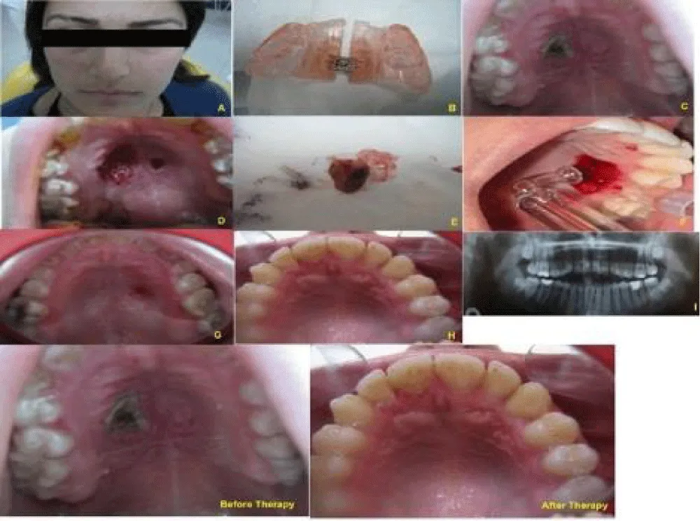
A) Clinical photograph of the patient’s profile
B) Photograph of orthodontic appliance
C) Preoperative clinical intraoral view
D) Clinical view at the time of surgery phase
E) Photograph of necrotic mass
F) Application of ozone
G) Four weeks after postoperative view following therapy
H) Postoperative clinical view three months after surgery
I) Panoramic radiograph view
Clinical findings: Intraoral examination revealed extensive necrotic lesions covered with dark yellowish fibrinous tissue, accompanied by surrounding erythema and ulceration on both the right and left sides of the hard palate (Figure 1C). The necrotic area on the palatal mucosa, measuring approximately 20 mm in diameter, was larger than the contralateral lesion. The involved palatal tissues appeared firmer and more sensitive on palpation compared with the adjacent healthy mucosa, and the patient reported severe pain in the affected regions. The lesion significantly interfered with speaking and eating functions, thereby negatively affecting the patient’s quality of life. Periodontal evaluation demonstrated gingival bleeding, edema, and erythematous changes associated with plaque accumulation around the affected area. Oral hygiene was considered inadequate, although periodontal probing depths were within physiological limits. Radiographic examination did not reveal any pathological findings (Figure I).
Diagnostic approach: Necrosis of the soft tissue after placement of the orthodontic appliance during maxillary expansion.
Therapeutic intervention and follow-up: The treatment approach included oral hygiene instruction, surgical debridement and excision of the necrotic soft tissue, ozone application, adjustment and polishing of the orthodontic appliance, and detailed patient guidance regarding postoperative care (Figure 1D, E and F).
Surgical procedure
Before the surgical procedure, extraoral antisepsis was achieved using a 10% povidone–iodine solution (Glividon®, Bikar Drug Ltd., Istanbul, Turkey), and the patient was instructed to rinse with 0.12% chlorhexidine digluconate mouthwash (Kloroben®, Drogsan Drug Ltd., Istanbul, Turkey) before administration of local anesthesia. Necrotic tissues were carefully removed using a surgical curette. Following meticulous mechanical debridement, the affected regions were gently irrigated with sterile physiological saline solution. Subsequently, relative isolation of the operative field was obtained, the areas were dried with sterile gauze, and ozone therapy was applied to the lesion sites.
Ozone application
Gaseous ozone was administered to the treated areas using an OzoneDTA device (OzoneDTA; Apoza Enterprise Co. Ltd., Taiwan) in accordance with the manufacturer’s recommendations. Ozone delivery was performed with a No. 3 probe at power level 9 for 60 seconds at each site. Gas ozone was applied three times for three consecutive days. Continuous aspiration was used throughout the procedure to minimize the risk of ozone inhalation.
Postoperatively, the patient was prescribed analgesic medication (Naproxen sodium 550 mg, every 12 hours for 5 days) and instructed to rinse twice daily with 0.12% chlorhexidine mouthwash (Kloroben®, Drogsan Drug Ltd., Istanbul, Turkey) for one week. She was also advised to avoid mechanical irritation or pressure on the surgical area during the healing period. In addition, oral hygiene instructions included brushing twice daily with a soft-bristled toothbrush for approximately 3 minutes each session, and a soft, non-irritating diet was recommended.
Complete soft tissue healing was observed four weeks after surgery (Figure G). At the three-month follow-up examination, the affected palatal region demonstrated a clinically healthy and normal appearance (Figure H). The patient continued to be monitored periodically for approximately two years postoperatively. Table 1 shows a summary of the clinical timeline, treatment interventions, and follow-up.
Patient perspective: The patient expressed understanding of the diagnosis and the recommended treatment of surgical excision and ozone therapy. She agreed to regular clinical monitoring for any changes in the lesion sites.
Patient consent: The present case report was conducted in accordance with the principles of the Declaration of Helsinki, and written informed consent was obtained from the patient for publication of the clinical findings and images.
Discussion
The present case involved oral soft tissue necrosis associated with the use of a tooth–tissue–borne orthodontic appliance (HAAS-TYPE expander), and the severity of the lesion appeared to increase due to prolonged and unrecognized tissue contact. Mechanical traumatic injuries within the oral cavity are generally localized in nature. Improperly fabricated dental appliances and defective restorations may lead to iatrogenic mechanical trauma to the oral tissues [2-4], as observed in this patient. As early as 1912, Greene Vardiman Black emphasized the close relationship between iatrogenic factors and periodontal tissue degeneration [5]. Although orthodontic treatment provides numerous well-documented benefits, including improvements in esthetics, phonation, mastication, swallowing function, periodontal condition, and patient self-confidence, orthodontic appliances may also lead to undesirable complications when adequate precautions and maintenance procedures are not implemented during treatment [6,7]. Reported adverse effects associated with orthodontic therapy include plaque accumulation, calculus formation, bleeding on probing, increased periodontal pocket depth, attachment loss, gingival recession, alveolar bone loss, enamel demineralization, external root resorption, tooth discoloration, pulpal devitalization, pain, hemorrhage, infection, asymmetric expansion, relapse, palatal ulceration, and tissue necrosis [6,7]. Several of these complications were evident in the present case.
Rapid maxillary expansion is a widely used orthodontic procedure for the management of transverse maxillary deficiency and posterior crossbite through enlargement of the maxillary dental arch [6,7]. The technique is considered effective, relatively simple to perform, and generally does not require extensive patient cooperation during appliance use [6]. Additional advantages include treatment predictability, long-term stability, efficiency, and increased maxillary arch perimeter. Despite its widespread clinical use, the literature contains limited information regarding severe soft tissue complications associated with RME therapy. In the current case, extensive necrosis affecting the palatal mucosa of the hard palate developed secondary to the expansion appliance and was managed successfully.
Several factors may contribute to periodontal and soft tissue complications during orthodontic treatment, including inadequate plaque control, poor patient compliance, limited oral hygiene ability, excessive or rapid appliance activation, defective or improperly fabricated orthodontic appliances, and insufficient patient motivation [6,7]. Therefore, careful appliance monitoring and strict oral hygiene maintenance are essential throughout treatment. In the present case, the complication was associated with a defectively manufactured appliance that caused continuous traumatic irritation of the palatal tissues.
Management of the lesion consisted of surgical removal of the necrotic tissue combined with correction and adjustment of the orthodontic appliance, resulting in uneventful healing and a favorable clinical outcome. Previous reports have similarly suggested that treatment of oral soft tissue necrosis generally involves excision of necrotic tissues together with elimination of the traumatic etiological factor [2]. Surgical debridement therefore appears to represent an effective therapeutic approach for mechanical traumatic injuries involving the oral soft tissues. In addition, ozone therapy has beneficial effects on the healing process, such as accelerating the epithelialization process, decreasing postoperative pain levels, and reducing patient morbidity. Previous studies have reported that ozone therapy in surgical applications exerts not only antimicrobial effects but also modulates the inflammatory response, enhances tissue oxygenation, and supports fibroblast activity and collagen synthesis [9,10]. In the present case, ozone was applied three times and accelerated the epithelialization process and reduced postoperative pain. Only a limited number of reports in the literature have described palatal soft tissue necrosis associated with orthodontic appliances. To the best of our knowledge, the present report is among the first to describe successful management of such a lesion related to rapid maxillary expansion therapy.
Conclusion
Traumatic lesions of the oral cavity may arise both in clinical dental settings and in everyday home environments. The professional competence, clinical experience, and current knowledge of dental practitioners are essential for reducing the likelihood of iatrogenic injuries. Moreover, many of these undesirable periodontal complications can be prevented through appropriate precautionary measures, careful monitoring, and timely intervention by both the orthodontist and the patient throughout treatment.
Patient consent
The patient’s parents/legal guardians provided informed consent for the therapeutic procedure and for the use of anonymized clinical data and photographs for scientific and educational purposes. Written consent is maintained in the authors’ records
Funding
This study received no specific grant from any funding agency in the public, commercial, or non-profit sectors.
Authors’ contributions
Alparslan Dilsiz diagnosed and managed the patient, conceived the idea of the case report, drafted the manuscript, and critically revised the manuscript.
Zeynep Tekin, Fevziye Bekdaş, Edanur Diri, and Şuheda Tak performed periodontal therapy and surgery procedure and ozone application. All authors read and approved the final version of the text.
- Holmstrup P, Plemons J, Meyle J. Non-plaque-induced gingival diseases. J Periodontol. 2018;89 Suppl 1:S28-S45. Available from: https://doi.org/10.1002/jper.17-0163
- Anura A. Traumatic oral mucosal lesions: a mini review and clinical update. Oral Health Dent Manag. 2014;13:254-259. PMID:24984629. Available from: https://pubmed.ncbi.nlm.nih.gov/24984629/
- Dilsiz A, Aydin T. Gingival recession associated with orthodontic treatment and root coverage. J Clin Exp Dent. 2010;2:e30-e33. Available from: https://www.medicinaoral.com/odo/volumenes/v2i1/jcedv2i1p30.pdf
- Dilsiz A, Aydin T. Self-inflicted gingival injury due to habitual fingernail scratching: a case report with a 1-year follow-up. Eur J Dent. 2009;3:150-154. PMID:19421397. Available from: https://pubmed.ncbi.nlm.nih.gov/19421397/
- Black A. Preventive treatment of periodontal diseases. Northwestern Dent J. 1912;10:60-73.
- Kurt G, Altuğ AT, Türker G, Kiliç B, Kiliç E, Alkan A. Effects of surgical and nonsurgical rapid maxillary expansion on palatal structures. J Craniofac Surg. 2017;28(3):775-780. Available from: https://doi.org/10.1097/scs.0000000000003468
- Pereira MD, Koga AF, Prado GPR, Ferreira LM. Complications from surgically assisted rapid maxillary expansion with HAAS and HYRAX expanders. J Craniofac Surg. 2018;29:275-278. Available from: https://doi.org/10.1097/scs.0000000000004079
- Prasad S, Nassar M, Azzam AY, Jose FG, Jamee M, Sliman RKA, et al. CaReL Guidelines: a consensus-based guidelines on case reports and literature review (CaReL). Barw Med J. 2024;2(2):13-19. Available from: https://www.barwmedical.com/index.php/BMJ/article/view/89
- Maglia DR, Souza BDAF, Visioli F. Efficacy of ozone therapy for oral mucosa wound healing: a systematic review and meta-analysis. Clin Oral Investig. 2024;28:490. Available from: https://doi.org/10.1007/s00784-024-05873-2
- Isler SC, Uraz A, Guler B, Ozdemir Y, Cula S, Cetiner D. Effects of laser photobiomodulation and ozone therapy on palatal epithelial wound healing and patient morbidity. Photomed Laser Surg. 2018;36:571-580. Available from: https://journals.sagepub.com/doi/abs/10.1089/pho.2018.4492

 Save to Mendeley
Article Alerts
Subscribe to our articles alerts and stay tuned.
 This work is licensed under a Creative Commons Attribution 4.0 International License.