Journal of Dental Problems and Solutions
A Rare Developmental Odontogenic Cyst: Lateral Periodontal Cyst and 16-Month Follow-Up: Case Report
1DDS, PhD. Professor, Department of Periodontology, Faculty of Dentistry, Atatürk University, Erzurum, Turkey
2DDS, Post Graduate Student. Department of Periodontology, Faculty of Dentistry, Atatürk University, Erzurum, Turkey
Author and article information
Cite this as
Dilsiz A, et al. A Rare Developmental Odontogenic Cyst: Lateral Periodontal Cyst and 16-Month Follow-Up: Case Report. J Dent Probl Solut. 2026; 13(2): 25-29. Available from: 10.17352/2394-8418.000138
Copyright License
© 2026 Dilsiz A, et al. This is an open-access article distributed under the terms of the Creative Commons Attribution License, which permits unrestricted use, distribution, and reproduction in any medium, provided the original author and source are credited.Aim: Although uncommon, Lateral Periodontal Cyst (LPC) represents an important entity in the differential diagnosis of lesions sharing comparable clinical and radiographic characteristics. The present case report describes the clinical presentation, diagnosis, treatment, and follow-up of a patient with LPC.
Case Report: A 41-year-old man was referred to the Department of Periodontology in February 2025 for the evaluation and management of a cystic lesion located in the left mandibular premolar–molar region. The provisional diagnosis of LPC, based on clinical and radiographic findings, was subsequently confirmed by histopathological examination following surgical enucleation. Treatment consisted of oral hygiene instruction, initial periodontal therapy, and complete excision of the lesion.
Results: The lesion was successfully managed through surgical periodontal treatment. Clinical and radiographic follow-up was carried out over 16 months. At the final follow-up visit, the surgical site exhibited satisfactory healing, with no evidence of recurrence and radiographic signs of bone regeneration.
Conclusion: The present case underscores the importance of integrating clinical, radiographic, and histopathological findings in the diagnosis of a lateral periodontal cyst. Histopathological examination remains indispensable for definitive diagnosis, while long-term follow-up is advisable to monitor healing and detect any possible recurrence.
Radiolucent lesions of the jaws comprise a broad spectrum of odontogenic and non-odontogenic cysts and tumors. Among these lesions, odontogenic cysts represent some of the most frequently encountered pathologies affecting the maxilla and mandible. Based on their etiology and pathogenesis, odontogenic cysts are generally categorized into two major groups: inflammatory and developmental cysts [1].
Although rare, Lateral Periodontal Cyst (LPC) represents an important developmental odontogenic lesion that should be considered when evaluating lesions with comparable clinical and radiographic presentations. It has been reported to account for fewer than 0.4% of all odontogenic cysts [2-4]. LPC most frequently develops adjacent to the lateral aspects of the roots of mandibular canine and premolar teeth, whereas occurrence confined to the molar region is relatively uncommon. Therefore, LPC should be considered among the developmental odontogenic cysts included in the differential diagnosis of radiolucent lesions arising in the intermolar area [4].
LPC is frequently detected incidentally during routine radiographic examinations and typically appears as a well-circumscribed oval or teardrop-shaped radiolucency situated between the roots of vital teeth. Owing to its radiographic appearance, it may occasionally be mistaken for a lesion of endodontic origin [5-7]. Although clinical and radiographic findings provide important diagnostic clues, definitive diagnosis relies on histopathological assessment. Microscopically, LPC is characterized by a thin epithelial lining containing focal epithelial plaques and glycogen-rich clear cells, which are considered remnants of odontogenic epithelium. The cyst lining usually consists of one to five layers of cuboidal or non-keratinized squamous epithelial cells with focal palisading. The exact histogenesis of LPC remains uncertain, and several theories have been proposed regarding its origin, including derivation from the reduced enamel epithelium, dental lamina remnants, or the epithelial rests of Malassez [2,3].
Surgical enucleation is generally regarded as the treatment of choice for LPC, although recurrence has been reported in a small number of cases [5,7]. Advanced imaging techniques, such as cone-beam computed tomography, may provide valuable information for treatment planning and facilitate a more precise surgical approach. Prompt management of suspected cystic lesions is important, particularly in older individuals, as it may help minimize postoperative complications and allow exclusion of potential malignant pathology. Early removal of LPCs therefore contributes to both accurate diagnosis and favorable clinical outcomes [4]. The present case highlights the clinical, radiographic, and histopathological characteristics of LPC and reinforces the importance of histopathological examination in establishing a definitive diagnosis. This case report was prepared in accordance with the CARE (CAse REport) guidelines [8].
Case report
Patient information: A 41-year-old male patient was referred to the Department of Periodontology in February 2025 for the assessment and management of a radiolucent lesion located adjacent to the mesial aspect of the mandibular left second premolar.
His medical history was noncontributory. He reported no systemic diseases, medication use, known allergies, or smoking habit. The patient sought evaluation after noticing a well-defined swelling in the left posterior mandibular region. The lesion was located beneath the mucosa and produced a localized prominence in the vestibular area.
Clinical Findings: Extraoral examination, including assessment of the facial profile, revealed no remarkable findings (Figure A). Intraoral examination demonstrated a localized swelling of the vestibular mucosa with largely normal overlying mucosal coloration in the left posterior mandibular region (Figure B). Palpation demonstrated a slight elevation without associated tenderness. Clinical inspection revealed a well-circumscribed, rounded swelling beneath the vestibular mucosa. The overlying mucosa appeared intact, with no evidence of erythema, ulceration, or discoloration. The surrounding gingival tissues exhibited normal morphology and color, without signs of gingival recession or enlargement. No periodontal pocketing was detected in the affected area. The adjacent teeth showed no mobility, and no diastema was present between teeth 35 and 36. Furthermore, the involved teeth were not tender to percussion, responded normally to thermal testing, and demonstrated vitality upon electric pulp testing. The patient was asymptomatic and reported no pain or discomfort.
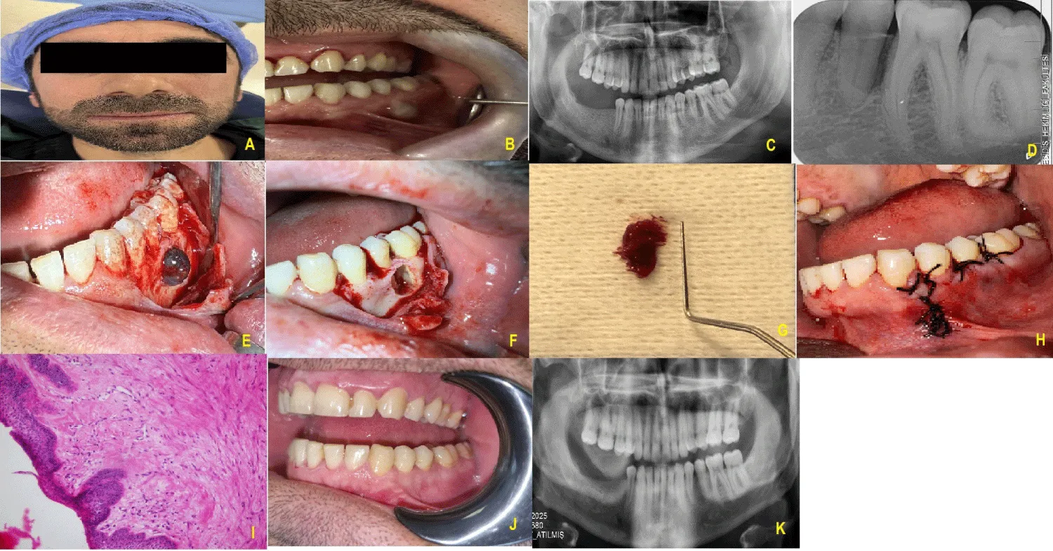
A) Front profile photograph of the patient during extraoral examination.
B) Vestibular swelling observed during intraoral examination.
C) Panoramic X-ray image.
D) Periapical X-ray image.
E) View of the cystic lesion exposed with the mucoperiosteal flap lifted after a sulcular incision in the left posterior region of the mandible.
F) View after the lesion was removed.
G) View of the cystic lesion
H) Postoperative intraoral suturing view.
I) Histopathological specimen; (×40 magnification, HE staining)
J) Postoperative intraoral examination image taken after 16 months.
K) Postoperative Panoramic X-ray image taken after16 months.
Panoramic radiography revealed a well-defined oval radiolucent lesion in the affected region. Periapical radiographic examination demonstrated a localized radiolucency associated with the area between teeth 35 and 36, accompanied by loss of lamina dura continuity. No evidence of root resorption was observed in the adjacent teeth (Figures C,D). Based on the clinical and radiographic findings, a provisional diagnosis of developmental LPC was established. Before surgical intervention, the patient underwent scaling and root planing and received detailed oral hygiene instructions. The patient was subsequently evaluated for diagnosis and treatment planning.
Diagnostic Approach: Based on clinical and radiographic findings, a provisional diagnosis of LPC was considered and confirmed by histopathological examination.
Therapeutic intervention and follow-up: Treatment consisted of oral hygiene instruction, initial periodontal therapy, and complete excision of the lesion.
Surgical procedure
Surgical treatment was carried out under local anesthesia using standard aseptic protocols. A sulcular incision extending from teeth 34 to 36 was performed, and a full-thickness mucoperiosteal flap was elevated. Following reflection of the flap, a cystic cavity lined by a distinct membrane was identified. Examination of the lesion revealed perforation of the vestibular cortical plate caused by expansion of the cystic wall (Figure E). Complete enucleation of the lesion was performed, and the surgical site was thoroughly irrigated with sterile saline solution (Figures F,G). The flap was repositioned and secured with 3-0 nonabsorbable silk sutures, followed by placement of a periodontal dressing (Figure H). Histopathological evaluation confirmed the diagnosis of LPC (Figure I). Correlation of the clinical, radiographic, and histopathological findings established the final diagnosis. Microscopically, LPC is characterized by a thin epithelial lining containing focal epithelial plaques and glycogen-rich clear cells, which are considered remnants of odontogenic epithelium. The cyst lining usually consists of one to five layers of cuboidal or non-keratinized squamous epithelial cells with focal palisading.
Postoperatively, analgesic medication was prescribed for 5 days at 12-hour intervals. The patient was instructed to rinse with 0.12% chlorhexidine mouthwash twice daily for 2 weeks and to avoid mechanical trauma to the surgical site. Toothbrushing was temporarily discontinued in the operated area during the healing period. Sutures were removed 15 days after surgery, and oral hygiene instructions were reinforced.
At the 4-week follow-up visit, complete soft-tissue healing was observed without any postoperative complications, and plaque control was satisfactory. The patient was subsequently monitored for 16 months. At the final follow-up examination, both clinical and radiographic assessments demonstrated uneventful healing, absence of recurrence, and evidence of bone regeneration in the treated area (Figures J,K).
Patient perspective: The patient expressed understanding of the diagnosis and the recommended treatment of surgical excision. He agreed to regular clinical monitoring for any changes in the lesion site.
Patient consent: This case report was conducted in accordance with the ethical principles of the Declaration of Helsinki (2013 revision). Written informed consent was obtained from the patient for publication of the clinical data and accompanying images.
Discussion
Lateral periodontal cyst is an uncommon developmental odontogenic cyst that arises along the lateral root surface of vital teeth within the alveolar bone. Because it is typically asymptomatic, the lesion is frequently identified incidentally during routine radiographic examinations. Consistent with previous reports, the patient in the present case did not experience pain or significant clinical symptoms. LPC predominantly affects adults and has been reported more frequently in males, particularly during the fifth and sixth decades of life. The mandibular premolar region is considered the most common site of occurrence, whereas involvement of the premolar–molar region is relatively uncommon [9,10]. The demographic and clinical characteristics observed in our patient are in agreement with those reported in the literature.
Radiographically, LPC generally appears as a small, well-circumscribed round or oval radiolucency, often surrounded by a thin sclerotic border. Alterations in the lamina dura and periodontal ligament space may also be present. In contrast to lesions of endodontic origin, the associated teeth usually remain vital. In the present case, the radiographic findings were compatible with those typically described for LPC. Nevertheless, radiographic and clinical findings alone are insufficient for definitive diagnosis. Histopathological evaluation remains the gold standard for confirming LPC and differentiating it from other lesions with similar clinical and radiographic appearances [10]. Previous studies have demonstrated that lesions initially presumed to be benign developmental cysts may occasionally exhibit unexpected histopathological characteristics, emphasizing the importance of microscopic examination [9-10]. Therefore, histopathological assessment was incorporated into the diagnostic process in the present case to establish an accurate diagnosis and guide appropriate treatment planning.
The differential diagnosis of LPC includes gingival cysts, radicular cysts, and odontogenic keratocysts. Gingival cysts share several clinical and epidemiological features with LPC; however, they are confined to soft tissues and generally lack radiographic findings [2,3]. Radicular cysts represent another important consideration because of their relatively high prevalence. Unlike LPC, these lesions are usually associated with non-vital teeth and are commonly related to pulpal necrosis [5,10]. Careful evaluation of tooth vitality together with radiographic characteristics is therefore essential to avoid unnecessary endodontic treatment. In the present case, comprehensive clinical and radiographic examinations supported the diagnosis of LPC.
Surgical enucleation is widely accepted as the standard treatment approach for LPC, and endodontic therapy is generally unnecessary when the associated teeth remain vital. Histopathological examination of the excised specimen not only confirms the diagnosis but also excludes the presence of other pathologic entities. Recurrence following surgical treatment is uncommon, although periodic follow-up is recommended [10]. In the present case, complete surgical excision was performed, the diagnosis was confirmed histopathologically, and the patient remained free of recurrence during the follow-up period.
Conclusion
Lateral periodontal cysts may mimic other odontogenic cystic lesions in both their clinical presentation and radiographic appearance, making histopathological evaluation essential for establishing a definitive diagnosis. Surgical enucleation remains an effective treatment modality, providing both diagnostic confirmation and favorable clinical outcomes, with recurrence reported only rarely. Given the uncommon nature of this lesion, documentation of additional well-characterized cases is valuable for increasing awareness among clinicians and improving the accuracy of differential diagnosis.
- Soluk-Tekkesin M, Wright JM. The World Health Organization classification of odontogenic lesions: a summary of the changes of the 2017 (4th) edition. Turk Patoloji Derg. 2018;34(1):1-18. Available from: https://doi.org/10.5146/tjpath.2017.01410
- Chrcanovic BR, Gomez RS. Gingival cyst of the adult, lateral periodontal cyst, and botryoid odontogenic cyst: an updated systematic review. Oral Dis. 2019;25(1):26-33. Available from: https://doi.org/10.1111/odi.12808
- Johnson NR, Gannon OM, Savage NW, Batstone MD. Frequency of odontogenic cysts and tumors: a systematic review. J Investig Clin Dent. 2014;5(1):9-14. Available from: https://doi.org/10.1111/jicd.12044
- Brooks JK, Portillo N, Bhatti AK, Parsa A, Basile JR. Lateral periodontal cyst in an interdental molar site: novel presentation in a geriatric patient. Gerodontology. 2025;42(1):124-128. Available from: https://doi.org/10.1111/ger.12759
- Dilsiz A, Gül Sevinc S. Odontogenic cysts of oral cavity: lateral periodontal cyst; a case report with a 14-year follow-up. J Dent Oral Biol. 2022;7(2):1193-1196.
- Formoso Senande MF, Figueiredo R, Berini Aytés L, Gay Escoda C. Lateral periodontal cysts: Available from: a retrospective study of 11 cases. Med Oral Patol Oral Cir Bucal. 2008;13(5):E313-E317. Available from: https://pubmed.ncbi.nlm.nih.gov/18449116/
- Ramalingam S, Alrayyes YF, Almutairi KB, Bello IO. Lateral periodontal cyst treated with enucleation and guided bone regeneration: a report of a case and a review of pertinent literature. Case Rep Dent. 2019;2019:4591019. Available from: https://doi.org/10.1155/2019/4591019
- Prasad S, Nassar M, Azzam AY, Jose FG, Jamee M, Sliman RKA, et al. CaReL guidelines: a consensus-based guidelines on case reports and literature review (CaReL). Barw Med J. 2024;2(2):13-19. Available from: https://www.barwmedical.com/index.php/BMJ/article/view/89
- Mendes RA, van der Waal I. An unusual clinicoradiographic presentation of a lateral periodontal cyst: report of two cases. Med Oral Patol Oral Cir Bucal. 2006;11(2):E185-E187. Available from: https://pubmed.ncbi.nlm.nih.gov/16505801/
- de Andrade M, Silva AP, de Moraes Ramos-Perez FM, Silva-Sousa YT, da Cruz Perez DE. Lateral periodontal cyst: report of case and review of the literature. Oral Maxillofac Surg. 2012;16(1):83-87. Available from: https://doi.org/10.1007/s10006-010-0257-2



 Save to Mendeley
Article Alerts
Subscribe to our articles alerts and stay tuned.
 This work is licensed under a Creative Commons Attribution 4.0 International License.