Journal of Dental Problems and Solutions
Human Dentinal Tubules alterations after desensitizing Dentifrices use: An ex vivo study
Rafael Lima Pedro1, Luise Gomes da Motta2 and Marcelo de Castro Costa3*
2Associated Professor, Dentistry, Department of Federal Fluminense University, Brazil
3Adjunt Professor, Pediatric Dentistry and Orthodontic, Department of Federal University of Rio de Janeiro, Brazil
Cite this as
Pedro RL, da Motta LG, de Castro Costa M (2017) Human Dentinal Tubules alterations after desensitizing Dentifrices use: An ex vivo study. J Dent Probl Solut 4(4): 076-081. DOI: 10.17352/2394-8418.000054Objective: Evaluated the effects of desensitizing dentifrices on dentinal tubule occlusion in human teeth thought qualitative and quantitative analysis.
Methods: Eighty dentin block were divided in eight groups, two controls (positive and negative) and six using desenstitizing dentifrices (Colgate Pro-alívio®, Colgate Tripla Ação®, Colgate Sensitivity®, Sensodyne Repair & Protect®, Sensodyne dentes sensíveis® and Cariax®) The specimens were storaged in natural saliva. A simulated toothbrushing machine were used corresponding 21 brushings weekly and used six cicles. For qualitative analysis Scanning Eletronic Microscopy (SEM) and Energy-Dispersive X-ray Epectroscopy (EDS) were used. Percentage of occluded tubules was obtained for quantitative analisys. The data were statistically analyzed by ANOVA and Tukey tests (p < 0.05).
Results: Colgate Pro-alívio®, Colgate Sensitivity®, Sensodyne Repair & Protect® and Sensodyne dentes sensíveis® showed better effect in occluded dentinal tubules (p<0.05), the slurry on dental surface were differents depending of active principle of samples and EDS showed presence of calcium and phosphorus in all groups.
Conclusion: All dentifrices were able to occluded dentinal tubules and Calcium and phosphorus were a regular found, however Cariax® andColgate Tripla Ação® had the worst results in quantitative analyses.
Introduction
Dentine hypersensitivity is a common dental problem caused by the exposure of dentinal tubules which allow the movement of intradentinal fluid leading to dentine hypersensitivity [1,2].
Many theories have been proposed to explain the dentin hypersensivity [3,4] as (a) odontoblasts and their processes act as dentinal receptor mechanisms; and (b) pulp nerves are stimulated by a hydrodynamic mechanism, and nerve impulses in the pulp are modulated by the release of certain polypeptides during pulp injury [3,4].
The hydrodynamic theory is widely accepted as the principal mechanism of action for this manifestation [5]. According to this theory, under certain chemical, thermal, tactile and osmotic stimuli the dentinal fluid moves inside the tubules indirectly stimulating the pulpal nerve endings, causing the sensation of pain. Nevertheless, the exact mechanism through which the dentinal fluid stimulates the nerve endings and causes pain is still unknown [5,6].
A viable treatment modality consists of desensitization by tubule occlusion, which decreases both dentine permeability and fluid movement, thereby reducing dentine sensitivity [6]. Thus, any substance that causes a decrease the dentinal fluid conductance by occluding the tubules is able to reduce clinical symptomatology of dentine hypersensitivity [7].
Various chemical agents have been used for mechanical formation of a natural smear layer by burnishing dentine induces tubule occlusion dentinal tubules occlusion and for dentine hypersensitivity treatment [6,8,9]. Introduction of topically applied compounds that form insoluble materials that precipitate in the tubules and on the surface, or that facilitate the formation of natural biological minerals, are also effective. Such compounds include abrasive particles, strontium or stannous salts, calcium phosphate, soluble oxalates and bioactive glasses [8-11].
In vivo studies have shown considerable decrease in hypersensitivity when teeth were brushed with desensitizing dentifrices [11-13]. However, the problem arises when attempts are made to determine the mode of action of active agents incorporated into these dentifrices.
Therefore, the aim of this study was to evaluate the effects of desensitizing dentifrices on dentinal tubule occlusion in human teeth thought qualitative and quantitative analysis.
Methodology
This was an in vitro study, using sound human dentin of permanent teeth for evalutation of dentin tubules occluding. This study was approved by the Ethics Committee of a Federal Institution.
Selection of experimental products
The dentifrices used for this research were those indicated for the treatment for dentin hypersensitivity with differents active ingredients reported for the manufactures. They are described in table 1.
Selection and preparation of the samples
Ten sound premolars teeth were select for this research. They were obtained from a Regulated Local Teeth Storage. These teeth were extracted for orthodontic incidation and passed through a visual inspection with a optical microscophy (DX 41 - Olympus, Tokyo, Japan), those with any type of racks were eliminated from this research.
After this pre-selection, the teeth were sectioned along their central axis, just below the cemento-enamel junction (CEJ) through a high precision metallographic cutter (Isomet 1000 precision saw, Buëhler, Lake Bluff, IL, USA), for tried to get the same numbers and orientations of dentinal tubules in all specimens. Only were used blocks until 2 mm above CEJ. These cuts were standardized measuring 2x2 mm, being checked by a digital caliper MPI/E-101 (Mytutoyo, Tokyo, Japan).
The specimens were then fixed with cyanoacrylate in plastic bases of 4x4 mm with the buccal surface exposed. It was then performed a standardized polishing on all specimens using water silicon carbide paper # 400 followed # 600 for a minute each, under water refrigeration using a polishing machine (Metaloghaphic Politriz - DPU 10, Struers, Denmark) to remove any surface contamination and facilitate the access to the dentin.
Storage of specimens
It was used a pool of human saliva in this research for storage the dentin blocks with the aim of simulated oral conditions. The saliva’ donnors were healthy and could not use any drugs in the last 1 mouth before. As fluoride could interfered in the final result in occluded dentinal tubules, all donnor were instructed to use a no fluoridated toothpaste (Malvatrikis Baby®) during the collection period. It was used a non-stimulated saliva with was frozen after the collect used during the all experimental period.
In order that tried to eliminated all possible vies, the dentin blocks of the same tooth passed through all dentifrices used in the experiment, beside controls groups, thus the specimens were divided into eight groups, each one containing ten specimens. One for each dentifrice used plus two controls groups, a negative control (G1) which it was applied to 37% phosphoric acid to remove any smear layer and alllowed to open dentinal tubules and a positive one (G2), which was not performed any experimental intervention beside the sample preparation. A total of eighty specimens were produced and used in this experiment.
The block of dentin were kept in separate containers throughout the research for there wasn’t any possibility of contamination amoung groups.
Experimental tests
The brushing of the specimens was performed with a Simulated Toothbrushing Machine (MEV2 (Odeme, Joaçaba, SC, Brasil) according to ISO/DTS 145692 [14].
Sixty soft brushes (Condor Plus, Condor ®) were used for the experimental They had 26 tufts of 0.25 mm diameter and 10 mm in height, it was used one for each specimen of the experimental groups.
This simulated toothbrushing machine worked at a frequency of 1400 cycles per time, corresponding to 21 brushings weekly (3 times daily for 7 days) for six week. The brushes moved 3.8 cm axially. A weight of 200 grams was attached on the rods of the machine to created a satisfactory brushing pressure [14].
All the dentifrices were dilluited in desonized water in a 1:1 ratio to simulate the action of the saliva in oral cavity during a normal brushing.
SEM analyses
After the last brushing session, specimens were sotred in a shaker machine for 2 hours, washed with desonized water na sputter coated with a thin gold palladium layer. Photomicrographs were taken with a Scanning Electron Microscopy (SEM) (JSM – 6510 L.V - OBE) from each dentinal surface at 1.000x and 5.000x magnification for qualitative analysis, and Energy-dispersive X-ray spectroscopy (EDS) were done as well for slurry evaluated . Thus quantitative analises was performed by obtained percentage of occluded tubules, by dividing the total numbers of occluded tubules by dividing the total numbers of occluded tubules by the total numbers of tubules in each photomicrography. This result was then multiplied by 100, to obtain the percentage of occluded tubules for each photography [15]. The datas were statistically analyzed by one-way Analysis of Variance (ANOVA) and Tukey test (p < 0.05) using SPSS 16.0 program.
Results
For quantitative results, table 2 display the mean percentage of occluded tubules in each group. Positive group showed almost all open dentinal tubules (4.6 ± 5,2), result similar to the negative group (11.3 ± 10.7).
Among groups that used desensitizing dentifrices, all specimens showed occluded dentinal tubules. However Colgate Pro-alívio®, Colgate Sensitivity®, Sensodyne Repair & Protect® and Sensodyne dentes sensíveis® showed better effect in occluded dentinal tubules (p<0.05).
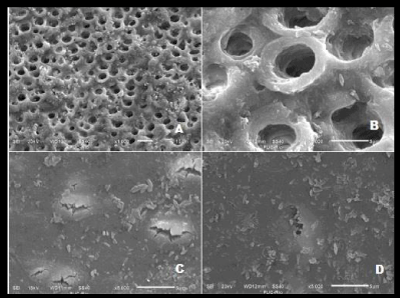
Analyzing the photomicrographs of SEM, the positive group showed almost all open tubules. While the negative group differed from the others by the presence of smear layer that partially occluded dentin tubules, but with a smoothly appearence. (Figure 1A-D).
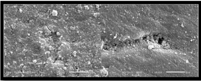
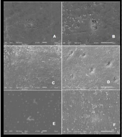
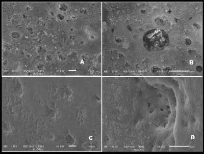
The Sensodyne Repair & Protect® showed the better result, however there were no difference significance compared with others desensitizing dentifrices (Figures 2A,B and 3A-F) with the exception of Colgate Tripla Ação® and Cariax® (Figure 4A-D).
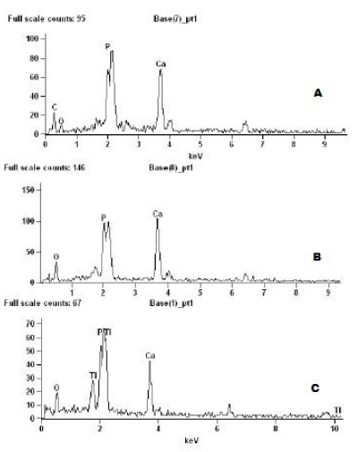
The EDS images shows similar results in all experimental groups (Figure 5A,B) with concentration of calcium and phosphorus. Some specimens shows slurry as titanium as Sensodyne dentes sensíveis® (5C). De figures of EDS of groups 4, 7 and 8 to demontrated similar founds in all groups.
The aspect of the slurry also differed among groups depending of active agent of the dentifrices. The slurry of Sensodyne Repair and Protect ® showed as a thick crystals bonded to the tooth surface and tubules as a barrier, thus in others dentifrices, these slurries showed more smoothly adhered with appearence to mineral reservoirs.
Discussion
Dentine hypersensitivity is the term used to describe common, painful condition of the permanent teeth; the etiology of which, however, is still poorly understood and the various mechanisms have been proposed to explain the development of dentinal hypersensitivity and treatment alternatives have been reviewed [16,17]. Most accepted of these is the hydrodynamic theory which was first explained by Gysi in 1900 and the experimental evidence for which was provided by Bränström [5]. According to this theory, the movement of dentinal fluid on stimulation with thermal, chemical, evaporative or electric stimulus is responsible for excitation of the underlying dentinal mechano-receptor resulting in sensitivity.
Many physiological and morphological studies have demonstrated that dentine hypersensitivity is more commum in cervical areas and results from open dentinal tubules at the surface being responsible for a higher dentin hypersensitivity by patients. The usuals causes for that ate abrasive habits, pacient’s age, maloclusion or other factors [6,18].
The difficulty found in treating for dentine hypersensitivity is expressed by the enormous number of techniques and therapeutic alternatives to relieve it [19]. This sensibility has been treated by a number of agents, which have been claimed to reduce pain by occluding dentinal tubules. However, other possible modes of action for desensitizing agents have been proposed, such as coagulating or precipitating constituents of tubular fluids, or by stimulating the formation of secondary dentine [17,20].
Several methods and materials have been tried to reduce dental sensitivity, ranging from home-use, over the counter products such as desensitizing mouthwashes, dentifrices or tray application foams to in-office application products such as varnishes, liners, restorative materials, dentinal adhesives iontophoresis procedures and more recently, lasers [19].
The home use products are most realistic and practical means of treating most patients with mild to moderate dentine hypersensitivity and they generally form the first step in routine management. Among these, desensitizing dentifrices have established themselves as principal home care therapeutic agents owing to the fact that they are readily and widely available, cost effective, simple to use and non-invasive and the habit of tooth brushing being almost universal [21].
However the exact way of each method acts for reducing sensitivity still shows undefined, thus this research tried to show in a vitro research, the effect of different agents in occluded dentinal tubules both qualitatively using SEM and EDS as quantitatively determining the percentage of tubules obliterated by each product tested as well changes in tooth surface and the arrangement of slurry on tooth, and their potential to produce secondary dentin or a mineral reserve.
The selection of dentifrices for this research were based in previous studies with proved the efficacy of active principles of the principal ingredients and the different modes of action of the different ingredients. These studies related for example that Arginine (Pro-Argine) is a sticky agent that physically occludes the tubules. The Calcium Sodium Phosphosilicate (Novamin) produces a form of apatite when in aqueous environment, an effect that is augmented by the presence of smear layer. The Potassium Nitrate, Potassium Citrate, Sodium Monofluorphosphate, Stronium Clhoride, and Sodium Potassium Phosphosilicate with the use, are deposited inside the dentinal tubules helping to obliterate them [1,3,8,12,13,22-27].
In order to try to avoid any bias that could affect the final result of this study, a new methodology was created from adaptations of similar studies [14,15] and was chosen to evaluate the results by various methods togheter such as SEM, EDS and quantification of tubules occluded in order to get a more reliable conclusion.
All blocks of dentin were obtained of the same region of the teeth, just below the cemento-enamel junction (CEJ). Thus, numbers and orientations of dentinal tubules could be standart. In addition, it was possible obtained from one tooth a number of samples enought for composing each group, experimental and controls, therefore each dentin block was able to be its own control for final analysis. In this study, the mean of occluded tubules of positive and negative group of each samples were similar than previous studies [15,27-30], so the results of dentifrices effects were truest results.
As fluoride could affect the occluded of tubules as previous studies demonstrated [25,31], it was chose storage the samples in natural saliva from donors that have not using fluoride dentifrices during the time of salive collection. This time was short to the point of not harming the donors.
Although some studies shows some substances as gold stantards in occluded tubules, specially Sodium Potassium Phosphosilicate [1,13,15,23,24,26,31,32], in our research all dentifrices were able to occluded dentinal tubules without any statistical difference. It could be seen at SEM images and qualitative analyses. The only exception was the dentifrices Colgate Tripla Ação® and Cariax®.
Thought SEM images it was possible to see the differences among the slurry formed from each dentifrice, althought there no statistical difference in the results of Sensodyne Repair and Protect®, it’s possible to see a solid mineral crystal formation on dental surface as well inside the dentinal tubules, while the others dentifrices showed a slurry disorganized on dentin surface as a mineral reserve and an aspect of occluded tubules by secondary dentin formation.
The EBS shows a presence of calcium and phosphorus in all samples, as others studies [15,33]. A substance usually found in this kind of study is a potassium [34,35]. As calcium, the potassium are able to be a reserver of minerals but potassium ions are thought to act by blocking synapses between nerve cells, thereby reducing nerve excitation and the associated pain [34,35]. In our study, the EDS couldn’t find potassium in the samples.
Although it could been found other substances not related by manufacturers in the bulla of dentifrices, as silica, titatium and carbon. These subtances have abrasives potencial and in theory are able to promove a superfial wear of dentin surface and then stimulated a formation of secondary dentin and occluded dentinal tubules [36,37]. While the titanium was a unexpected finding, once its acts in remineralization process mostly [38,39]. Probably the titanium was present in the dental structure instead dentifrices.
Therefore, the results of this research clarify the different effects on dental surface and slurry formation associating to differents principal active dependind of formulation of dentifrices. Taking this into consideration, researchs in vivo could transpose these findings allowing a more resolutive treatment for dentin hipersensitivity.
Conclusion
The Groups G4, G5, G6 e G7 Sensodyne Dentes Sensíveis® were statistically better on occluded dentinal tubules. The EDS showed that calcium and phosphorus were a regular found in all samples analyzed and SEM images demonstrated a formation of a slurry occludding tubules and stimuling secondary dentine.
- Orchardson R, Gillam DG (2006) Managing dentin hypersensitivity. Journal of the American Dental Association (1939) 137: 990-998. Link: https://goo.gl/gVukw6
- Gillam DG, Mordan NJ, Newman HN (1997) The Dentin Disc surface: a plausible model for dentin physiology and dentin sensitivity evaluation. Advances in dental research 11: 487-501. Link: https://goo.gl/oxb7vd
- Addy M, Dowell P (1983) Dentine hypersensitivity--a review. Clinical and in vitro evaluation of treatment agents. Journal of clinical periodontology 10: 351-363. Link: https://goo.gl/s14Vui
- Dababneh RH, Khouri AT, Addy M (1999) Dentine hypersensitivity - an enigma? A review of terminology, mechanisms, aetiology and management. British dental journal 187: 606-611. Link: https://goo.gl/FhzgGC
- Brannstrom M, Linden LA, Johnson G (1968) Movement of dentinal and pulpal fluid caused by clinical procedures. Journal of dental research. 47: 679-682. Link: https://goo.gl/PxNKvo
- Pashley DH (1986) Dentin permeability, dentin sensitivity, and treatment through tubule occlusion. Journal of endodontics 12: 465-474. Link: https://goo.gl/YSiU4N
- Markowitz K, Pashley DH (2008) Discovering new treatments for sensitive teeth: the long path from biology to therapy. Journal of oral rehabilitation. 35: 300-315. Link: https://goo.gl/syYHGY
- Suge T, Ishikawa K, Kawasaki A, Yoshiyama M, Asaoka K, et al. (1995) Effects of fluoride on the calcium phosphate precipitation method for dentinal tubule occlusion. Journal of dental research. 74: 1079-1085. Link: https://goo.gl/2ixcrs
- Kerns DG, Scheidt MJ, Pashley DH, Horner JA, Strong SL, et al. (1991) Dentinal tubule occlusion and root hypersensitivity. Journal of periodontology. 62: 421-428. Link: https://goo.gl/Yn9C5H
- Wang Z, Jiang T, Sauro S, Wang Y, Thompson I, et al. (2011) Dentine remineralization induced by two bioactive glasses developed for air abrasion purposes. Journal of dentistry. 39: 746-756. Link: https://goo.gl/NXC7ni
- Pereira JC, Segala AD, Gillam DG (2005) Effect of desensitizing agents on the hydraulic conductance of human dentin subjected to different surface pre-treatments--an in vitro study. Dent Mater 21: 129-138. Link: https://goo.gl/n23byM
- Pashley DH, O'Meara JA, Kepler EE, Galloway SE, Thompson SM, et al. (1984) Dentin permeability. Effects of desensitizing dentifrices in vitro. Journal of periodontology 55: 522-525. Link: https://goo.gl/NBokPQ
- Gandolfi MG, Silvia F, H PD, Gasparotto G, Carlo P (2008) Calcium silicate coating derived from Portland cement as treatment for hypersensitive dentine. Journal of dentistry. 36: 565-578. Link: https://goo.gl/q6a9eC
- Richmond R, Macfarlane TV, McCord JF (2004) An evaluation of the surface changes in PMMA biomaterial formulations as a result of toothbrush/dentifrice abrasion. Dent Mater 20: 124-132. Link: https://goo.gl/oU14Vy
- Arrais CA, Micheloni CD, Giannini M, Chan DC (2003) Occluding effect of dentifrices on dentinal tubules. Journal of dentistry. 31: 577-584. Link: https://goo.gl/2hQGxK
- Addy M (2005) Tooth brushing, tooth wear and dentine hypersensitivity--are they associated? International dental journal 55(4 Suppl 1): 261-267. Link: https://goo.gl/xCVQUM
- Berman LH (1985) Dentinal sensation and hypersensitivity. A review of mechanisms and treatment alternatives. Journal of periodontology 56: 216-222. Link: https://goo.gl/M9fhJh
- Absi EG, Addy M, Adams D (1987) Dentine hypersensitivity. A study of the patency of dentinal tubules in sensitive and non-sensitive cervical dentine. Journal of clinical periodontology 14: 280-284. Link: https://goo.gl/jvcCSo
- Porto IC, Andrade AK, Montes MA (2009) Diagnosis and treatment of dentinal hypersensitivity. Journal of oral science 51: 323-332. Link: https://goo.gl/Dkwk3i
- Bergenholtz G, Jontell M, Tuttle A, Knutsson G (1993) Inhibition of serum albumin flux across exposed dentine following conditioning with GLUMA primer, glutaraldehyde or potassium oxalates. Journal of dentistry 21: 220-227. Link: https://goo.gl/xzcfUd
- Curro FA (1990) Tooth hypersensitivity in the spectrum of pain. Dental clinics of North America. 34: 429-437. Link: https://goo.gl/DBVDL8
- Prati C, Venturi L, Valdre G, Mongiorgi R (2002) Dentin morphology and permeability after brushing with different toothpastes in the presence and absence of smear layer. Journal of periodontology. 73: 183-190. Link: https://goo.gl/xpDmTX
- Orsini G, Procaccini M, Manzoli L, Sparabombe S, Tiriduzzi P, et al. (2013) A 3-day randomized clinical trial to investigate the desensitizing properties of three dentifrices. Journal of periodontology 84: e65-73. Link: https://goo.gl/eU8aJL
- Kakodkar G, Lavania A, Ataide Ide N (2013) An In vitro SEM Study on the Effect of Bleaching Gel Enriched with NovaMin on Whitening of Teeth and Dentinal Tubule Occlusion. J Clin Diagn Res. 7: 3032-3035. Link: https://goo.gl/YWrU9Y
- West N, Newcombe RG, Hughes N, Mason S, Maggio B, et al. (2013) A 3-day randomised clinical study investigating the efficacy of two toothpastes, designed to occlude dentine tubules, for the treatment of dentine hypersensitivity. Journal of dentistry. 41: 187-194. Link: https://goo.gl/NnCeEV
- Fu D, Pei D, Huang C, Liu Y, Du X, et al. (2013) Effect of desensitising paste containing 8% arginine and calcium carbonate on biofilm formation of Streptococcus mutans in vitro. Journal of dentistry 41: 619-627. Link: https://goo.gl/7HWuVY
- Farmakis ETR, Kozyrakis K, Khabbaz MG, Schoop U, Beer F, Moritz A (2012) In vitro evaluation of dentin tubule occlusion by Denshield and Neodymium-doped yttrium-aluminum-garnet laser irradiation. J Endod 38: 662-666. Link: https://goo.gl/K8tRQy
- Fosse G, Saele PK, Eide R (1992) Numerical density and distributional pattern of dentin tubules. Acta odontologica Scandinavica 50(4): 201-210. Link: https://goo.gl/FaokUb
- Komabayashi T, Nonomura G, Watanabe LG, Marshall GW Jr, Marshall SJ (2008) Dentin tubule numerical density variations below the CEJ. Journal of dentistry. 36: 953-958. Link: https://goo.gl/aXwwtk
- Garberoglio R, Brannstrom M (1976) Scanning electron microscopic investigation of human dentinal tubules. Archives of oral biology 21: 355-362. Link: https://goo.gl/BH1VYe
- Prabhakar AR, Manojkumar AJ, Basappa N (2013) In vitro remineralization of enamel subsurface lesions and assessment of dentine tubule occlusion from NaF dentifrices with and without calcium. Journal of the Indian Society of Pedodontics and Preventive Dentistry 31: 29-35. Link: https://goo.gl/cLcwV6
- Acharya AB, Surve SM, Thakur SL (2013) A clinical study of the effect of calcium sodium phosphosilicate on dentin hypersensitivity. Journal of clinical and experimental dentistry 5: e18-22. Link: https://goo.gl/ShmUzH
- Earl JS, Topping N, Elle J, Langford RM, Greenspan DC (2011) Physical and chemical characterization of the surface layers formed on dentin following treatment with a fluoridated toothpaste containing NovaMin. The Journal of clinical dentistry. 22: 68-73. Link: https://goo.gl/nJYC12
- Markowitz K, Bilotto G, Kim S (1991) Decreasing intradental nerve activity in the cat with potassium and divalent cations. Archives of oral biology 36: 1-7. Link: https://goo.gl/n9M6UE
- Peacock JM, Orchardson R (1995) Effects of potassium ions on action potential conduction in A- and C-fibers of rat spinal nerves. Journal of dental research 74: 634-641. Link: https://goo.gl/AaWx5j
- West NX, Hooper SM, O'Sullivan D, Hughes N, North M, et al. (2012) In situ randomised trial investigating abrasive effects of two desensitising toothpastes on dentine with acidic challenge prior to brushing. Journal of dentistry. 40: 77-85. Link: https://goo.gl/NTXmjK
- Macdonald E, North A, Maggio B, Sufi F, Mason S, et al. (2010) Clinical study investigating abrasive effects of three toothpastes and water in an in situ model. Journal of dentistry 38: 509-516. Link: https://goo.gl/PS98VA
- Kazemi RB, Sen BH, Spangberg LS (1999) Permeability changes of dentine treated with titanium tetrafluoride. Journal of dentistry 27: 531-538. Link: https://goo.gl/BjmAZV
- Nassur C, Alexandria AK, Pomarico L, de Sousa VP, Cabral LM, et al. (2013) Characterization of a new TiF(4) and beta-cyclodextrin inclusion complex and its in vitro evaluation on inhibiting enamel demineralization. Archives of oral biology 58: 239-247. Link: https://goo.gl/uRZNyQ
Article Alerts
Subscribe to our articles alerts and stay tuned.
 This work is licensed under a Creative Commons Attribution 4.0 International License.

 Save to Mendeley
