Journal of Dental Problems and Solutions
A cephalometric study of the tongue position in class III patients
Wilfredo Molina Wills1* and Vanessa Rodriguez2
2Private Practice, Venezuela
Cite this as
Wills WM, Rodriguez V (2019) A cephalometric study of the tongue position in class III patients. J Dent Probl Solut 6(2): 028-031. DOI: 10.17352/2394-8418.000069Aims: The objective of this study was to evaluate the tongue position during deglutition with respect to the occlusal plane in class III patients with teeth in centric occlusion.
Methods: 24 lateral cephalometric radiographs of a group of 30 class III patients were randomly selected and divided into two
Groups: The first group for class III patients with open bite and the second group for class III patients with inverted overbite. The angles formed by the plane of the tongue and the occlusal plane obtained in all the samples were compared between the two studied groups. The T-test was used to evaluate the differences among studied angles. Results: The average of the anterior angles in patients with inverted overbite was lower than the average of the anterior-lower angles in class III patients with open bite. Anterior-lower angles with values between 24.17 and 34.08 were observed for class III with inverted overbite. T test of equality of means 6.661 significance, the angles were significantly different (p< .001) in both studied cases.
Conclusion: Tongue posture is significantly different during deglutition in class III with open bite and inverted overbite.
Introduction
Tongue is the largest organ of the oral cavity [1]. The posture, size and shape of the tongue are of significance for dent alveolar structures [2]. It has been showed that form; position and function of the tongue have some direct relationship with some types of oro-dental morphopathologics [3].
The tongue have probably influence by modifications in oro-dental environment, especially of the mandible [4]. It appears that a range of physiologic, pathologic, and mechanism factors can influence growth [5]. Although it has been that a close form and function relationship exists.
In previous studies have been reported the influence or not of the tongue size on the perimeter of the mandible dental arch in class III patients [6]. In the same way, it has been mentioned in class III patients with open bite has concave profile, a long lower facial height and mandible protrusion [7].
The purpose of this study was to evaluate the position of the tongue in relationship with the occlusal plane during swallowing with teeth in centric occlusion in class III patients.
Material and Methods
Lateral cephalometric radiographs with the x-ray beam perpendicular to the patient’s sagittal plane in natural head position to standardize orientation of the head that is reproducible for each individual were taken. This head position is a common method of orientation for cephalometric radiography [8,9]. Twenty-four lateral cephalometric radiographs of a group of 30 class III patients were randomly selected. These cephalogram were realized during deglutition with teeth at centric occlusion with the same operator. The selected cephalometric radiographs were divided into two groups: The first group for class III patients with open bite and the second group for class III patients with inverted overbite.
The angles formed by the plane of the tongue and the occlusal plane obtained in all the samples of the first group were compared with the same angles of the twelve samples of Group 2.
Clinical characteristics
Patients selected for this study had a growth pattern related to a dentofacial deformity with mandibular prognathism in relation to the maxilla. The selected cases had serious maxillomandibular discrepancies and typical class III profile.
All subjects met the following inclusion criteria: patients with molars and canines in Angle’s class III relation in open bite and inverted overbite were selected.
Individuals with class III profile.
Exclusion criteria: class III unilateral or incisors relation edge to edge cases were not selected.
Craniofacial deformities
Deviation of mandible
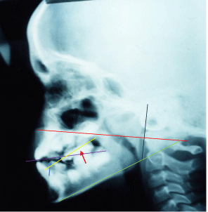
As marker for radiographic analysis of the position of the tip and dorsum of the tongue, a thin line of barium sulphate was used on the surface of the dorsum and tip of the tongue.
All radiographs were taken from the same x-ray machine with tongue position during deglutition. Lateral cephalograms were traced upon acetate paper. The exposure time for each film was of 9 seconds.
Tongue cephalometric points
-Tongue tip (TT)
-Highest point of the dorsum on the tongue (HDP)
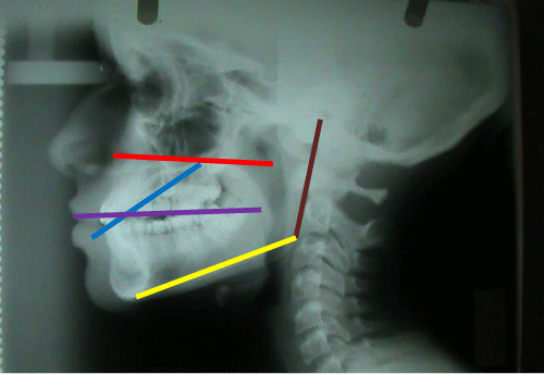
Planes and parameters used (Figure 1).
-Occlusal plane (line connecting first molars contact point and the middle point of canine overbite.
-Tongue tip to highest point on the tongue (line connecting TT and HDP).
-Plane ramus tangent.
-Mandibular plane.
-Palatal plane.
The palatal and occlusal planes were used to determine the presence or not of hyper divergent growth pattern in the studied cases.
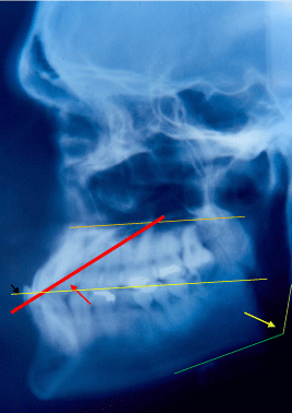
Angles (Figure 2)
-Anterior- lower angle
-Posterior- upper angle
-Goniac angle.
Student’s t-test for independent samples was used to measure the statistical significance between the tongue angles during deglutition in class III patients with inverted overbite and open bite.
To carry out this study, the patients approved all the procedures.
Ethical contents
This study was approved by ethics committee of the school of dentists of Merida state in Venezuela.
All the issues of the Helsinki Declaration related to research in human subjects were observed.
Results
Twelve cephalograms were classified as class III with open bite for 50 % of the studied cases and twelve cephalograms for 50 % of the cases with class III and inverted overbite table 1.
Table 1- f= (frequency). The major percentage of the 24 studied cases, the tip of the tongue was located at the level of the first premolar during deglutition (13 cases for a 54.36% of all studied patients) figure 3.
Table 2- 58.33% of the cases showed goniac angles between 121 and 129.
Table 3- 11 of the 12 cases with inverted overbite showed goniac angles within normal values, being the intermediate value of 120.1
Table 4 - Anterior-lower and posterior-upper angles in class III patients with inverted overbite. The average of the anterior angles in patients with inverted overbite was lower than the average of posterior angle of the tongue observed in the same studied cases.
Table 5 – In 83.33% of the studied cases, the anterior-lower angle was higher in class III patients with open bite.
Statistical results
Anterior-lower angles showed a 24.17 STD deviation 3.639 for class III with inverted overbite, and a 34.08 STD deviation 3.655 for class III with anterior open bite, f 0.076. The student test showed that the difference between the studied angles in class III cases was statistically significant (p< .001)
Discussion
The soft tissue in the oro-facial area formed by lips, floor of the mouth, tongue and soft palate are the determinants of the functional space. The role of the tongue in positioning of dent-alveolar structures has been emphasized. The growth, posture or functions of tongue are of significance [1]. This study suggests that position of tongue is related to skeletal morphology and can act differently depending of the sagittal skeletal pattern of each subject.
It is currently accepted the influence of tongue size on the perimeter of the mandibular dental arch 6. In the cases evaluated in this investigation on class III and open bite, it was possible to observe the tip of the tongue at the level of the first premolars with centric occlusion during deglutition. In studies carried out on the position of the tongue in class III, it has been reported an advanced position in class III patients with inverted over bite [10]. In this study, it was observed that the tongue occupies a more advanced position in class III with inverted overbite than in those cases with open bite. Similarly, a smaller angle was also observed between the tip of the tongue and the occlusion plane in cases of inverted overbite and class III that in those cases with open bite and class III during swallowing.
Due to the findings of this study, it is possible to suggest that the position and thrust of the tongue in class III patients may be related to the bite type. But more studies are needed to clarify this issue.
Conclusion
The present study has shown that:
1-Variations in the tongue position were evident between individuals with class III malocclussion with open bite and inverted overbite with respect to the occlusal plane. The anterior and posterior angles between the plane of the tongue and the occlusal plane were significantly different in the groups studied.
- Peat JH (1968) A cephalometric study of tongue position. Am J Orthod 54: 339–351. Link: https://bit.ly/2XU5ymD
- Rakosi T (1978) London: Wolf Medical Publication Limited; An atlas and manual of cephalometric radiography 96–98. Link: https://bit.ly/2Y4LEVl
- Fishman LS (1969) Postural and dimensional changes in the tongue from rest position to occlusion. Angle Orthod 39: 109–131969. Link: https://bit.ly/2Z1C02I
- Nanda RS, Dandajena TC, Nanda R (2009) Biomechanics strategies for nonextraction Class II malocclusions. In: Nanda R, editor. Biomechanics and esthetic strategies in clinical orthodontics. 1st ed. St. Louis: Missouiri, Elsevier 177. Link:
- Jhon Mew (2015) The influence of the tongue on dentofacial growth, Angle orthodontist 85: 4. Link: https://bit.ly/2YZzl9V
- Tamari K, Shimizu K, Ichinose M, Nakata S, Takahama Y (1991) Relationship between tongue volume and lower dental arch sizes, American journal of orthodontics and dentofacial orthopedics 100: 453-458. Link: https://bit.ly/2LtCPhT
- Togawa R, Shoichiro L, Shouichi M (2010) Skeletal Class III and open bite treated with bilateral sagittal split osteotomy and molar intrusion using titanium screws.The angle orthodontist 80: 1176-1184. Link: https://bit.ly/2JOdTOU
- Weber Diana W, Fallis Drew W, Parker Mark D (2013) Three dimensional reproducibility of natural head position . American journal of orthodontics and dentofacial orthopedics 143: 738-744. Link: https://bit.ly/2XT9KD6
- Naveen B, Jeetinder S, Gurmeet G, Monika G, Gurpreet K (2012) Reliability of natural head position in orthodontic diagnosis. A cephalometric study.Contemporary clinical dentistry 3: 180-183. Link: https://bit.ly/2M3XdFF
- Kalgotra S, Mustag M (2016) Position of tongue in skeletal class II and class III, A cefalometric study. journal of medical sciences 15: 33-38. Link: https://bit.ly/2LqtyqV
Article Alerts
Subscribe to our articles alerts and stay tuned.
 This work is licensed under a Creative Commons Attribution 4.0 International License.


 Save to Mendeley
