Journal of Dental Problems and Solutions
Treatment and failure of amalgam restoration analyzed according to class of restoration
AO Olaleye1* and OP Shaba2
2Associate Professor, Departement of Restorative Dentistry, College of Medical Sciences, University of Lagos, Nigeria
Cite this as
Olaleye AO, Shaba OP (2020) Treatment and failure of amalgam restoration analyzed according to class of restoration. J Dent Probl Solut 7(2): 084-089. DOI: 10.17352/2394-8418.000090Aims: This is a cross sectional; longitudinal retrospective study to find our reasons for placement of amalgam restoration at a Teaching Hospital in Nigeria and the most common classes of amalgam cavity prepared for amalgam restoration.
Material & methods: The record of patients also offended the dental centre of hospital in Nigeria was used for this study and all II records were centralized to separate those that attended the Conservation Dental Clinic for placement were recalled for a cross-examination and comparison with the records.
Results: Out of the 431 patients recalled, two hundred and seventy seven turned up (64.3%).
Two thousand and ninety four restoration were placed in regular attendees with classes I,II & V Accounting for 60.08%, 36.77% and 3.16% of all restorations placed respectively, primary caries accounted for 74.1% of all restoration placed, fractured restoration 16.1 % defective margins 3.7%; secondary caries 2.8% dislodged restoration 1.2 %;overcharging amalgam restoration 0.4% cervical abrasion 1.3 % and other reasons which include attrition, iatrogenic preparation accounted for 0.3%.
The reasons given for failure in the pooled study was seen repeating itself in that order in Class I and II.
Discussion: Class I restorations was the most commonly placed restoration followed by class II and class V restoration, the most common cause of failure in this study in all the classes of restoration was fractured amalgam restoration and the percentage is much higher for class II restorations. This may be due to the high masticatory load it is subjected to as a result of the cultural diet effect practiced in this environment, whereas in other studies carried out in the Caucasian region and other developed economies, secondary caries form the major reason for placement of restoration.
Clinical significance: Amalgam fillings are the most commonly performed restoration when treating caries but data in the developing countries on amalgam is sparse and dearth. It is of importance to know the longevity, the failure pattern, shortcoming of the restoration and to find out if the dates in developed countries tally with developing world for analysis and comparison.
Conclusion: Dietary habit may be a major reason in failure of amalgam restoration and it is important to note that cultural background may be a deciding factor in the types of failure seen.
The problem of over diagnosis of carious lesion may also play a part in primary caries especially in the Teaching Hospital/Dental College unlike what is seen in General Dental centres or Hospital, because Dental students and resident doctors in training are involved in the clerking and treatment of patients.
Introduction
Treatment of dental caries has been the preoccupation of dentists since the 17th century when it was discovered that vitriol can be dissolved in strong acids and when mercury was added to produce a filling material. Several materials have been tried between the years 1601 and 1896 when the first commercial alloy rich in silver was produced by G.V black [1].
Although some authors had equated restoring a tooth to “countdown” to extraction, it is still necessary to critically look into some of the problems encountered during the restoration of a tooth and various factors that will and affect the placement and success of the restoration2.
Since the inception of amalgam unfortunately no material has been able to withstand the test of time and the rigours of requirements needed for the constant stress and pressure found in oral environment. It is imperative that various factors that could seemingly affect the success of amalgam restoration should be critically evaluated and analyzed
For almost two centuries, dental amalgam has been an established material of choice for the treatment of dental caries in cases of virgin caries, lost or failed restorations in the posterior teeth. The biochemical and clinical properties, ease of application and use, serviceability of dental amalgam has made the material widely acceptable and relevant despite the various wars against it. It therefore remains the material of choice for most routine operative procedures in posterior teeth [1,2].
Recent random clinical trial showed that all the various systems side effects associated with the use of amalgam restorations using clinical based parameters were not linked with amalgam restoration thus showing that the use of amalgam in restoring teeth are safe [3-5].
Analysis of treatment and failure patterns seen in amalgam would expose the limitations and consequently the area that needs improvement and it would create the necessary awareness required so that the operations, could be informed of the circumstances that will affect the life of the fillings.
The mostly adduced reasons for the use of amalgam are but not limited to less technique sensitivity, superior mechanical properties higher survival good resistance to wear, excellent lead bearing properties and its convenient application [6-8].
This type of study can be complicated by lack of records on type of materials used, lack of uniform criteria for decisions to place and replace restorations, variations in decision-making between different clinicians [9] limited number of restorations, selection of patients, loss of patients [10,11]. A study [12] gave the most common reasons for restoration failure in decreasing order of proximal overhang, recurrent caries and food impaction.
Very few studies analyzed failure patterns seen according to classes of restoration. A study [13] reported that secondary carries was the reasons mostly cited for placement of amalgam restoration while another study [14] discussed the class distribution of the various reason given for replacement of amalgam restorations.
The aim of the study is therefore to analyze the reason given for replacement of amalgam restorations in classes I, II and V in Nigeria.
Materials and methods
A retrospective study of records of restorations placed can be used to determine the reasons for placement and replacement of amalgam restorations provided all the treatment carried out are properly and accurately recorded.
This study were records of patients between 1979 and 1992 in University College Hospital, Ibadan and are deemed regular attendees, if the patient attended the clinic for at least once in a year for a period of 5 years.
The records used in this study had proper documentation of all treatments carried out, only regular attendances were included in the study and all the patients involved in this study are at least 16 years old as at the time of first attendance.
As at the time the treatments were carried out the methods of caries detection was by visual & tactile perception, periapical & bitewing radiographs.
The reasons given for placement of amalgam restoration were recorded and because the records were self-explanatory it is possible sometimes to have more than one reason why an amalgam restoration failed. However, during collation and analysis of the data major criteria were set out in order of importance which are:
1. Primary caries
2. Fracture restoration
3. Secondary caries
4. Marginal defects/Ditching
5. Dislodged restoration
6. Overhanging amalgam restoration
7. Cervical abrasion
8. Fractured tooth
9. Dentinal sensitivity/Exposure due to attrition
7 – 9 were classified as others.
Any failure occurring as a result of more than one criteria would be regrouped according to the order of importance. The records were compiled according to class of restoration and each class is analysed according to the criteria given for placement and replacement of amalgam restoration.
Results
A total of two thousand and ninety-four amalgam restorations (2094) were placed in the study.
One thousand two hundred and fifty-eight (1,258) class I amalgam restoration were involved, while class II were seven hundred and seventy (770) and class V amalgam restoration was sixty six (66) which accounted for 60.01%M 36.8% and 3.1% respectively (Table 1).
Primary caries was the reason given for replacement of one thousand and five hundred and fifty two amalgam restoration (1552) which accounted for 74.1% of all restorations placed in this study and in the total primary placement was one thousand five hundred and eighty six restorations (1586) which formed 75.7% of all restorations placed in this study. Replacement formed 24.3% of all restorations which translated to five hundred and eight restorations (508) (Table 2)
The class I amalgam fillings included complex restorations such as Buccal/Lingual/Palatal extension while some involved extensive cuspal restorations while none was reinforced with pins.
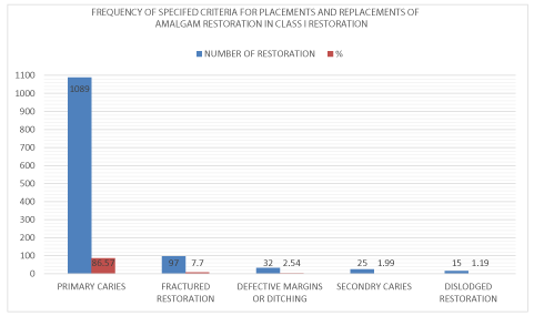
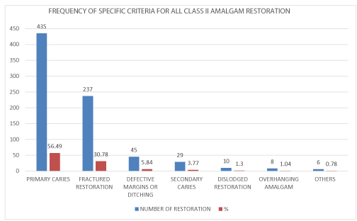
Primary placement accounted for 86.6% (1094) of all class I amalgam restorations while replacement of amalgam restorations accounted for 13.4% which translated to one hundred and sixty-nine restorations (Table 3). Primary caries was the reason given for placement of four hundred and thirty-five (435) amalgam restorations accounting for 56.4% of all the class II restoration, however total primary placement accounted for 57.27% which is four hundred and forty-one amalgam restoration (441).
Fractured restoration was the reason given for placement of two hundred and thirty seven restoration (237) which was 30.8% defective margin was 5.8% (45 amalgam restorations), secondary caries was 3.8% (twenty nine restorations); dislodged restorations was 1.3% and (ten restorations) overhanging amalgam restorations was 1.0% (6 amalgam restorations) and other reason such as fractured, cusp, occlusal attritions and iatrogenic preparations accounted for 0.8% (Table 4).
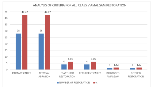
There was a total of sixty six class V amalgam restoration out of which 28 were placed due to primary caries, cervical abrasion was 28(42.4%) each while fractured restoration and recurrent caries accounted for 6.1%(4) each. Only one amalgam restoration each was recorded for dislodged and marginal ditching respectively. Primary placement (caries and cervical abrasion) amounted to 5656 restorations which formed 84.8% of all class V restoration.
Discussion
This study was a retrospective longitudinal study employing the use of records in the case file of already treated patients which will show only the reasons the treatment failed not the factors causing the failure. The cross-sectional part of the study was to find out the authenticity of the record of treatment done. This is because various factors that could determine the failure of restorations vary from type of materials used at any given time especially in this environment where materials are imported. This will in turn be shaped by content & composition of the materials; patient factors such as oral hygiene & habits, dietary habits; Operator's efficiency & abilities. Due to the fact that there are varying degrees of abilities of different operators, it is assumed that the outcome of the results would be smoothen outEven though there are several factors that could impact the success or otherwise of amalgam which is outside the scope of this study, however, it has been known that some problems which were identified as factors that could influence the success of amalgam restoration included lack of information on the type of materials used, oral hygiene status of patient, oral habits of the patients involved and the operators’ efficiency, however, the varying operator abilities involved would go a long way in smoothening out operator deficiencies [8-11,13-16].
Various studies [14-16] carried out in the past showed that primary caries accounted for about 25-29% of all restorations done whereas this study showed ‘a higher rate of 74.6% for primary caries treatment which is an indication that more than four out of every five restorations placed in this environment was due to primary caries.
Although it is also known that the number of caries diagnosed could be exaggerated especially pits and fissures caries thus leading to what is called over-diagnosis of caries, more so, in cases of dental students being involved in examination and diagnosis of patient in a setting of the Teaching Hospital. Ability to know when to treat a carious lesion is dependent the practitioners understanding of the pathogenesis of dental caries vis-à-vis its application to clinical treatment.
There is a dearth of reports on clinical studies that had analysed the relative percentages of primary caries according to the different classes, however, there was a study [10] that described factors causing replacement in relation to different classes.
It could be affirmed that at this stage of our development, recurrent caries is the least of the problems confronting amalgam restorations in Nigeria, rather it is the preponderance of fractured restoration.
Fractured restoration was the commonest cause of classes I and II restorations failure in Nigeria accounting for 62.4% of all failures or replacement of amalgams restoration unlike in a study [16] in Pakistan which accountant for 47% of the restoration replaced due to bulk fracture whereas in another study [17] only 4.7% was adduced to bulk fracture The cavity design & size of various lesion cannot explain the huge data of fractured restoration which was found in this study compared to several studies carried out in developed economies of Europe, America and some Asian countries. Not following the principles of resistance & retention forms will not adequately take care of the disparity seen in the number of failures arising from fractured restorations. So it is easier to conclude that something else must have contributed in this high rate of fractured restoration which was not reported in any studies. Therefore, the reason for such high rate in restoration fracture may lie in the patient's factor which may be explainable in the dietary habits seen in this environment.
In addition, the operators involved are resident doctors undergoing residency programs at different stages & dental students which has been shown in a study that abilities of these different operators were seen to even out.
Defective margins of restorations accounted for 3.2% of all restoration while replacement formed 14.4% of all replacement whereas in the study [17] conducted in Pakistan only 6% accounted for marginal defect/ditching. The latter condition of marginal defects has been variously linked to the old conventional amalgam with a preponderance of gamma – 2 phase. This becomes more important when it is viewed against the background that all alloys used in the country were imported and most of the times, the dealers and agents importing such were ignorant of properties and specifications of such alloys. It is also true that such alloys would be cheaper than the new copper alloys as the demand for such alloys in the developed countries has been greatly reduced or non- existent.
Fractured restorations had been linked to a general dietary habit found in Nigerians and the study showed [19] that this habit is commoner in females. Studies [20-22] have shown that alloys containing high gamma – 2 phase may contribute to a large extent to the degree of ditching or defective marginal deteriorations seen. However, the proposition by an author [20] that this might ultimately be responsible for recurrent caries cannot be extended to this result because the level of recurrent carries seen in this environment is very low.
Even though the factors responsible for the frequency of amalgam replacement were identical for classes I & II, it is observed that the percentage of failure seen in class II amalgam restoration was higher than in class I. One hundred and sixty-nine (169) amalgam restoration were replaced in class II and the percentage of failure attributed to the fact fractured restoration in class 1. This might not be unconnected to the fact that class I restorations are usually contained within walls of sound tooth tissue surrounding the restorations thus preventing fracture even with the habits noticed in Nigeria whereas the class II restoration was not totally enclosed by tooth tissue. It is therefore expected that most fractures would be located at the isthmus or along the proximal box.
In this study, most cavities are very large and deep before patients reported for treatment and none of these restorations were aided with pins for retention. It is also debatable as there are controversies surrounding the longevity or otherwise of retention pins for large cavities [23-26].
Although only 2.4% of all class II amalgam replaced failed because of cervical/overhanging restoration unlike a study [18] whose Figures 1-3 was 5% it is still essential that operators must imbibe the habit of proper preparation, take adequate, and the use of wedges to protect overflow into the cervical areas. It would also be a good practice if the operator if the operator routinely takes a post-operative periodical x ray of the restoration in order to rule out the problem of cervical overhang of cavity of cavity of amalgam restorations.
A study [22] has shown that a small cervical overhang of amalgam may lead to accumulation of plague followed by bacterial invasion and ultimately may result in recurrent caries. When considering replacement of amalgam restorations, the recurrent caries noted in class I (14.79%) was higher than those seen in class 11(8.81%) whereas in other studies [17,21] recurrent caries accounted for 55% and 65% of all class II replacements. Another study [25] concluded that 20-24% of restoration failure was due to secondary caries the reason for this is not clear but this may be due to the fact that in this environment, an operator may consider saving a large, deep tooth with a class I cavity but not an extensive class II carious tooth in doing this, a temporary dressing of zinc phosphate cement with a lining of calcium hydroxide (Dycal) may be placed. This is in the hope that calcific dentine lesions allowing inflammatory exudates to drain unlike in a class I which will behave like a closed lesion. It is also possible that some of the failures that were diagnosed as fractured restorations may have initially been due to recurrent caries ab initio.
Improper condensation could have left behind a feather edge of amalgam at the cavosurface margin. The flaking off of this edge coupled with contraction noted in amalgam may be responsible for some of the diching effect noticed.
Lack of proper finishing like proper carving, burnishing, polishing and dietary habits may be responsible for the high rate obtained in some studies which is a high on 17%, this study however, recorded a very low rate of margin failure of 3.72%.
Conclusion
Primary caries formed the largest percentages of reasons for amalgam replacement which fractured restoration was noted as the commonest reason for amalgam replacement in this study.
There is the need to educate and re-orientate patients as regards their dietary habits so that the life of the fillings may be prolonged.
Careful selection of the type of dental alloy used is essential as the orders ones which contain high level of gamma – 2 phase may have had a contrary effect on the survival of these restorations.
The use of post-operative radiographs will also go a long way in reducing failure while it is very vital for operators to pay careful attention to operative techniques such as the width of isthmus in proximal cavities.
Clinical significance
It is necessary that data for amalgam restoration be collected as regards distribution and failure patterns, longevity and factors affecting the success or failure of the restoration, more so, with the current amalgam starting in some developed countries in Europe. The distribution of pattern and failure will impact positively on the cost benefits of the restoration and ultimately lead to research whether on there is any racial differences on placement and replacement of amalgam restoration.
- Greener EH (1979) Amalgam-Yesterday, Today and Tomorrow. Oper Dent 4: 24-35. Link: https://bit.ly/31GbK1a
- Mjor IA, Jokstand A, Quist V (1990) longevity of Posterior Restorations. Int Dent J 40: 11-17. Link: https://bit.ly/2IWV1jJ
- DeRouen TA, Martin MD, Leroux BG, Townes BD, Woods JS, et al. (2006) Neurobehavioral effects of dental amalgam in children: a randomized clinical trial. JAMA 295: 1784-1792. Link: https://bit.ly/3orwPpU
- Bellinger DC, Trachtenberg F, Barregard L, Tavares M, Cernichiari E, et al. (2006) Neuro-psychological and renal effects of dental amalgam in children: a randomized clinical trial. JAMA 295: 1775-1783. Link: https://bit.ly/3jza5AB
- Nomann NA, Polan MAA, Jan CM, Rashid F, Taleb A (2013) Amalgam and Composite restoration in posterior teeth Bangladesh Journal of Dental Research and Education 03: 30-35. Link: https://bit.ly/35xvtS1
- Shenoy A (2008) Is it the end of the read for dental amalgam. A critical review. J Cons Dent 11: 99-107. Link: https://bit.ly/35DsaZH
- Robertson TM, Heyman HO, Swift EJ (2006) Sturdevant Art and Science of Operative Dentistry, 5th ed. Chicago: Mosby 5: 353-426.
- Summit JB, Robbinss JW, Hilton TJ, Schwarts RS, Santos J (2006) Fundamental of Operative Dentistry. A contemporary Approach, 3rd ed. Philadelphia: Quintessence 56:106.
- Nuttal NM, Elderton RJ (1983) The nature of restorative dental treatment decisions. British Dental Journal 154: 363-365. Link: https://go.nature.com/2HzCZ6W
- Maryniuk GA (1984) In search of treatment longevity. A 30 year perspective. Journal of the American Dental Association 109: 739-744. Link: https://bit.ly/35DsaZo
- Widdop FT (1983) Better restorative dentiary in the eighties. Australian Dental Journal 28: 13-21. Link: https://bit.ly/35xw1Hz
- Davari AR, Ezodini F, Daneshkazemi AR, Tabar MA (2009) A Clinical evaluation on Class II amalgam restorations failure at Yazid. Journal of Qavin University of Medical Sciences 12: 56-62. Link: https://bit.ly/2HzDyO5
- Forss H, Widstrom E (2003) Reasons for restorative therapy and longevity of restorations in adults. Journal Acta Odontologica Scandinavica 62: 82-86. Link: https://bit.ly/2J1VCk9
- Klausner LH, Green TG, Charbeneau GT (1987) Placements and replacements of amalgam restorations. A challenge for the profession. Oper Dent 12: 105-112. Link: https://bit.ly/35Aojw9
- Mjor IA, Toffenetti F (1992) Placement and replacement of amalgam restorations in Italy. Oper Dent 17: 70-73. Link: https://bit.ly/35C390D
- Mjor IA (1981) Placement and replacements of restorations. Operative Dentistry 6: 49-54.
- Shah SA, Khan M, Saleem M (2010) Replacement of amalgam restorations: A Study. Pakistan Oral Dental Journal 30: 237-243. Link: https://bit.ly/37EgvMN
- Ahmed H, Mujeeb F, Rashid S, Hossein T (2010) Reasons for the failure of class I and II amalgam restoration. Pakistan Oral and Dental Journal 35: 509-512.
- Olaleye AO (1997) Longevity and failure patterns of amalgam restoration at the University College Hospital, Ibadan Nigeria 1979-1992. Fellowship of West African College of Surgeons Thesis. WACS Oct.
- Mahler DB, Terkia LG, Van Eysden J (1973) Marginal feature of amalgam restorations. Journal of Dental Research 52: 823-827. Link: https://bit.ly/2FXNJLs
- Osborne JW, Binon PP, Gale EN (1980) Dental amalgam: Clinical behaviour up to eight years Operative Dentistry 5: 24-28.
- Mjor IA, Esperik S (1980) Assessment of variables in Clinical studies of amalgam restorations. J Dent Res 59: 1511–1515. Link: https://bit.ly/37LPNBN
- Sturdevant CM, Roberson TM (1995) The Art and Science of Operative Dentistry, Mosby: University of Michigan. Link: https://bit.ly/37HmK29
- Sarabi N, Mogaddas MJ, Ghaboolian T (2008) One year clinical assessent of calls II amalgam restorations. J Mashhad Dent School 32: 123-128. Link: https://bit.ly/31L5q8N
- Maxhood A, Sidique B, Iqbal A, Khattak O (2004) A cross-sectional analysis of amalgam restoration. Pakistan Oral Dentistry Journal 24: 155-156. Link: https://bit.ly/3kujStj
- Palotie U, Verkalati M (2012) Reasons for replacement of restorations: dentist perception. J Act Odont Scand 70: 485-490. Link: https://bit.ly/34vsY3j
Article Alerts
Subscribe to our articles alerts and stay tuned.
 This work is licensed under a Creative Commons Attribution 4.0 International License.


 Save to Mendeley
