Annals of Musculoskeletal Medicine
Perioperative hyperglycemia and postoperative periprosthetic joint infection (PJI) after total knee and hip arthroplasty
Yuki Maeda1*, Nobuo Nakamura1 and Nobuhiko Sugano2
2Department of Orthopaedic Medical Engineering, Osaka University Graduate School of Medicine, Suita city, Japan
Cite this as
Maeda Y, Nakamura N, Sugano N (2018) Perioperative hyperglycemia and postoperative periprosthetic joint infection (PJI) after total knee and hip arthroplasty. Ann Musculoskelet Med 2(1): 001-006. DOI: 10.17352/amm.000011Background: The quantitative relationship based on infection after total joint arthroplasty and peri-operative blood glucose levels are not fully discussed.
Hypothesis: The purpose of this study was to find the relationship between peri-operative hyperglycemia and infection after total joint arthroplasty by suggesting a cut-off value over which were the risk factor for postoperative infection.
Materials and Methods: A total of 234 patients (209 women, 25 men) who underwent total knee or hip arthroplasty from October 2012 to March 2014 met the inclusion criteria. There were 78 total knee arthroplasty (TKA) and 158 total hip arthroplasty (THA) cases. Of all, 33 had a history of diabetes mellitus (DM). We had followed up for a minimum of 1year postoperatively. The blood glucose levels for two days postoperatively before each meal and the fasting blood glucose (FBG) levels at 1, 2 and 3 weeks after surgery in the morning were measured and compared between the infected and non-infected groups.
Results: Higher preoperative HbA1c levels over 7.0 mg/dL, the blood glucose levels at two days over 200 mg/dL, and the FBG levels at one week over 140 mg/dL was higher infection rate after TJA than any other factors.
Discussions: FBG levels should be monitored cautiously and we treated tight to maintain the blood glucose level under such cut-offs.
Introduction
Peri-prosthetic joint infection (PJI) is not only a serious individual health problem requiring long-term use of antibiotics or sometimes staged-revision surgery, but also a serious public health problem increasing national healthcare cost [1,2] as well. Diabetes mellitus (DM) is known to be one of the risk factors for PJI and its prevalence is on an increasing trend in the general population [3]. There are some reports showing the relationship between postoperative glucose levels and postoperative infections in the field of cardiothoracic surgery [4-7]. These studies suggested that postoperative hyperglycemia might be one of the risk factors for postoperative infection or wound complications in the cardiothoracic surgery. Therefore, we hypothesized that post-operative hyperglycemia might be also one of the risk factors of PJI. However, the quantitative relationship between the peri-operative blood glucose levels and PJI has not been fully established.
The aim of this study is to find the relationship between peri-operative hyperglycemia and PJI.
Materials and Methods
This retrospective cohort study was approved by our institutional review board. A total of 234 patients (209 women, 25 men) underwent TKA or THA from October 2012 to March 2014 at our institution. Patients were excluded if they underwent revision surgery. There were 78 patients who underwent TKA and 156 patients who underwent THA. There were 32 one stage bilateral and 46 unilateral TKA cases, and 14 one stage bilateral and 142 unilateral THA cases. Table 1 shows baseline data for all patients. The diagnosis in TKA patients was osteoarthritis in 75 patients and rheumatoid arthritis in 3 patients. The diagnosis in THA patients was osteoarthritis in 149 patients, osteonecrosis of the femoral head in 4 patients, femoral neck fracture in 1 patient, rheumatoid arthritis in 1 patient and epiphyseal dysplasia in one patient. The mean preoperative C-reactive protein levels were 0.25±0.5 mg/dL (range; 0 to 4.34 mg/dL). All patients were followed up for a minimum of 1year postoperatively.
Of the 234 patients, 32 (14.0%) had a history of type II DM before surgery. Preoperatively, the fasting blood glucose (FBG) levels and plasma hemoglobin A1c (HbA1c) were measured within two months before surgery. Three patients (9.4%) of the 32 DM patients required insulin control of diabetes, 24 patients (75.0%) in oral medicine and 5 patients in diet only. Table 2 shows the diabetes-related characteristics.
All operations were performed in the same ISO class 5 room (ISO; the International Organization for Standardization) with laminar airflow. All TKA were undertaken using a midline incision and a para-patella approach. 112 THAs were undertaken using posterior approaches and the remaining 44 THAs were undertaken using antero-lateral approaches. 231 of 236 surgeries were performed under general and epidural anesthesia at our institution. The mean surgical times in both THA and TKA were 111.1±39.0 minutes. The mean tourniquet times in TKA were 99.6±34.6 minutes. After surgery, we used closed suction drains and retained for less than 24hours. Every patient received intravenous antibiotics (cefazolin) for two days postoperatively. Every patient restarted to eat next morning after the operations.
We measured the blood glucose levels for two days postoperatively before each meal for all patients who underwent TKA and THA using self-monitoring of blood glucose (SMBG) meters (ARKRAY, Co., Ltd., Tokyo, Japan). We took their blood and measured the fasting blood glucose (FBG) levels at 1,2 and 3 weeks after operations. We used the rapid-acting insulin according to our protocol of sliding scale insulin when the blood glucose levels before each meal were over 200 mg/dL for two days postoperatively. We used 4 units of the rapid-acting insulin from 200 to 250 mg/dL, 6 units from 250 to 280 mg/dL, 8 units from 280 to 300 mg/dL, and 10 units over 300 mg/dL.
PJI was diagnosed based on the criteria for defining a surgical site infection (SSI) [8]. For data analysis, first, the patterns of the blood glucose levels for these two days and FBG levels at 1, 2 and 3 weeks after operation were identified for each patient, and the blood glucose levels were compared between the infected and non-infected groups. Second, the risk factors of PJI were investigated.
All statistical analyses were performed in Ekuseru-Toukei 2012 (Social Survey Research Information Co., Ltd., Tokyo, Japan). Correlations were considered significant when the p value was less than 0.05. Correlations among the comparison of the background between the infected group and the non-infected group were determined using Wilcoxon rank sum test and Fisher’s exact test. Correlations among the blood glucose levels between in the infected group and in the non-infected group were also determined using Wilcoxon rank sum test and Fisher’s exact test.
Results
The overall incidence of PJI among the 234 patients was 1.2% (n=3). All three PJI (two women, one men), occurred after TKA. One woman underwent TKA of the right side due to the primary osteoarthritis. She had required insulin control of diabetes. On 64th days after the operation, her knee was swollen and became red and inflamed. Her fluid samples were negative on culture, but we made a diagnosis of PJI in view of the abscess like white cloudy fluids, elevated the C-reactive protein and erythrocyte sedimentation rate levels. Open debridement with retention of the implant was undergone once. Finally, PJI was under control. Another woman underwent bilateral TKA due to the primary osteoarthritis. She had required oral medicine control of diabetes. On 137th days after the operation, her right knee was swollen and became red and inflamed. Her fluid samples were positive on culture, Streptococcus agalactiae. Open debridement with retention of the implant was also undergone. Finally, PJI was under control. One man underwent unilateral TKA due to the rheumatoid arthritis. He had no history of DM or other complications. He took 5mg prednisolone per day. On 30th days after the operation, his left knee was swollen, and sinus tract was communicated with the prosthesis. His fluid samples were positive on culture, Staphylococcus Aureus. We made a diagnosis of PJI in view of the organisms isolated from an aseptically obtained culture and the existence of the sinus tract. Open debridement with retention of the implant was also undergone twice, but the infection was not under control. Finally, the amputation was required for the treatment of an infected TKA implant with life-threatening systemic sepsis.
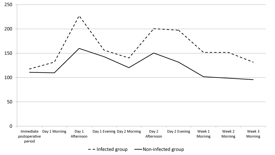
Table 3 shows that the comparison of the background between the infected group and the non-infected group. In this study, the rate of PJI was significantly higher in patients with higher preoperative FBG values in TKA patients. Blood glucose levels for the two postoperative days tended to be higher in the infected than in the non-infected group, although the differences were not statistically significant (Figure 1). FBG levels at 1 week after operation also tended to be higher in the infected than the non-infected group. In particular, the blood glucose levels in the afternoon of Day 1 were 227.0±8.5 mg/dL in the infected group and 159.4±46.6 mg/dL in the non-infected group, but there was no significant difference. (p=0.06, Wilcoxon rank sum test).
We compared the likelihood of PJI in patients with blood glucose levels before each meal in two days postoperatively and with FBG levels at one week postoperatively. In this study, the rate of PJI was also higher when the postoperative maximum blood glucose levels before each meal were over 200 mg/dL in the two days postoperatively and the FBG levels were over 140 mg/dL at one week after operation. (p=0.02 and 0.006, respectively, Fisher’s exact test) (Tables 4,5). When we compared infection rates for patients with maximum blood glucose levels over 200 mg/dL in the two days postoperatively and the FBG levels over 140 mg/dL at one week after operation with others, the infection rate was 22.2% (2/9 patients) in the former compared to 0.4% (1/226 patients) in the latter. This was a significant difference. (p=0.004, Fisher’s exact test) (Table 6).
Discussion
The number of DM patients who need to undergo THA or TKA have been increasing [9]. As many literatures have stated the associations between the postoperative infections and DM or a high level of HbA1c in the field of cardiothoracic surgery [4-7], it was also reported that the patients that underwent total joint arthroplasty with DM had a significantly higher risk of postoperative PJI than that of the patients without DM [10-15]. For example, Hwang et al. reported that the patients with the preoperative blood glucose levels >200 mg/dL and HbA1c levels >8.0 mg/dl were at the risk for wound complications after total knee arthroplasty [13]. Tarabichi et al. reported that the HbA1c level over 7.7 mg/dL was associated with a higher risk of postoperative PJI [15]. Although it has been reported that it is important to control the postoperative glucose levels to decrease the rate of PJI [10], there are few reports about the association with hyperglycemia and PJI. This motivated us to study the postoperative blood glucose levels after TJA.
The highlight of our study was first that the peri-operative blood glucose values at Day 1 and 2 were tended to be higher in infected patients than those of the non-infected group, and the average blood glucose levels increased to about 200 mg/dL in the afternoon of Day 1 and 2 in the infected group. It also showed that PJI patients were higher blood glucose level at 1 week after operation than not-infected patients.
Two reports about the relationship between the hyperglycemia and the postoperative PJI were introduced as follows [14,16]. Stryker et al. [14], reported that patients with a mean postoperative glucose >200 mg/dL are at the risk for wound complications after total joint arthroplasty. Mraovic et al. reported the perioperative blood glucose levels at Day 1 morning in infected patients who underwent TJA were significantly higher than that in not-infected patients [16]. Mravoic also reported the glucose values at Day 1 morning over 200mg/dL was a significant risk factor for infection with an over two-fold increased rate of the infection comparing with the glucose levels under 200ml/dL [16].
Compared with these literatures we introduced, the blood glucose levels in the afternoon were higher than that in the morning and evening in the infected group from the figure in our study. Moreover, there was only one patient with the blood glucose levels over 200 mg/dL at Day 1 morning and that patient was not infected. From those results, we hypothesized that it would be more important to measure the blood glucose levels in the afternoon than in the morning. Our study suggested that the postoperative blood glucose level over 200 mg/dL in the afternoon at Day 1 and 2 was a predictive factor of PJI.
Most investigators rarely pay attention to the glucose level after one week postoperatively although they pay attention to the glucose levels within a few days postoperatively in many literatures [4,5,12,14,16]. However, the FBG level at 1 week of more than 140 mg/dL might be associated with a higher risk of a postoperative PJI from our results.
Though many literatures have stated the associations between the postoperative infections and a high level of HbA1c [4-7,14-16], HbA1c values had a possibility of inaccurate data with patients suffered from anemia and hemoglobinopathies due to abnormal red blood cell turnover [14]. It might be better to monitor HbA1c levels preoperatively and to check the blood glucose levels postoperatively to know the risk of PJI. We recommend that we may have to monitor the glucose levels for more weeks when the blood glucose levels were at Day 1 and 2 over 200 mg/dL and the FBG levels at 1 week over 140 mg/dL.
The reasons the hyperglycemia was affected the increasing to the infected patients had been reported that acute and short-term hyperglycemia affects all major components of innate immunity and impairs the ability of the host to combat infection [17]. Hyperglycemia included reduced neutrophil activity (e.g. chemotaxis, formation of reactive oxygen species, phagocytosis of bacteria) [17]. To reduce SSI, we had thought to have to control the postoperative blood glucose levels strictly before [18]. However, in Nice Sugar Study [19], it was reported that intensive glucose control increased mortality among adults in the ICU: a blood glucose target of 180 mg/dL or less resulted in lower mortality than did a target of 81 to 108 mg/dL. It was concluded conventional glucose control, a blood glucose target of 180 to 200 mg/dL, decreased mortality. From our results and Nice Sugar Study, we had monitored and controlled the perioperative blood glucose levels less than 200 mg/dL in view of the mortality and SSI. We considered the reasonable target of blood glucose levels less than 200 mg/dl postoperatively from the perspective of lower mortality and infection.
There were some limitations in our study. First, we did not do a multivariate analysis because of the small number of the subjects. So, this was a preliminary study. Second, we performed the retrospective study of data from our hospital record. Our study may be subject to selection bias. The percentage of THA was much more than that of TKA in our study. And, the most patients were female and low BMI in our cohort. The infection rate may change if even the ratio of THA and TKA or the ratio of sex or high BMI were changed. However, we believe that selection bias was reduced by consecutive enrollment of the study patients.
In the future, a larger size of patients included in infected patients will be needed in this study. Further study will be needed. Moreover, blood glucose is usually measured at fixed time points, but this does not enable continuous assessment of postoperative fluctuations. To measure postoperative fluctuations in blood glucose will needed in the future.
The conclusions of the present study suggest that it is important to monitor blood glucose levels, especially in patients with postoperative blood glucose levels more than 200 mg/dL in two days after operation and postoperative blood glucose levels more than 140 mg/dL at 1week after operation.
- McAlister FA, Majumdar SR, Blitz S, Rowe BH, Romney J, et al. (2005) The relation between hyperglycemia and outcomes in 2471 patients admitted to the hospital with community-acquired pneumonia. Diabetes Care 28: 810-815. Link: https://goo.gl/xxYVMm
- Kurup H, Thomas M (2013) Orthopaedics and Diabetes. Acta Orthop Belg 79: 483-487. Link: https://goo.gl/ZyEbaE
- Jamsen E, Nevalainen P, Eskelinen A, Huotari K, Kalliovalkama J, et al. (2012) Obesity, diabetes, and preoperative hyperglycemia as predictors of periprosthetic joint infection: a single-center analysis of 7181 primary hip and knee replacements for osteoarthritis. J Bone Joint Surg Am 94: e101. Link: https://goo.gl/U7V4Ka
- Zerr KJ, Furnary AP, Grunkemeier GL, Bookin S, Kanhere V, et.al. (1997) Glucose control lowers the risk of wound infection in diabetics after open heart operations. Ann Thorac Surg 63: 356-361. Link: https://goo.gl/jHLwaB
- Latham R, Lancaster AD, Covington JF, Pirolo JS, Thomas CS Jr (2001) The association of Diabetes and glucose control with surgical-site infections among cardiothoracic surgery patients. Infect Control Hosp Epidemiol 22: 607-612. Link: https://goo.gl/hyM16r
- Vuorisalo S, Haukipuro K, Pokela R, Syrjala H (1998) Risk features for surgical-site infections in coronary artery bypass surgery. Infect Control Hosp Epidemiol 19: 240-247. Link: https://goo.gl/VTx5wS
- Borger MA, Rao V, Weisel RD, Ivanov J, Cohen G, et al. (1998) Deep sternal wound infection: risk factors and outcomes. Ann Thorac Surg 65: 1050-1056. Link: https://goo.gl/VStC9Q
- Mangram AJ, Horan TC, Pearson ML, Silver LC, Jarvis WR (1999) Guideline for Prevention of Surgical Site Infection, 1999. Centers for Disease Control and Prevention (CDC) Hospital Infection Control Practices Advisory Committee. Am J Infect Control 27: 97-132. Link: https://goo.gl/GLzLR1
- Boyle JP, Honeycutt AA, Narayan KM, Hoerger TJ, Geiss LS, et al. (2001) Projection of diabetes burden through 2050: impact of changing demography and disease prevalence in the U.S. Diabetes Care 24: 1936-1940. Link: https://goo.gl/yURTZt
- Tsang ST, Gaston P (2013) Adverse peri-operative outcomes following elective total hip replacement in diabetes mellitus: a systematic review and meta-analysis of cohort studies. Bone Joint J 95: 1474-1479. Link: https://goo.gl/rB8gQf
- Iorio R, Williams KM, Marcantonio AJ, Specht LM, Tilzey JF, et.al. (2012) Diabetes mellitus, Hemoglobin A1C, and the incidence of total joint arthroplasty infection. J. Arthroplasty 27: 726-729. Link: https://goo.gl/awBnY3
- Han HS, Kang SB (2013) Relations between long term glycemic control and postoperative wound and infectious complications after total knee arthroplasty in type 2 diabetics. Clin Orthop Surg 5: 118-123. Link: https://goo.gl/SJAgZ1
- Hwang JS, Kim SJ, Bamne AB, Na YG, Kim TK (2015) Do glycemic markers predict occurrence of complications after total knee arthroplasty in patients with diabetes? Clin Orthop Relat Res 473: 1726-1731. Link: https://goo.gl/xp5io2
- Stryker LS, Abdel MP, Morrey ME, Kor DJ, Morrey BF, et al. (2013) Elevated Postoperative blood glucose and Preoperative Hemoglobin A1C are associated with increased wound complications following total joint arthroplasty. J Bone Joint Surg Am 95: 808-814. Link: https://goo.gl/wjKAcQ
- Tarabichi M, Shohat N, Kheir MM, Adelani M, David Brigati D, et al. (2017) Determining the threshold for HbA1c as a predictor for adverse outcomes after total joint arthroplasty: A Multicenter, Retrospective Study. J Arthroplasty 32: S263-S267. Link: https://goo.gl/nc4QWH
- Mraovic B, Suh D, Jacovides C, Parvizi (2011) Perioperative hyperglycemia and postoperative infection after lower limb arthroplasty. J Diabetes Sci Technol 5: 412-418. Link: https://goo.gl/4EQuhG
- Turina M, Fry DE, Polk HC Jr (2005) Acute hyperglycemia and the innate immune system: Clinical, cellular, and molecular aspects. Crit Care Med 33: 1624-1633. Link: https://goo.gl/sr16Sy
- van den Berghe G, Wouters P, Weekers F, Verwaest C, Bruyninckx F, et.al. (2001) Intensive insulin therapy in critically ill patients. N Engl J Med 345: 1359-1367. Link: https://goo.gl/1UDCTL
- NICE-SUGAR STUDY investigators (2009) Intensive versus Conventional Glucose control in Critically III Patients. N Engl J Med 360: 1283-1297. Link: https://goo.gl/jjwrKz
Article Alerts
Subscribe to our articles alerts and stay tuned.
 This work is licensed under a Creative Commons Attribution 4.0 International License.


 Save to Mendeley
