Annals of Musculoskeletal Medicine
In Vivo Human Gastrocnemius Architecture With Changing Joint Angle at Rest and During Graded Isometric Contraction of Normal and Weak Muscle
Yuri Koryak*
Cite this as
oryak Y (2020) In Vivo Human Gastrocnemius Architecture With Changing Joint Angle at Rest and During Graded Isometric Contraction of Normal and Weak Muscle. Ann Musculoskelet Med 4(1): 010-014. DOI: 10.17352/amm.000021Architectural properties of the triceps surae muscles complex were determined In Vivo for thirty subjects. These subjects were assigned to two groups. The first group of subjects consisted of 8 healthy men and the second group of subjects was composed of 22 patients with motor disorders. The ankle was positioned at -15 ° (dorsiflexion), and 0 ° (neutral anatomical position), and 15 °, and 30 ° (plantarflexion), with the knee set at 120 °and with an angle in the ankle joint of 90 °. At each position, longitudinal ultrasonic images of the Medial (MG) and Lateral (LG) Gastrocnemius and Soleus (SOL) muscles were obtained while the subject was relaxed (passive) and performed 50 % maximal voluntary isometric plantar flexion (active), from which fascicle Lengths (L) and angles (Θ) with respect to the aponeuroses were determined. From the ultrasonic image, it was observed that and Θ changed during an isometric contraction of the triceps surae muscle. Changes in L and were expressed as a function of relative torque. The Θ change was not identical for the three muscles. The fascicle Θ of MG demonstrated the greatest variation in three muscles. The effects of activation and relaxation positions were significant in all three muscles. The differences in MG fascicle Θ because of changes in ankle positions were significant among control and patients both in the passive and active conditions. Fascicle Θ of LG and SOL not differed among control and patient in the relaxation condition but not in the activation condition. For LG, and SOL ol fascicle Θ were changes were larger in control with the patients. The mean values fascicle Θ of MG, LG, and SOL an isometric contraction (50 % MVC) in the control groups increased by 60 %, 41 %, and 41 %, respectively; in the patient groups were a smaller increase, by 28 %, 26 %, and 36 %, respectively. Different lengths and angles of fascicles, and their changes bу contraction by patients and normal subjects, might bе related to differences in force-producing capabilities of the muscles and elastic characteristics of tendons and aponeuroses.
New & noteworthy
The present work was the first to combine measuring the fiber length and pennation angle (ultrasound imaging) as main determinants of mechanical force production and the muscle function patients and healthy human. Elderly age is not the reason for reducing the contractile properties of the calf muscle according to the study of ultrasound architecture of the muscle. Changes in muscle architecture are due to disease and physical inactivity. Studying skeletal muscle architecture is important in understanding the pathological changes caused by diseases or physical inactivity. These results will help in the early stages of the disease, as well as in the treatment and rehabilitation of the patient to determine the prognosis of the functional state of the muscle and the amount of possible physical exertion.
Introduction
A number of studies have documented that the microgravity environment encountered during spaceflight or simulated by using models of weightlessness induces alterations in skeletal muscle function [1-13]. In the absence of weight-bearing activity, strength loss is the most evident consequence of atrophy. Moreover, showed that muscle strength decreased during bed-rest or immersion and suggested that the loss of strength [4,5] was due primary to muscle atrophy [6,7]. Muscle atrophy has been shown to be pronounced in the lower limb muscles [8], and it has often been observed that the reduction of strength is greater than that of muscle size [8,9]. Therefore, these observed changes following a period of immobilization may cause changes in fascicle length.
Most skeletal muscles in humans are more or less pennated [10], in which muscle fibres are arranged at an angle with respect to the line of action of the muscle. The angle of muscle fibres with respect to the tendon action line is an important functional characteristic of the muscle. A large pennation angle allows more contractile material to be placed along the tendon increasing the muscle’s force generating capacity, results in a less efficient force transmission through the tendon and results in a reduced fibre length, compromising shortening velocity and excursion range [11]. Pennation angle changes proportionally as a function of isometric contraction intensity.
Muscle architecture, i.e. geometrical arrangement of fibres within a muscle, has been shown to have a substantial influence on the force-generating capabilities of the muscle [12]. This angulation (pennation angle) has been shown to affect force transmission from muscle fibres to tendon, and hence muscle force generation [10,12]. The architecture of a skeletal muscle is an important determinant of its functional characteristics [10]. Human muscle architecture may be studied noninvasively In Vivo both at rest and during muscle contraction, by using real-time ultrasonography [12]. Indeed, several investigators [13] have demonstrated that during isometric contractions muscle architecture undergoes remarkable changes. Changes in fiber length by contraction are thus expressed as fascicle length changes.
In an attempt to improve our understanding of In Vivo changes of muscle architecture, modern imaging techniques have been used [12-14]. Real-time ultrasonography enables In Vivo muscle scanning and offers promise for a realistic determination of changes in muscle architecture [12]. Real-time Ultrasonic (US) measurements were taken in the present study in the Triceps Surae (TS) muscle in healthy man and patients. In this report, we have studied by means of ultrasonography the relationships between architectural parameters [lengths fascicles (L), and pennation angles (Θ) of fascicles, and Muscle Thickness (MT)] and level of force exerted in highly-subjects and patient with consequences of cerebral palsy. The purpose of our research work was to determine In Vivo changes in pennation angle and fibre length in each muscle of the triceps surae complex [medial (MG) and lateral (LG) gastrocnemius and soleus (SOL) muscles], both at rest and moment produced voluntarily during an isometric ankle plantarflexion. We employed real-time ultrasonography to visualize fascicles In Vivo.
Methods
Subjects
Thirty subjects participated in this study. These subjects were assigned to two groups. The first group of subjects consisted of 8 healthy men (average age: 52 ± 3.6 years), and the second group of subjects was composed of 22 patients men and women (average age: 55 ± 3.4 years) with motor disorders caused: sharp infringement of brain blood circulation and its consequence - hemiparesis, consequences of cerebral palsy, myelopathy, radiculopathy on the background of osteochondrosis of the spine, obliterating atherosclerosis of the vessels of the lower extremities, and hypodynamia on the background trauma of shins. Prior to the experiment, the details and possible risks of the protocols were explained to the subjects and written informed consent was obtained from each of them. The study was approved by the Medical Ethics Committee is accordance with the Helsinki Declaration. Subjects were familiarized with all the testing procedures 1 week prior to the baseline tests.
Ankle extension torque
Participants were seated upright in Isokinetic Dynamometer Biodex (Biodex System 4 PRO™, Biodex Medical Systems, Shirley, New York, USA) chair with their trunk positioned and secured to the seatback with waist and shoulder belts to ensure consistent positioning and minimal movement. The hips were positioned to 90 ° with the knee and the knee joint was positioned at 120 ° and the ankle was in the 90 ° joint angle. The lateral malleolus of the right foot was aligned with the axis of rotation on the Biodex dynamometer. The foot was fastened to the footplate with inelastic straps that were firmly secured behind and on the underside of the footplate to prevent heel lift. When heel lift occurred or torque did not return to baseline protocol was stopped and repeated after 3–5-min rest. Subjects performed three sets of four repetitions of maximal isokinetic ankle extension at an angular velocity of 0°/s-1 with a 2-min resting period between contractions unless the third trial exceeded one of the two first ones by more than 10 %. In that case an additional trial was performed. The participants were instructed to grip the side handles to help stabilize the trunk.
Ultrasound scanning
The longitudinal US images of the MG and LG muscles were obtained at 30 % proximal level of lower leg (the distance between the lateral malleolus of the fibula and the lateral condyle of the tibia), and soleus (SOL) muscles were obtained at 50 % of the distance between the poplitealcrease and the center of the lateral malleolus the using the B-mode ultrasound apparatus Sonoline Elegra (Sonoline Elegra, Siemens, Germany). Briefly, the measurements were carried out while the subjects stood with their weight evenly distributed between both legs. The mediolateral widths of the MG and LG muscles were determined by ultrasound over the skin surface, and the position of one-half of the width was used as a measurement site. The echoes from interspaces of fascicles and from the superficial and deep aponeurosis were visualized [15].
A real-time B-mode US apparatus (Sonoline Elegra, Siemens, Germany) with a 7.5-MHz linear-array probe (with 80-mm scanning length) was used to obtain sagittal images of the GM, GL and SOL, at rest and at 50 % of plantarflexor MVC at the neutral ankle position. A transducer with a 7.5-MHz linear-array probe was placed perpendicular to the tissue interface. A marker (⬇) was placed between the skin and the ultrasonic probe as the landmark to confirm that the probe did not move during measurements. The scanning head was coated with water-soluble transmission gel, which provided acoustic contact without depressing the dermal surface. The subcutaneous adipose tissue-muscle interface and the muscle-bone interface were identified from the ultrasonic image, and the distance from the adipose tissue-muscle interface to the muscle-bone interface was adopted as representative of muscle thickness. All measurements were performed on the right leg. During all measurements, subjects were instructed to relax their leg muscles. Each subject’s right foot was firmly attached to a dynamometer (Cybex® II, USA). The subjects were verbally encouraged to perform static contractions with the ankle plantarflexor with a maximum possible effort. Three contractions were performed by a 1 min rest interval between bouts and the highest value was considered the Maximum Voluntary Contraction (MVC). US images were obtained during the experimental trial on the previously determined stronger leg. Each subject was then asked to maintain the stronger leg contractions for at least 2-3 s at the neutral ankle position.
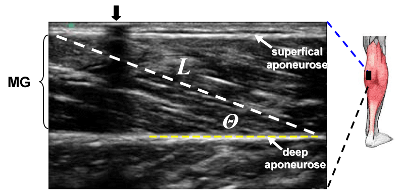
The fascicle pennation angle (Θ) was measured from the angles between the echo of the deep aponeurosis of each muscle and interspaces among the fascicles of that muscle [12,13]. The length of fascicles (L) across the deep and superficial aponeurosis was measured as a straight line between the insertion points of the fascicle, onto the aponeurosis of the muscle [16,16]. The MG and LG muscle fascicles generally extended off the ultrasound scan window and it was therefore necessary to estimate part of the fascicular trajectory. Using digitizing software, the visible portion of the fascicle was measured and the remaining part was estimated assuming linear continuation of the fascicle and aponeurosis in the proximal direction. Previously reported that the order of error associated with this method of fascicle length estimation is ~4 % [17].
Muscle Thickness (MT) was measured as the distance between the superficial and the deep aponeuroses echoes, and L at rest was measured as the length of the line drawn along the echoes parallel to the fascicles from the deep up to the superficial [13] Figure 1.
US images were calculated by US-system, using program Magic View 300 (Siemens) with archiving the data by Sienet (Siemens, Germany). In each muscle the average of the five images was used for data analysis.
Statistical analysis
Data were checked for normality of distribution using the Kolmogorov–Smirnov test. Independent-sample Student t-tests were used to test for baseline differences between the two groups for all reported variables. Paired-sample Student t-tests were used to test for differences the two groups. A 2×2 factorial analysis of variance was used to test for differences between groups (healthy men and patients). Data are reported as means ±SD unless otherwise stated; the level of significance was accepted at p < 0.05.
Results
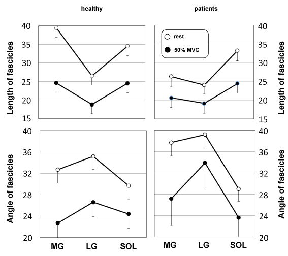
The US findings in the patient groups were compared to of a control group. From the US image, it was observed that L and Θ changed during in the passive condition and specially the active condition. The degree of L change was not identical for the three muscles The effects relaxation and an isometric contraction of the triceps surae muscle (50 % MVC) on L were significant for MG and LG and there was also a significant interaction between control and patients in these muscles (Figure 2). In other words, in MG and LG, changes in L changes were larger with the SOL. In the active condition, L of MG were not different between control and patients, although in the passive condition the difference was significant. The L of SOL were not different.
From the US image, it was observed that and Θ changed during an isometric contraction of the TS muscle. Changes in L and were expressed as a function of relative torque. The Θ change was not identical for the three muscles. The fascicle Θ of MG demonstrated the greatest variation in three muscles. The effects of activation and relaxation positions were significant in all three muscles. The differences in MG fascicle Θ because of changes in ankle positions were significant among control and patients both in the passive and active conditions. Fascicle Θ of LG and SOL not differed among control and patient in the relaxation condition but not in the activation condition. For LG, and SOL ol fascicle Θ were changes were larger in control with the patients. The mean values fascicle Θ of MG, LG, and SOL an isometric contraction (50 % MVC) in the control groups increased by 60 %, 41 %, and 41 %, respectively; in the patient groups were a smaller increase, by 28 %, 26 %, and 36 %, respectively (Figure 2).
MT of MG and LG were not significantly greater in control than in patients, but in SOL were loss than in control. Changes in MT were expressed as a function of relative torque. TM of MG at was no significantly different between rest and 50 % MVC. In contrast, MT of LG and SOL increased from rest to MVC by 21.9 % (p < 0.05) and 17.9 % (p < 0.05), respectively. In MG and SOL patients was not significantly different either between different imaged. TM LG and SOL were not significantly greater in patients by 10.7 % and 3.6 %, respectively, but MG was decreased by 3.6 %.
Discussion
Our results show that the US image method applied is valid and reliable for assessing the size of a large, individual, human locomotor muscle. This method can provide information on cross-section area changes along the entire muscle length in response to training, disuse or as a spaceflights.
Internal architecture of the TS complex (MG) was altered. Both fascicle length and pennation angle were reduced after in patients groups, this strongly suggests a loss of both in-series and in-parallel sarcomeres, respectively. This observation is in agreement with previous findings in disuse conditions [9,18-20]. The functional consequence of the decreased fascicle length was a reduced shortening during contraction. It is necessary to note, that at some patients with motor disorders, having restriction of mobility, the normal ultrasonic architecture of muscles was marked. It is possible to assume, that disuse of a muscle is not the unique factor influencing on the ultrasonic architecture of muscles. Studying of architecture of muscles at patients with various motor disorders allows to understand better the intimate processes in healthy persons under influence of various factors, including microgravitation, long space flights, where restriction of impellent activity takes place, despite of using preventive measures, and experiments on animals demonstrate the development of a muscular atrophy in conditions of microgravitation.
In conclusion, this study shows for the first time that in patients with motor impairment under conditions of performing a functional load, changes in muscle architecture differ from healthy subjects. The present data suggest that the architecture and contractile capacity of human pennate muscle are interrelated, In Vivo. It is assumed that some of the noted features of muscle architecture due to disease and physical inactivity require further study. Moreover, it can be assumed that the conditions of weightlessness - physical inactivity, can also be the cause of such changes in the muscle system. Studying skeletal muscle architecture is important in understanding the pathological changes caused by diseases or physical inactivity. These results will help in the early stages of the disease, as well as in the treatment and rehabilitation of the patient to determine the prognosis of the functional state of the muscle and the amount of possible physical exertion.
The method of ultrasound scanning of muscles is a highly informative and accessible method for assessing the architecture of skeletal muscles in humans and can be used in combination with other methods to assess the functional state of muscles and to study the mechanisms that determine changes under the influence of various factors, and a comparative study of muscle architecture in patients with different motor disorders is important for understanding the processes occurring in healthy individuals under the influence of various factors, including microgravity, sports training, with various diseases and during treatment and rehabilitation.
The author thank N. Mironov, Director of Clinical Hospital № 1 President Medical Center, and his staff in rendering assistance in carrying out the given research. I expresses special gratitude Mrs. M. Kuz’mina M.D., Ph.D., for the executed ultrasonic researches (Chief of Department of ultrasonic diagnostics N. Vit’ko, M.D., Dr.Sci., and Head of Branch of ultrasonic diagnostics V. Nasarenko, M.D., Ph.D).
Finally, the author thank of all volunteers who participated in this study.
Grants
The study is supported by the Russian Academy of Sciences (Grant 63.1).
- Cavagna GA, Heglund NC, Taylor CR (1977) Mechanical work in terrestrial locomotion: two basic mechanisms for minimizing energy expenditure. Amer J Physiol 233: R243-R261. Link: https://bit.ly/39FkMOY
- Yu K (2001) Electrically evoked and voluntary properties of the human triceps surae muscle: effects of long-term spaceflights. Acta Physiol Pharmacol Bulg 26: 21-27. Link: https://bit.ly/30cBVMS
- Yu K, Siconolfi SF, Kozlovskaya IB, Gilbert JH, Layne ChS (1997) Maximal voluntary (MVC), tetanic (Po) and single twitch (Pt) contractions before & after space flight. Faseb J A1408.
- Yu K (2002) Surface action potential and contractile properties of the human triceps surae muscle: effect of “dry” water immersion. Exp Physiolog 87: 101-111. Link: https://bit.ly/2P7sXtY
- Yu K (2003) Contractile properties and fatiguability of the human triceps surae muscle after exposure to simulated weightlessness. Basic Motor Control to Functional Recovery III.
(ed. N. Gantchev), Varna, Univ. Press 369-380. - Duchateau J, Hainaut K (1987) Electrical and mechanical changes in immobilized human muscle.
J Appl Physiol 62: 2168-2173. Link: https://bit.ly/3gfDAqD - Kubo K, Akima H, Kouzaki M, Ito M, Kawakami Y, et al. (2000) Changes in the elastic properties of tendon structures following 20 days bed-rest in humans. Eur J Appl Physiol 83: 463-468. Link: https://bit.ly/2DjElAb
- LeBlanc A, Gogia P, Schneider V, Krebs J, Schonfeld E, et al. (1988) Calf muscle area and strength changes after five weeks of horizontal bed rest. Amer J Sports Med 16: 624-629. Link: https://bit.ly/2BIurrk
- Suzuki Y, Murakami T, Haruna Y, Murakami T, Haruna Y, et al. (1994) Effects of 10 and 20 days bed rest on leg muscle mass and strength in young subjects. Acta Physiol Scand 616: 5-18. Link: https://bit.ly/39JVejJ
- Gans С, Bock WJ (1965) The functional significance of muscle architecture - a theoretical analysis. Ergeb Anat Entwicklungsgesch 38: 115-142. Link: https://bit.ly/3f9CriU
- Gans С (1982) Fiber architecture and muscle function. Exerc Sport Sci Rev (Teijung R.T., ed.), Franklin Institute Press< Phuladelphia 10: 160-207. Link: https://bit.ly/39GdIS2
- Kawakami Y, Abe T, Fukunaga T (1993) Muscle-fiber pennation angles are greater in hypertrophied than in normal muscles. J Appl Physiol 74: 2740-2744. Link: https://bit.ly/2D69IhN
- Fukunaga T, Ichinose Y, Ito M, Kawakami Y, Fukashiro S (1997) Determination of fascicle length and pennation in a contracting human muscle In Vivo. J Appl Physiol 82: 354-358. Link: https://bit.ly/39JJlKC
- Narici MV, Binzoni T, Hiltbrand E. Fasel J, Terrier F, et al. (1996) In Vivo human gastrocnemius architecture with changing joint angle at rest and during graded isometric contraction. J Physiol 496: 287-297. Link: https://bit.ly/39IHGF2
- Kumagi K, Abe T, Brechue WE, Ryushi T, Takano S, et al. (2000) Sprint performance is related to muscle fascicle length in male 100-m sprints. J Appl Physiol 88: 811-816. Link: https://bit.ly/2DivHlF
- Abe T, Kumagai K, Brechue WF (2000) Fascicle length of leg muscle is greater in sprinters than distance runners. Med Sci Sports Exerc 32: 1125-1129. Link: https://bit.ly/3ffytFt
- Reeves ND, Narici MV, Maganaris CN (2004) Effect of resistance training on skeletal muscle-specific force in elderly humans. J Appl Physiol 96: 885-892. Link: https://bit.ly/2D42bAc
- Akima H, Kubo K, Kanehisa H, Suzuki Y, Gunji A, et al. (2000) Leg-press resistance training during 20 days of 6˚ head-down-tilt bed rest prevents muscle deconditioning. Eur J Appl Physiol 82: 30-38. Link: https://bit.ly/2CWTLe5
- Fukunaga T, Roy RR, Shellock FG. Hodgson JA, Day MK, et al. (1992) Physiological cross-sectional area of human leg muscles based on magnetic resonance imaging. Orthop Res 10: 928-934. Link: https://bit.ly/3gj09un
- Yu K (2002) “Dry” immersion induces neural and contractile adaptations in the human triceps surae muscle. Environ Med 46: 17-27. Link: https://bit.ly/2Pc3bEM
Article Alerts
Subscribe to our articles alerts and stay tuned.
 This work is licensed under a Creative Commons Attribution 4.0 International License.


 Save to Mendeley
