Archives of Otolaryngology and Rhinology
Profile of Compressive Goiters
Ndiaye C*, Thiam A, Seck SN, Pilor N, Ndour MF, Sylla MS, Maiga S, Ahmed H, Mbaye A, Ndour N, Diallo MB and Tall A
Department of Otolaryngology-Head and Neck Surgery, National University Hospital of Fann, Senegal
Cite this as
Ndiaye C, Thiam A, Seck SN, Pilor N, Ndour MF, Sylla MS, et al. Profile of Compressive Goiters. Arch Otolaryngol Rhinol. 2025;11(1):001-004. Available from: 10.17352/2455-1759.000158Copyright
© 2025 Ndiaye C, et al. This is an open-access article distributed under the terms of the Creative Commons Attribution License, which permits unrestricted use, distribution, and reproduction in any medium, provided the original author and source are credited.Introduction: Goiter is a localized or diffuse hypertrophy of the thyroid gland. During its evolution, this hypertrophy can lead to compression of neighboring organs, a condition known as compressive goiter. This is a rare entity in thyroid pathology, and few studies have been carried out. Many studies have been carried out on thyroid pathologies in general, but few have focused on compressive goiter.
This study aimed to outline the epidemiological, clinical, therapeutic, and evolutionary profile of compressive goiters.
Material and methods: This was a retrospective study from January 2012 to December 2021 of all patients diagnosed and operated on for compressive goiter in the ENT department of FANN. The parameters studied were age, sex, family history of goiter, signs of compression, type of thyroidectomy, histology, and postoperative course.
Results: We identified 65 patients with compressive goiter. The sex ratio was 0.12 (m/f). A family history of goiter was found in 23.07% of cases. A cervicothoracic CT scan was performed in 26.15% of cases.
Total thyroidectomy was performed in all our patients, and we noted 18.47% complications during post-operative follow-up. Anatomopathological data showed a benign goiter in 70.48% of our series.
Conclusion: Compressive goiter is a rare thyroid disease that can be life-threatening because of compression. Total thyroidectomy remains the treatment of choice.
Introduction
Goiter is a localized or diffuse hypertrophy of the thyroid gland [1]. During its evolution, this hypertrophy can lead to compression of neighboring organs, a condition known as compressive goiter. This is a rare entity in thyroid pathology, and few studies have been carried out. The literature often reports clinical cases [2,3].
In our context, lack of information and resources, and reliance on traditional medicine, all contribute to delaying the need for consultation. The consequence is the development of voluminous goiters leading to compression of nearby organs. This situation motivated us to study the clinical, therapeutic, and histological aspects of compressive goiters.
Materials and methods
This was a retrospective, descriptive study over ten years from January 2012 to December 2021 of all patients diagnosed and operated on for compressive goiter in the ENT department of the FANN University Hospital. All patients underwent surgery. The indication for surgery was not a malignant cytology but compressive complaints. The parameters studied were age, sex, history of familial goiter, signs of compression, type of thyroidectomy, histology of the surgical specimen, and complications. The analysis was done by comparisons of proportions using Excel software.
Results
Socio-demographic data
During this period, 436 patients underwent goiter surgery, 65 of whom (14.90%) showed signs of compression.
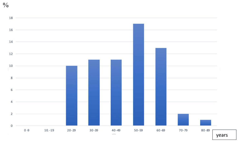
The mean age was 39, with extremes of 21 and 83. The 50-59 age group accounted for 17%.
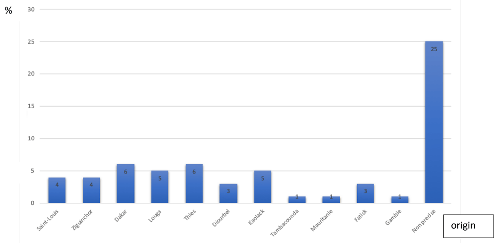
Figure 1 shows the age distribution. The sex ratio was 0.12 (m/f). A family history of goiter was found in 23.07% of cases. Our study population originates from the capital and other regions and is distributed as shown in Figure 2.
Clinical data
Dyspnea was present in 53.85% of patients, followed by dysphagia (52.30%), as shown in Table 1.
Thyroid tests were performed in all patients. The distribution according to hormonal status is shown in Table 2.
In terms of imaging, a chest X-ray was performed in 41.54% of cases. Cervical ultrasonography was performed in 86.15% of patients and revealed a multinodular goiter in 58.4%.
Cervicothoracic CT scans were performed in 26.15% of patients.
Fine-needle aspiration cytology (FNAC) was performed in 41.53% of cases and revealed a benign follicular lesion in 34.63%. The results of FNAC are shown in Table 3.
Treatment data
Tracheostomy was performed in 09 patients (9.22%).
All patients underwent total thyroidectomy in our series, and 06 (six) patients underwent the next dissection at the same time.
Anatomopathological data showed a benign goiter in 70.48% of our series. Among cancers, 50% were papillary carcinomas (Table 4).
During post-operative follow-up, we noted 18.47% complications, including 2 cases of hematoma, 5 cases of hypocalcemia, 3 cases of unilateral recurrent paralysis, and 2 cases of death.
Discussion
Compressive goiter accounted for 14.90% of thyroid pathologies operated on in FANN's ENT department. A study carried out by Diedhiou, et al. [4] on thyroidectomies in another structure in the capital in 2021 found a frequency of compressive goiter of 9.8%. In Morocco, in the CHU Ibn Rochd in Casablanca, a study conducted by Benbakh, et al. [5] in 2014 found a frequency of 4.29%. This difference in prevalence could be explained by the delay in consultation and under-medicalization characteristic of developing countries. Added to this is the fear of surgery, as well as certain beliefs in some parts of the country, where the goiter is considered a criterion of beauty. All these factors favor a long evolution of the goiter, leading to significant volume gain and compression. In our series of compressive goiters, the average duration of evolution was 3 years.
The mean age of our patients was 48 years, with extremes of 21 and 83 years, with a clear female predominance in line with the data in the literature [2,4,6-8].
This confirms the high prevalence of thyroid disease in women in their forties. There is no difference between the profile of patients with compressive goiter and patients with thyroid pathology in general.
Regarding previous history, we found that 23.07% of patients had a family history of goiter. Indeed, similar results were found in Ndiaye's study [9], with a notion of familial goiter in 24.32%. In the literature, we often find a familial predisposition. [8,9].
We note that these results are classic data from the literature and fall within the framework of goiters in general. However, they do not allow us to predict the compression of neighboring organs. The compressive character could be linked to the duration of evolution, which is the consequence of a delay in consultation.
Goiters can take a long time to develop, as they remain asymptomatic for a long time. In our study, the duration ranged from 2 months to 20 years, with an average of 3 years. For Benbakh, et al. [5], the average duration was 5 years. We have also found an average duration ranging from 8 to 10 years, or even longer [6,9]. This discrepancy depends on the histological type of goiter, the patient's tolerance of compression, the level of access to care, and the patient's level of information.
Among the signs of compression due to goiters, the most frequent is that of the respiratory tract, resulting in dyspnea. Dyspnea is potentially serious, as it can be life-threatening. In our series, the rate was 53.85%. In the literature, we found a much higher prevalence of dyspnea in our series, with percentages exceeding 80%, as shown in Table 5 [6,9,10]. In the face of this respiratory distress, it is important to distinguish tracheal dyspnea from laryngeal dyspnea. Tracheal dyspnea results from direct compression of the trachea, whereas laryngeal dyspnea is linked to damage to the recurrent nerves. Consequently, a compressive goiter associated with laryngeal dyspnea would be highly suggestive of malignancy. Nevertheless, the presence of signs of compression is not predictive of malignancy. This is confirmed by our study, in which the goiter was benign in 79.48% of cases.
Ultrasound plays a central role in confirming this malignancy. Ultrasound confirms the thyroid nature of the cervical swelling and the presence of suspicious nodules but is not the most effective imaging technique for assessing the relationship between the thyroid and adjacent organs [7,11-14].
For a better assessment of anatomical relationships and confirmation of compressive nature, a cervicothoracic CT scan is the appropriate imaging modality. In addition to determining the degree of compression, it can also be used to visualize the mediastinal extension of the goiter, as part of the search for the plunging nature of the goiter. In our study, only 26.15% of patients underwent a cervicothoracic CT scan. This rate seems low compared with the literature. It is linked to the fact that our patients come to us in an emergency, requiring admission to the operating theatre for decompression thyroidectomy. CT scanning is essential for the diagnosis and management of compressive goiters. Showing relationships and extensions, enables better planning of surgery, thus reducing postoperative morbidity. Most authors report optimal sensitivity for this examination in their series [15,16].
In our series, where 73.85% were operated on without imaging, we had 18.47% complications. The morbidity of compressive goiter surgery varies in the literature from 4% to 12%, including recurrent nerve injury, hypoparathyroidism, and respiratory complications linked to a compressive hematoma or tracheomalacia, a rare complication [17].
Magnetic resonance imaging is the most effective test for diagnosing compressive goiter. It enables the extensions and relationships between the goiter, the tracheoesophageal axis, and the vascular axes to be clearly defined [7].
All patients underwent total thyroidectomy. In our context, we perform primary endoscopy before total thyroidectomy to rule out associated hypopharyngeal cancer. Indeed, hypopharyngeal cancer is the leading cancer of the upper aerodigestive tract in Senegal [18].
Complications associated with surgery for a compressive goiter are essentially similar to those of standard thyroid surgery. These include bleeding, damage to the recurrent laryngeal nerve, and temporary or permanent hypoparathyroidism [19]. In our study, transient hypocalcemia was one of the most common complications, accounting for 7.69%. The prevalence of postoperative hypocalcemia was higher for some authors, ranging from 20% to 37% [20-22].
Laryngeal paralysis was encountered in 4.62% of patients, and in some authors varied between 3 and 8.2% [4-6]. In our study, 3.08% of patients had a hematoma, in contrast to the study by Benbakh, et al. [5] in which no hematoma was observed postoperatively.
We recorded 2 cases of death (3.07%). These deaths occurred in patients with anaplastic carcinoma and were not related either to the compression exerted by the goiter itself or to the surgery. These deaths were linked to the natural evolution of anaplastic carcinomas, which have a very poor prognosis [23].
The incidence of these complications is slightly higher in patients with compressive goiter. Surgery requires extensive dissection, and the parathyroid glands may be difficult to identify due to the large size of the goiter.
Prophylactic tracheostomy was performed in 6 patients (9.22%). These results are superior to those found in the literature, with a tracheostomy rate of almost zero [24,25]. Prophylactic tracheostomy was performed in cases of tracheomalacia resulting from the long compression of the goiter on the trachea, but also to avoid asphyxia in cases of compressive hematoma. This can be explained by the fact that in our context, following surgery, patients are admitted to the hospital in the department and not to intensive care where closer monitoring could be carried out.
Conclusion
Compressive goiter is a rare entity among nodular goiters. It shares the same socio-demographic features as other thyroid pathologies. It can be life-threatening due to signs of compression. Compressive nature is not predictive of malignancy. The treatment of choice remains total thyroidectomy wherever possible. However, surgery is more prone to complications.
- Agoda-koussema LK, Adjenou K, Amana B, Goeh-akue KE, Djagnikpo O, Gbinu K, et al. Ultrasound aspects of thyroid gland anomalies, apropos of 134 ca. J Sci Res Univ Lomé. 2008;10. Available from: https://www.ajol.info/index.php/jrsul/article/view/52502
- Clemencot A, Jnan M, Koszutski M. Acute airway obstruction: A case of compressive thyroid goiter. Eur J Emerg Resusc. 2018;30:38-40. Available from: https://doi.org/10.1016/j.jeurea.2018.04.001
- Anajar S, Coulibaly K, Tahiri I, Taali L, Hajjij A, Saadi M, et al. Emergency management of a large compressive goiter: a case report. Pan Afr Med J. 2022;41(256):265. Available from: https://pmc.ncbi.nlm.nih.gov/articles/PMC9187980/
- Diédhiou D, Thioye MM, Sow D, Ndour MA, Diallo IM, Halim C, et al. Thyroidectomy at the Abass Ndao Hospital Center: clinical profiles, indications and results in 706 cases. Afr J Intern Med. 2021;8(2):37-43. Available from: https://www.scirp.org/reference/referencespapers?referenceid=3174379
- Benbakh M, Bouchared N, Elboussaadani A, Rouadi S, Roual A, Mahtar M. Compressive goiters. French Ann Otolaryngol Head Neck Pathol. 2014;131:120. Available from: https://doi.org/10.1016/j.aforl.2014.07.249
- Rios A, Rodriguez JM, Canteras MJ, Galindo PJ, Tebar FJ, parrilla P. Surgical Management of Multinodular Goiter With Compression Symptoms. Arch Surg. 2005;140:49-53. Available from: https://doi.org/10.1001/archsurg.140.1.49
- Zainine R, El Aoud C, Bachraoui R, Beltaief N, Sahtout S, Besbes G. Diving Goiters. About 43 cases. Medical Tunisia. 2011;89(11):860-865. Available from: https://latunisiemedicale.com/pdf/vol89_N%C2%B011_n10.pdf
- Sagna Y, Wend Pagnangdé AHB, Gninkoun JC, Traoré I, El Hanane O, Adji F. Epidemiological profile of toxic multinodular goiters in Fez (Morocco). Health Sci Dis. 2024;25(2):91-94. Available from: https://www.hsd-fmsb.org/index.php/hsd/article/view/5276
- Ndiaye M. Study of compressive goiters in 74 cases collected at the ENT and head and neck surgery department of the Aristide Le Dantec hospital. Thesis Med. 2011;N 34.
- Moumen M, Mehhane M, Kadiri B, Mawfiz H, Elfares F. Compressive goiter, A propos of 80 cases. J Chir. 1989;126(10):521-6. Available from: https://pubmed.ncbi.nlm.nih.gov/2592460/
- Makeieff M, Marlier F, Khudjadze M, Garrel R, Crampette L, Guerrier B. Diving goiters. About 212 cases. Ann Chir. 2000;125:18-25. Available from: http://dx.doi.org/10.1016/S0001-4001(00)00117-3
- Sethom A, Brahem H, Ouni H. The plunging goitres. About 15 cases. J Tun Orl. 2005;14:21-24.
- Andre P, Berginiat N, Doreau P, Triaureau G, Berginiat M. Spontaneous hemorrhage in intrathoracic goiter causing respiratory arrest. J Eur. 1999;3:124-7.
- Jennings A. Evaluation of substernal goiters using computed tomography and MR imaging. Endocrinol Metab Clin North Am. 2001;30:401-14. Available from: https://doi.org/10.1016/s0889-8529(05)70192-4
- Rodriguez JM, Hernandez Q, Pinero A, Ortiz S, Soria T, Ramirez P, et al. Substernal goiter: clinical experience of 72 cases. Ann Oto Rhinol Laryngol. 1999;108:501-4. Available from: https://doi.org/10.1177/000348949910800515
- Shah PJ, Bright T, Singh SS, Lang CM, Pyragius MD, Malycha P, et al. Large retrosternal goiter: A diagnostic and management dilemma. Heart Lung Circ. 2006;15:151-2. Available from: https://doi.org/10.1016/j.hlc.2005.10.011
- Zaghre N, Kaba MK, Bargo CR, Bambara CN, Nao EEM, Sereme M, et al. Historical compressive and plunging goiter: About 1 case. Health Sci Dis. 2024;25(3):99-101. Available from: https://doi.org/10.5281/hsd.v25i3.5393
- Pangpoukne Sando, Badiane AS, Edimo LEF, Ba NFK, Rachidou H, Diallo M, et al. Primary oto-rhino-laryngological cancers: Epidemiological, clinical and therapeutic aspects about 231 cases in Sénégal. World J Adv Res Rev. 2023;20(02):474-477. Available from: https://doi.org/10.30574/wjarr.2023.20.2.2301
- Koumaré AK, Sissoko F, Ongoiba N, Bereté S, Traoré Diop AK, Bagayogo TB, et al. Benign goiters in surgery in Mali. E-memorial Nat Acad Surg. 2002;1(4):1-6. Available from: https://e-memoire.academie-chirurgie.fr/ememoires/005_2002_1_4_01x06.pdf
- Robert J, Mariéthoz S, Pache JC, Bertin D, Caulfield A, Murith N, et al. Short and long-term results of total vs. subtotal thyroidectomies in the surgical treatment of Graves’ disease. Swiss Surg. 2001;7:20-4. Available from: https://doi.org/10.1024/1023-9332.7.1.20
- PappalardO G, Guadalaxara A, Frattaroli FM, Tilomei G, Falaschi P. Total compared with subtotal thyroidectomy in benign nodular disease: personal series and review of published reports. Eur J Surg. 1998;164:501-6. Available from: https://doi.org/10.1080/110241598750005840
- Razack MS, Lore JM Jr, Lippes HA, Schaefer DP, Rassael H. Total thyroidectomy for Graves’ disease. Head Neck. 1997;19(5):378-83. Available from: https://doi.org/10.1002/(SICI)1097-0347(199708)19:5%3C378::AID-HED3%3E3.0.CO;2-X
- Chiacchio S, Lorenzoni A, Boni G, Rubello D, Elisei R, mariani G. Anaplastic thyroid cancer: prevalence, diagnosis and treatment. Minerva Endocrinol. 2008;33(4):341-57. Available from: https://europepmc.org/article/med/18923370
- Ouoba K, Sano D, Wandaogo A, Drabo Y, Cisse R, Sanou A, Soudre BR. Thyroidectomy complications (about 104 cases at Ouagadougou H.U.C). Cah ORL. 1998;33(3):178-82. Available from: https://pascal-francis.inist.fr/vibad/index.php?action=getRecordDetail&idt=2330198
- Sow ML, Benchekroun A, Cherbonnel G, Toure CT, Toure P, Diop A. Compressive goiters: Dakar experience of 65 observations. Dakar Médical. 1982;27(1):10-18. Available from: https://pascal-francis.inist.fr/vibad/index.php?action=getRecordDetail&idt=PASCAL82X0273381
Article Alerts
Subscribe to our articles alerts and stay tuned.
 This work is licensed under a Creative Commons Attribution 4.0 International License.


 Save to Mendeley
