Archives of Otolaryngology and Rhinology
Woakes’ syndrome: Report of a rare case
Amar Hazwan1, Shi Nee T1,2* and Syed Zaifullah Syed Hamzah1,3
2Department of Otorhinolaryngology - Head and Neck Surgery, KPJ Healthcare University College, Nilai, Negeri Sembilan, Malaysia
3Department of Otorhinolaryngology - Head and Neck Surgery,Oriental Melaka Straits Medical Centre, Pusat Perubatan Klebang, 75200 Melaka, Malaysia
Cite this as
Hazwan A, Shi Nee T, Syed Hamzah SZ (2019) Woakes’ syndrome: Report of a rare case. Arch Otolaryngol Rhinol 5(3): 062-064. DOI: 10.17352/2455-1759.000099Woakes’ syndrome, first described by Woakes in 1885, also better known as ethmoiditis, is a rare condition causing disfigured facial appearance by extensive nasal polyposis growth in the nasal cavity and paranasal sinuses. The etiology remains poorly understood. Severe nasal polyposis can exert chronic pressure and eventually cause deformities of the nose and face. In this case report, we would like to present how our patient presentation, treatment and outcome of the disease.
Introduction
Chronic rhinosinusitis (CRS) with or without nasal polyp is one of the most prevalent disease affecting worldwide [1]. This disease cause a significant impact n a person quality of life, with the severity of symptoms comparable to those with debilitating diseases such as diabetes mellitus, congestive heart failure, chronic obstructive pulmonary disease [2]. Woakes’ syndrome, first described by Woakes in 1885 which is also better known as ethmoiditis, is a rare condition causing disfigured facial appearance by extensive nasal polyposis growth in the nasal cavity and paranasal sinuses [3]. It was further elaborated by Société Française de Laryngology withe the following characteristics; deformed nasal pyramid due to hypertrophic process, ethmoiditis, childhood disease of bilateral nasal polyps in the middle meatus and failure in treatment with recurrences [3,4].
According to PubMed search, there were only 11 reported case with the keyword, Woakes’ syndrome from the year 1960 till 2016. In this case report, we would like to present our patient presentation, treatment and outcome of the disease.
Case Report
A 44 years old gentleman presented to Otorhinolaryngology (ORL) Clinic Hospital Tawau with complaint of left nasal blockage which gradually progressed to bilateral nasal blockage. Patient also presented with persistent nasal discharge on the left side. Patient has hyposmia and noticed his left nose become enlarged causing disfigurement of his facial feature. There were no history of impairment of hearing, double vision, or neck swelling.No history of facial pain, allergy, trauma or family history of malignancy.
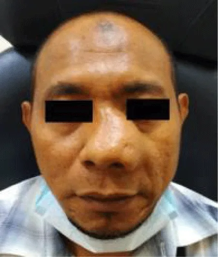
Upon examination, there is distinct broadening and enlargement of the left nasal pyramid. Anterior rhinoscopy revealed polypoid mass filling the entire nasal cavity with enlargement of the nose. External appearance showed nose enlargement with no distinctive border of the left nose. The tip of the nose is deviated to the right (Figure 1). Nasoendoscopy over the left nose, unable to performed due to enlarged mass occupying entire nasal cavity. Nasoendoscopy of the right nose revealed deviated nasal septum to the right causing the obstruction, the osteomeatal complex of right nasal cavity is clear from polyps and no discharge seen. Post nasal space showed polyp from left nasal cavity extends to the posterior choana and occupying the whole nasopharynnx. Oral cavity examination was unremarkable.
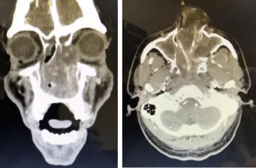
Contrasted-Enhanced Computed Topography(CT) scan of paranasal sinuses showed soft tissue opacity of the left and right nasal cavities, choana and nasopharyngeal space, bilateral maxillary sinuses, ethmoid sinuses, left sphenoid and left frontal sinus pushing nasal septum to the right (Figure 2). There were no bony erosion or calcification seen.
Nasal biopsy was taken in the outpatient clinic which revealed bening inflammatory polyp. We proceeded with functional endoscopic sinus surgery (FESS) later on. Intra-operatively noted that, left nasal mass originates from posterior edge of left osteomeatal complex, inferior to lamina papyreceae, extend posteriorly to nasopharynx, anteriorly to nares, superiorly to the base of skull. Left medial maxillary antrostomy was done.
Microscopy report came back as inflammatory benign nasal polyp. Patient was follow up in outpatient clinic. During his visit, patient did not complain of nasal blockage anymore and could breathe through the nose. No evidence of recurrence upon follow up for 1year. Patient was then planned for septorhinoplasty for reconstruction,however patient refused the operation.
Discussion
Woakes first described a form of necrotising ethmoiditis and mucous polyps to the medical society of the London in 1885. He also reported a ‘broadening of the bridge of the nose’ in these causes that he found to be occurring ‘sometimes’ [5]. The syndrome was later described by Appaix and Robert in 1924 as having following characteristics; bilateral nasal polyps in the middle meatus beginning during childhood;ethmoiditis;hypertropic process with nasal pyramid deformation;therapeutic failure followed by repeated and rapid recurrence [6]. Kellerhals and De Uthemann defined Woakes' syndrome in 1979 as the broadening of the nose, frontal sinus aplasia, bronchiectasis, and dyscrinia (production of highly viscous mucus) [7]. In recent years, Woakes syndrome has usually been characterized as severe recurrent nasal polyps with consecutive destruction of the nasal pyramid leading to the broadening of the nose due to chronic pressure of the polyps [3]. Nevertheless, there are also report that consider nasal polyps and facial deformities may be the only clinical features [8].
Most cases of Woakes syndrome seen to occur in children and young adults due to plasticity of the developing and growing bony facial structures. However there are few cases of adult onset have also been reported [3,8,9]. Although the etiology of nasal polyps remains unclear, they are associated with allergies, asthma, infection, cystic fibrosis, and aspirin sensitivity [8].
Our patient did not have any relevant past or family medical history, including allergies, chronic sinusitis, other than nasal polyps. . Although it is rare and our patient did only partially meet the criteria for Woakes syndrome, we diagnosed this case as Woakes syndrome as the deformities of the external nose and face is secondary to nasal polyps and associated with ethmoiditis based on CT scan.
Functional Endoscopic Sinus Surgery (FESS) is a gold standard in managing nasal polyps. The aim is to restore normal sinus ventilation and drainage, remove the polyps or other tissue obstructing the osteomeatal complex. As in this case Medial Maxillary Antrostomy (MMA) and excision of the mass was adequate to improve nasal passage and OMC ventilation. To address the nasal deformity, septorhinoplasty was performed to restore nasal function by maximizing the nasal airflow and to improve cosmetic appearance [10]. However a simple facial digital compression without osteotomies had also be done to improve cosmetic appearance [8].
Conclusion
Severe nasal polyposis can exert chronic pressure to cause deformities of the nose and face. Nasal polyps and facial deformities may be the only clinical features of Woakes syndrome. FESS can be done to manage the nasal polyp with or without addressing the facial deformity.
The authors would like to express gratidude to the patient who allow verbal consent for the pictures taken in making this case report visually explanatory.
- Chaaban MR, Walsh EM, Woodworth BA (2013) Epidemiology and differential diagnosis of nasal polyps. Am J Rhinol Allergy 27: 473–478. Link: http://bit.ly/2G8Vmfd
- Fokkens WJ, Lund VJ, Mullol J (2012) EPOS 2012: European Position Paper on Rhinosinusitis and Nasal Polyps 2012. A summary for otorhinolaryngologists. Rhinology 50:1–12. Link: http://bit.ly/2NBCPhG
- Caversaccio M, Baumann A, Helbling A (2007) Woakes’ Syndrome and Albinism. Auris Nasus Larynx 34: 245-248. Link: http://bit.ly/2NCahEP
- Loof MD, Leenheer ED, Holtappels G, Bachert C (2016) Cytokine Profile of Nasal and Middle Ear Polyps in A Patient with Woakes' syndrome and Eosinophilic Otitis Media. BMJ Case Rep. Link: http://bit.ly/2FW7iAK
- Woakes E (1885) Necrotising Ethmoiditis and Mucous Polyps. Lancet 61: 619.
- Appaix A, Robert J (1953) Deforming, Recurrent Nasal Polyposis of The Young; Woakes’ disease. Rev Laryngol Otol Rhinol (Bord) 74: 216-254. Link: http://bit.ly/2XJ43qv
- Kellerhals B, De Uthemann B (1979) Woakes’ Syndrome: The Problem of Infantile Nasal Polyps. International Journal of Pediatric Otorhinolaryngology 1: 79-85. Link: http://bit.ly/2S7EhqN
- Schoenenberger U, Tasman A (2015) Adult-Onset Woakes’ Syndrome: Report of A Rare Case. Case Rep Otolaryngology 2015. Link: http://bit.ly/2YDoNwS
- Ueda M, Hashikawa K, Iwayama T, Terash H (2017) Rhinoplasty via The Midface Degloving Approach for Nasal Deformity due to Nasal Polyps: A Case Report of Woakes’ Syndrome. Oral and Maxillofacial Surgery Cases 3: 64-69. Link: http://bit.ly/2XJJ3Qt
- Wardani RS, Mayangsari ID, Koento T, Amouzegar E (2014) Laporan Kasus Woakes syndrome. Oto Rhino Laryngologica Indonesiana 44: 76-82. Link: http://bit.ly/2XJJhHj
Article Alerts
Subscribe to our articles alerts and stay tuned.
 This work is licensed under a Creative Commons Attribution 4.0 International License.


 Save to Mendeley
