Archives of Otolaryngology and Rhinology
Positional changes in the hyoid bone, hypopharynx, cervical vertebrae, and Cranium before and after correction of glosso-larynx (CGL)
Susumu Mukai*
Cite this as
Mukai S (2018) Positional changes in the hyoid bone, hypopharynx, cervical vertebrae, and Cranium before and after correction of glosso-larynx (CGL). Arch Otolaryngol Rhinol 4(2): 037-043. DOI: 10.17352/2455-1759.000073Introduction: Respiratory inhibition by ankyloglossia with deviation of the epiglottis and larynx (ADEL) was thought to be caused by tongue muscles. But to explain this effect by the tongue muscles alone was unreasonable. Thorough investigations of ADEL by head and neck X-rays were performed. The surveillance revealed the existence of an important organ necessary in the neck to separate air and food as well as to regulate respiration.
Methods: Changes in the vertebrae, cranium, hyoid bone, and the width of the hypopharynx were compared by simple lateral X-radiographs before and after correction of the glosso-larynx (CGL) in 116 adults.
Results: The face was held straight, the hypopharynx was widened, and the hyoid bone moved about half the size of a vertebra down and forward.
Discussion: This study concluded that an organ in the neck that separates air and food as well as regulates respiration is necessary. The structure of the organ might consist of the styloid process, the hyoid bone, and a part of the trachea. It must perform up and down movements at any time. The structure was named “hyoid bone composition (HBC)”. The HBC originates from the 2nd gill, and belongs to the respiratory system. The first role of the HBC is protection of the respiratory system. Upper force to the HBC inhibits respiration and vice versa.
In humans there is a constant upper force to the HBC by the tongue muscles. This condition is thought to be ADEL. CGL is a surgery in which the genioglossus muscles are cut and the force to the HBC is released. As a result, respiration is increased and symptoms and signs of ADEL are ameliorated.
Conclusion: The HBC is necessary in the human neck. It moves down and forward after CGL. Respiration increases by movement of the HBC. The face is also held straight by changes in the center of gravity. (309 words).
Introduction
From A. Paré (1510-1590) through the Renaissance to recent years, an important procedure to promote newborn nursing was to cut the central ventral side of the tongue and separate its tip as far as the genioglossus muscles (GGms) from the floor of oral cavity. It was done whether a baby had a lingual frenum or not by midwives at the Baptismal. Suckling difficulties, vomiting during suckling, colicky cries, shallow sleep and/or awakening by minute slight sounds, cold extremities, swollen abdomen, and other problems encountered in newborn care disappeared just after the surgery. This condition has been called ankyloglossia. Ankyloglossia is accompanied by deviation of the epiglottis and larynx, so it has been renamed “ankyloglossia with deviation of the epiglottis and larynx (ADEL)”. ADEL inhibits respiration in babies as well as infants and adults. Symptoms and signs of ADEL have been observed at all ages [1-11].
A successful completion of the operation for ADEL is needed not only to separate the tip of the tongue as far as the GGms but it is also necessary to cut several bundles of GGms. This surgery has been designated as correction of the glosso-larynx (CGL). The epiglottis and larynx stand straight toward the choanal cavity after CGL. CGL not only increases respiration but also ameliorates various symptoms and signs of ADEL. In addition, it widens the hypopharynx and changes the position of the cranium [12-18].
That these signs and symptoms were ameliorated indicated that ADEL is a condition caused by inhibition of respiration. It was first thought that the tongue muscles caused inhibition of respiration, but the fact that several symptoms were ameliorated could not be explained by changes in the tongue muscles alone.
Both positional and angle changes in the hypopharynx, hyoid bone, vertebrae, and cranium were compared before and after CGL by X-ray (XP) images.
Both results from this study and clinical research on ADEL caused the author to deduce that an important organ is necessary in the neck to separate air and food as well as to regulate respiration. The organ must be composed of structures centered with the hyoid bone not the larynx alone. Provisionally it is named “hyoid bone composition (HBC)”. The characteristics of the HBC are discussed in this study. ADEL and ameliorations after CGL can be clarified by examination of the HBC.
Methods
Cases
Studied were 116 cases (68 females and 48 males) with age distribution from 14 to 74 years (average 40 y, SD, 13.9 y; females, 39 y; males, 41 y). They received CGL because of complaints related to symptoms and signs of ADEL. This study was done with the consent of the patients after a full explanation of the aim of the research was provided.
X-ray imaging (XP)
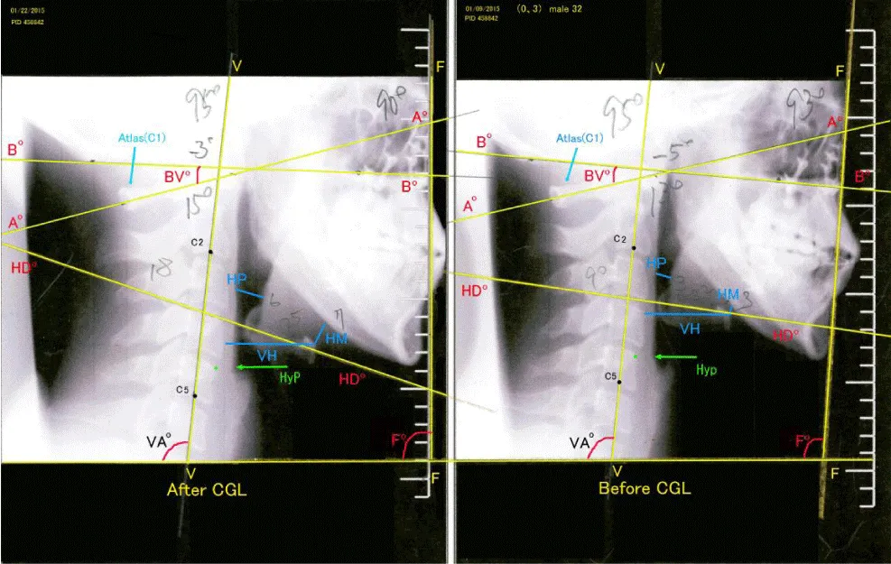
XP method: The study participants sat facing a mirror with their usual natural posture. A vertical line was drawn in the center of the mirror. After the head of the individual was positioned vertically, XP images were taken from right to left. XP images were taken twice, before and after CGL. These images were captured by a digital CCD imaging sensor NAOMI 6 (NAOMI RF SYSTEM lab.). The pre-CGL and post-CGL images were compared (Figure 1).
Observations
The following items A)〜C) were measured before and after CGL:
A) Hypopharyngeal space and distance from the hyoid bone to vertebrae (Figure 2)
1) Width of the narrowest hypopharyngeal space (HP)
2) Distance from the body of the hyoid bone to the lower margin of the jaw (HM).
3) Distance from the body of the hyoid bone to the ventral margin of the vertebrae (VH).
B) Angles of the cranium, vertebrae, hyoid bone, and face (Figures 3a,b)
4) Angle of the base of the cranium (Bº-Bº).
Angle of a line drawn from the lower end of the mastoid to the bottom of the maxillary sinus.
5) Angle of the posterior tubercle of atlas (Aº-Aº).
Angle of lines drawn from the center of the posterior arch of atlas to the center of the posterior tubercle (C1).
6) Angles (BVº) made by lines (Bº-Bº) and (Aº-Aº).
7) Incline of lines drawn from the hyoid bone (HDº-HDº).
8) Angles of the vertebrae (VAº).
Inclines of lines drawn from the center of the 2nd vertebra to the center of the 5th vertebra (VAº).
9) Inclines of the face (Fº): angles of lines from the uppermost orbital cavity to the outer and undermost piriform aperture.
C) Location of the cervical vertebrae at the same level as the hyoid bone and margin of the hypopharynx
10) Location of the horizontal line that crosses from the center of the hyoid bone to the vertebrae (VH).
11) Location of the hypopharyngeal space at the vertebrae (HyP; green)
Microsoft Excel v.14.7.1 and StatView 1-4-5 were employed for statistical analyses.
Results
Hyoid bone and hypopharynx (Figure 2)
1) Width of the narrowest hypopharyngeal space (HP)
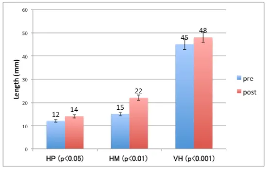
Average HP before and after CGL was 12 mm and 14 mm, respectively. There was a statistically significant difference observed between the two (P<0.05).
2) Distance from the center of the hyoid bone to the lower margin of the jaw (HM).
Average of the HM before and after CGL was 15 mm and 22 mm, respectively. A significant difference was observed (p<0.01).
3) Distance from center of the hyoid bone to the horizontal margin of the vertebrae (VH).
Average VH before and after CGL was 45 mm and 48 mm, respectively.
A significant difference was observed between the two (p<0.001).
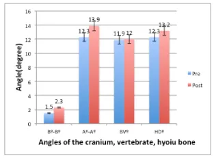
Angles of the cranium, vertebrae, hyoid bone, and face (Figure 3a).
4) Angle of the base of the cranium (Bº-Bº).
Average horizontal angle of the cranium (Bº-Bº) before and after CGL was -1.5º and -2.3º, respectively. There was no statistically significant difference between the two measurements.
5) Angle of the posterior tubercle of atlas (Aº-Aº).
Average (Aº-Aº) before and after CGL was 12.8º and 13.9º, respectively. Although there was a 1.1º increase after CGL, there was no statistically significant difference between the two measurements.
6) Angles (BVº) made by lines Bº-Bº and Aº-Aº.
Average BVº before and after CGL was 11.9º and 12.0º, respectively. There was no significant difference between the two.
7) Horizontal angle of lines drawn for the hyoid bone (HDº-HDº).
Gradient of the hyoid bone (HDº) before and after CGL was 12.2º and 13.2º, respectively. No statistically significant difference was observed between the two.
8) Inclines of vertebrae (VAº) (Figure 3b).
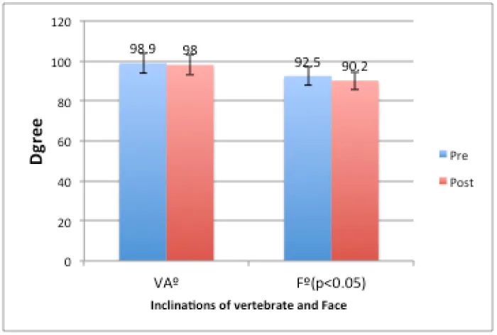
Horizontal angle of lines drawn from the center of the 2nd vertebra to the center of the 5th vertebra (VAº). Average incline of VAº before and after CGL was 98.9º and 98.0º, respectively. There was no significant difference between the two measurements.
9) Incline of the face (Fº) (Figure 3b).
Average Fº before and after CGL was 92.5º and 90.9º, respectively. A statistically significant difference was observed between the two (p<0.05).
Location of the vertebrae
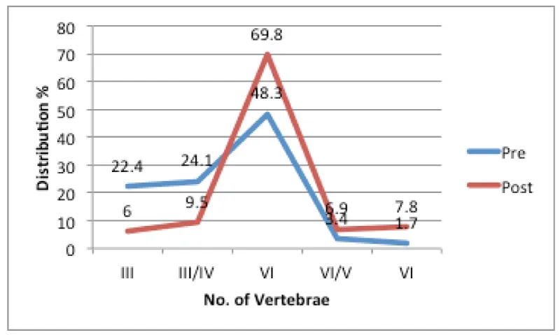
10) Distribution of the location of the horizontal lines that cross from the center of the hyoid bone to the vertebrae (VH). (Figure 4).
Distribution of the VH before CGL was the 3rd vertebra in 26 cases (22.4%), between the 3rd and 4th vertebrae in 28 cases (24.1%), 4th vertebra in 56 cases (48.3%), between the 4th and 5th vertebrae in 4 cases (3.4%), and the 5th vertebra in 2 cases (1.7%).
Distribution of the VH after CGL was the 3rd vertebra in 7 cases (6.0%), between the 3rd and 4th vertebrae in 11 cases (9.5%), the 4th vertebra in 81 cases (69.8%), between the 4th and 5th vertebrae in 8 cases (6.9%), and the 5th vertebra in 9 cases (7.8%).
Average distribution of the location of VH before and after CGL was the upper part of the 4th vertebra and the lower part of the 4th vertebra, respectively.
There was a statistically significant difference in the average distribution of the locations before and after CGL (χ2p<0.05).
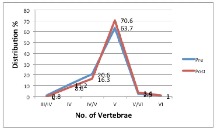
11) Height of the hypopharynx at various vertebrae (HyP: green) (Figure 5).
Average height distribution of the HyP before CGL was between the 3rd and 4th vertebrae in 1 case (0.9%), 4th vertebra in 13 cases (11.2%), between the 4th and 5th vertebrae in 24 cases (20.7%), the 5th vertebra in 74 cases (63.8%), between the 5th and 6th vertebrae in 3 cases (2.6%), and the 6th vertebra in 1 case (0.9%).
Average height distribution of the HyP after CGL was between the 3rd and 4th vertebrae in 0 cases (0.0%), 4th vertebra in 10 cases (8.6%), between the 4th and 5th vertebrae in 19 cases (16.4%), the 5th vertebra in 82 cases (70.7%), between the 5th and 6th vertebrae in 4 cases (3.4%), and the 6th vertebra in 1 case (0.9%).
There was no statistically significant difference in distribution before and after CGL.
Discussion
Hypopharyngeal space and distance from the hyoid bone to vertebrae
1) Width of the narrowest hypopharyngeal space (HP)
2) Distance from the body of the hyoid bone to the lower margin of the jaw (HM).
3) Distance from the body of the hyoid bone to the ventral margin of the vertebrae (VH).
All of the above three values were increased after CGL. It was confirmed in this study that with widening of the hyoid space, the hyoid bone moves down and forward, as has already been reported [12-14].
Angles for the cranium, vertebrae, hyoid bone, and face
4) Angle of the base of the cranium (Bº-Bº).
5) Angle of the posterior tubercle of atlas (Aº-Aº).
6) Angles (BVº) that were made by the line for (Bº-Bº) and (Aº-Aº).
Although there were no statistical differences in the three measured angles before and after CGL, the average BVº before and after CGL was 11.9º and 12.0º, respectively.
It was thought that the angles between the cranium and vertebrae might have a constant value.
7) Inclines of vertebrae (VAº).
There was no statistical difference before and after CGL in the horizontal angle of lines drawn from the center of the 2nd vertebra to the center of the 5th vertebra (VAº).
8) Horizontal angle of lines drawn for the hyoid bone (HDº-HDº).
There was no significant difference observed between angles examined before and after CGL, suggesting that the angle was nearly the same before and after CGL.
9) Incline of the face (Fº).
There was a statistical difference in the vertical angle of faces of study participants. Before and after CGL, the faces leaned slightly downward (92.5º) while after the surgery the faces were held almost straight (90.9º). Many cases said that their first impressions after surgery were that they could see objects clearer than before surgery [19-20]. These impressions indicate that study participants could observe objects straight ahead with both eyes. This difference suggests that the center of gravity of the cranium was moved by the CGL.
Location of vertebrae
10) The most prominent finding in this study was a change in the location of the hyoid bone in relation to the vertebrae after CGL. The hyoid bone descended at a rate of approximately half of one vertebra, reaching the middle of 4th vertebrate and had moved 7 mm forward.
It was already reported that an important role of the cranium is cooling of the brain center through the nasal cavities [21,22,23]. Besides cooling of the brain, the facial portion of the cranium is specialized for taking in air and food by the nose and mouth, respectively, which then pass through the neck. The air and food are strictly and accurately sorted so that the former goes to the airway and the latter to the esophagus.
A structure might be built in the neck for this purpose, in that by controlling the respiratory function the airway and alimentary tract are kept separate. The hyoid bone might play a principal rôle in this structure. It is connected to both stylohyoid ligaments by the lessor horns on the upper body of the hyoid bone. The stylohyoid ligaments unite both styloid processes at the base of the cranium that developed from the second branchial arch.
The digastric muscles (ms), mylohyoid ms, and stylohyoid ms are attached to the upper part of the hyoid bone. A relatively wide ligamental membrane is attached to the upper half of the hyoid bone and the GGms are attached to the membrane. At the center of the ligamental membrane is a vertical ligamental membrane. The GGms are spread like a fan, which forms the shape of the tongue. The center of the tongue is separated right and left by the vertical ligamental membrane (lingual septum). There are no connections to vessels and nerves on both sides of the tongue by the lingual septum [24].
Under the surface of the hyoid bone the thyrohyoid ligaments hang down like drapes and are connected with thyroid cartilage. Thyroid muscle groups are attached to the cartilage. A cricoid cartilage is connected under the thyroid cartilage. The epiglottis and larynx exist within the thyro-crico cartilage. The trachea is connected with the cricoid cartilage. The HBC starts from both styloid processes which are connected to the hyoid bone by the stylohyoid ligaments at both (right and left) little horns of the hyoid bone. It then ends at the thyro-crico cartilage. The direction of the forces on the hyoid bone are clearly divided into two, upward and downward. As a result the HBC has mobility. It can be said that the HBC has a specific role as an organ in the neck.
When eating begins, food is taken into the mouth and mastication is begun. At this time the muscles of the mouth, tongue, and pharynx begin contractions. The HBC moves upwards. At the same time, respiration begins to be inhibited. When swallowing starts, respiration stops and the HBC shrinks and moves to the uppermost position. The vocal folds shut and the epiglottis covers the larynx. The respiratory tract probably shrinks, too. A mass of food divide into two portions pass through the hypopharynx and it enter into the esophagus. Inspiration and expiration are completely inhibited at this moment. While the HBC receives an upward force, respiration is inhibited.
The matter that is swallowed passes into the esophageal ostium. No sooner has the swallowed matter passed the esophageal ostium, the HBC goes downward and its length recovers. At the same time the posterior cricoid muscles begin to activate. At the time of rebreathing after swallowing, the airway is thought to be wider than at the start of mastication. Widening of the ala nasi is observable synchronously with post cricoid muscle activities [25-29].
During the opposite of swallowing, vomiting starts from the abdominal reflexes [30]. The esophagus opens and the contents of the alimentary tract reflux into the mouth. The HBC ascends at the highest point during this occasion and the tongue is almost pushed out from the mouth and drooling occurs. Both inspiration and expiration are completely impossible in this situation as well. The HBC rises upward and shuts down the respiratory tract during abdominal straining at evacuation and parturition. These closings of the respiratory tract by the HBC are to protect the lower respiratory tract as well as protect from collapse of the intra-thoracic organs.
During respiration the muscles both inside the HBC and the larynx are activated. When a person sniffs, both ala nasi and vocal folds open and narrow with the sniffing rhythms. Tone controls in wind instruments depend upon the opening of the vocal folds. Beautiful tones and vibrato are played by narrowed vocal folds with minute movements of laryngeal muscles. However, these laryngeal spontaneous muscle movements are not perceptible by the players themselves. These conditions suggest that humans lack a feedback circuit from the muscles of the respiratory tract in the HBC. It is probable that the HBC lacks perception of movements of its own muscles [31-37].
CGL is a surgery in which several bundles of GGms are cut. Increases in respiratory rates, VC, and FEV1.0% were observed after this procedure. In this study, movements of the hyoid bone were shown to be down and forward. This observation at the same time revealed that the HBC moved the same way. The locational change in the vertebra in relation to the hyoid bone was about half of one vertebra both forward and down.
It was first reported that the increase in respiration after CGL was caused by inhibition by the tongue muscles. But these changes are not explained by the tongue muscles alone. Hypopharyngeal spaces and the upper trachea were expanded after CGL. Widening of airways was shown. When the HBC moves down, respiration accelerates and the airway widens.
It is natural that the expansion of the respiratory tract, the increase in FEV1.0%, and the clearer sounds of voices observed after CGL are caused by the downward shift of the HBC, which widens the airway. These conditions facilitated the respiratory function [12-14,18].
On the contrary, it can be said that a higher position of the HBC inhibits respiration. Also, it might be thought that excess attachments of the tongue muscle to the hyoid bone resulted in pulling up the HBC. The higher position of the HBC inhibits respiration. ADEL might be caused by a higher position of the HBC. It can be said that ADEL is caused by the excess attachment of the tongue muscles to the hyoid bone, especially those of the GGms. This condition pulls up the HBC and, as a result, respiration is depressed.
There were no statistical differences between before and after CGL in the location of the hypopharynx in relation to vertebrae. This means there was no positional change in the esophageal ostium. The ADEL does not involve the esophagus.
Conclusion
1. In the human neck there must be an organ that precisely discriminates movement of air and foods from the nose and mouth as well as controls respiration.
2. The hyoid bone plays a principal rôle in this construction. It is connected to both stylohyoid ligaments. The stylohyoid ligaments connect to the lessor horns on the upper body of the hyoid bone. The stylohyoid ligaments unite both stylohyoid processes at the base of the cranium that developed from the second branchial arch. It was named the hyoid bone composition (HBC)
3. The HBC originates from both styloid processes and it ends at the thyro-crico cartilage.
4. The HBC belongs to the respiratory system and its most important role is to protect the respiratory systems.
5. HBC must perform up and down movements at anytime. The HBC depresses respiration when the forces pull it up. It accelerates respirations with downward forces.
6. As the HBC in humans is pulled up by the excess attachment of the tongue muscles to the hyoid bone, respiration is inhibited.
7. It is probable that the HBC lacks perception of movements of its own muscles.
8. The multiple symptoms and signs of ADEL do not originate from the tongue itself.
9. The CGL is a surgery that releases the upward forces on the HBC.
10. The face was held straight by the change in the center of gravity of the cranium as the result of CGL.
Conclusion
Office rhinology is evolving and with increased surgeon comfort in performing office rhinology procedures, the gold standard MSA procedure (with the described potential advantages compared to the maxillary BSD procedure) could have a useful role to play if added to the surgeons “office rhinology” armamentarium in selected patients. There may be times based on the needs and goals of the surgeon that the MSA would be a preferred choice over the maxillary BSD, and the surgeon can have confidence the procedure could easily be performed in the office setting versus the operating room with very low patient morbidity and rapid recovery.
- Paré A. De l'empeschement et retraction de la langve In: Malgaigne JF, ed. Amboise Paré; Oevres complétes; Réimpression de l'édition de Paris (1840-1841). Genéve: Slatkine Reprinte. 1970: 455-456.
- Mukai M (2010) a father of a modern medicine and a doctor of a barber. The Japan French Medical Association bulletin 75: 5.
- Baudelocque JL. A system of midwifery I, II, III: translated from the French, by J Heath. London: ECCO, 1781.
- Baudelocque JL. Principes sur l'art des accouchements, par demandes et réponses, en faveur des élèves sages-femmes de la campagne (1775), sepième édution. Paris: Germer-Baillière, 1839.
- Loux F. Le jeune enfant et son corps dans la médecine traditionnelle. Paris: Flammarion. 1978: 126-129.
- Mukai S, Mukai C, Asaoka K, Nagasugi S, Ogiyam M. Postoperative Changes in Infant with ADEL (in Japanese with English abstract). Zetuyuchalushou Kenkyuukai Kaiho (Annals ADEL) 1991; 1: 35-45.
- Mukai S, Mukai C, Asaoka K (1991) Ankyloglossia with deviation of the epiglottis and larynx. The Annals of otology, rhinology & laryngology Supplement. 100: 3-20. Link: https://goo.gl/LVSjx8
- Mukai S. History of the ankyloglossia. Zetuyuchakusho Kenkyuukai Kaiho (Annals ADEL) (In Japanese) 1991; 1:1-5.
- Mukai S, Mukai C, Asaoka K (1993) Congenital ankyloglossia with deviation of the epiglottis and larynx: symptoms and respiratory function in adult. Ann Otol Rhinol Laryngol. 102: 620-624. Link: https://goo.gl/juYnyW
- Mukai S (2016) Changes of Breastfeeding and of Breast Before and after Surgeries on the Ankyloglossia with Deviation of the Epiglottis and Larynx (ADEL). Health. 8: 868-886. Link: https://goo.gl/CgDF1Z
- Mukai S, Sato M, Taguchi K, Fukuchi Y (2016) Changes of Breastfeeding and of Breast Before and after Surgeries on the Ankyloglossia with Deviation of the Epiglottis and Larynx (ADEL). J Nursing Healthcare. 1-8. Link: https://goo.gl/DTUufL
- Mukai S, Mukai C, Nagasugi S (1995) Changes in respiratory rate following correction of ADEL (Japanese with English abstract). Zetuyuchakushou Kenkyuukai Kaiho (Annals ADEL). 5: 1-9.
- Mukai S, Mukai C, Nagasugi S (1996) Digit sucking starts with respiratory insufficiency (in Japanese with English abstract). Zetuyuchakucho Kenkyukai Kaiho (Annals ADEL). 1: 76-80.
- Mukai S (1998) Ankyloglossia with Deviation of the Epiglottis and Larynx (ADEL) (In Japanese). Zetuyuchaksho Kenkyuukai Kaiho. 8: 1-53.
- Nitta M, Mukai S, Mukai C (2000) Locational changes between hyoid bone and cranium after correction of glosso-larynx (CGL) (Japanese with English abstract). Pract Otol (Kyoto). 93: 43-47.
- Nitta M, Mukai S, Mukai C (2000) The expansion of the hypopharynx by correction of glosso-larynx. Psychiatry and Clinical Neurosciences. 54: 344-345. Link: https://goo.gl/paoBax
- Mukai S, Nitta M (2002) Correction of the glosso-larynx (CGL) and resultant positional changes of the hyoid bone and cranium. Act Otolaryngol (Sweden). 122: 644-650. Link: https://goo.gl/qsf2PH
- Yamamoto I, Mukai S, Nakagawa K, H. O, Yamada Y (2011) Analysis of hypopharyngeal airway space after correction of glosso-Larynx (CGL) by both cephalometry and compute tomography. Nihon Zetuychakusho Gakkai Kaishi (Annals ADEL). 17: 13-18.
- Mukai S (2015) Human nose might be involved in cooling of the brain. International J Clinical Medicine. 6: 482-486. Link: https://goo.gl/9WH6Ai
- Mukai S (1992) Reports after sugeries of ADEL; subjects reports of feelings after correction of glosso larynx (CGL) (In Japanese). Zetuyuchaksho Kenkyuukai Kaiho. 2: 1-91.
- Mukai S (1994) Changes after correction of the glosso larynx (CGL) that seen in the reports after the CGL. Zetuyuchakushou Gakkai Kaishi (Annl ADEL). 4: 1-154.
- Mukai S (2017) Amelioration of sleep by expansion of the nasal cavity: Roles of facial skull and nasal catiry (In Japanese). Zetuyuchakushou Kenkyuukai Kaiho (Annals ADEL). 21: 25-33.
- Mukai S (2017) Amelioration of sleep by expansion of the nasal cavity: A role of the nose. Annals of Clinical Otolaryngology. 18: 1-6.
- Rouvière H (1924) Os hyoîde, in Anatomie humaine. tête et cou, tome 1 Anatomie humaine. Paris: Masson. 114-117, 473-477.
- Yoshida T (1979) Electricmyographic and X-ray investigations of normal deglutition (Japanese with English abstract). Ji-Bi. 25: 834-872.
- Shin T (1994) Relationship between swallowing and respration (In Japanese). Jibi to Rhinsho. supple 1: 332-363.
- Hayashi I, Hayashi Y, Uno I, Fujiwara Y, Takahashi H (1997) Correlation between the submental surface electromyogram and the thyrohyoid or the geniohyoid electromyograms (In Japanese with English abstract). Jibi to Rhinsho. 43: 666-672.
- Umezaki T (2007) Nervous system of swallowing (In Japanese). Kouji Nohkino Kenkyu. 27: 215-221.
- Satoda T (2013) Understanding of mechanisms of eating, swallowing and aspiration by CG and mechanical model (In Japanese) Tokyo: Ishiyaku Shuppan Ltd.
- Ganong W (2016) Nonchemical influences on respiration; Respiratory components of visceral reflexes In: Ganong W, ed. Review of medical physiology, 25th ed. New York, Chicago: Lange Medical Book. 662-664.
- Mukai S (1988) Vocal cord movements during sniff (Jananese with English abstract). Nihon Jibiinkoka Gakkai kaiho 91: 2089-2095. Link: https://goo.gl/Y5ZypV
- Mukai S, Minegishi S (1987) Vibrato were played by the larynx. Flatus 4: 7-24.
- Mukai S (1989) Vocal cord adduction while sniffing. Arch Otolaryngol Head Neck Surg. 115: 1362-1366. Link: https://goo.gl/jUf9kq
- Mukai S, Mukai C, Suzuki I (1989) Dysosmia with vocal cord paralysis during sniff, abstract 740 14th World Congres of Otorhinolaryngology Head and Neck Surgery. Madrid, Spain. 214.
- Mukai S (1989) Four cases of dysosmia with no vocal cords adduction during sniff (laryngeal dysosmia). (Japanese with English abstract). Nihon Jibiinkoka Gakkai kaiho. 92: 1211-1219.
- Mukai S (1989) Laryngeal movements during wind instruments play (Japanese with English abstract). Nihon Jibiinkoka Gakkai kaiho. 92: 260-270. Link: https://goo.gl/uhhVfp
- Mukai S (1990) Relationship between playing wind instruments and movements of vocal folds; objective approach to sounds (In Japanese). Igaku no Ayumi. 125.

Article Alerts
Subscribe to our articles alerts and stay tuned.
 This work is licensed under a Creative Commons Attribution 4.0 International License.
 Save to Mendeley
